Journal List > Korean J Gastroenterol > v.78(3) > 1147560

Acute Abdominal Pain due to Accessory Splenic Infarction in an Adult: A Case Report

Abstract

Accessory spleens are common congenital anatomic variations that are usually asymptomatic. On the other hand, they can be clinically significant if complicated by hemorrhage, torsion, or infarction. This paper describes a case of an infarcted accessory spleen in a 30-year-old male who presented with abdominal pain. Abdominal CT and MRI revealed an isolated mass, 4.5 cm in size, in the perisplenic area. An infarcted accessory spleen was suspected. The patient underwent laparoscopic accessory splenectomy. Histopathology identified the mass as splenic tissue that had undergone ischemic necrosis. A definitive diagnosis of an infarcted accessory spleen was made, and the patient was discharged on day 5 after surgery symptom-free.

INTRODUCTION

The spleen develops from the primitive mesoderm during the 5th week of embryologic life, wherein the splenic primordium grows out from the dorsal mesogastrium and rotates from the midline to the left side of the abdomen while acquiring its vascular and lymphoid tissues.1 Various congenital malformations can occur during these developmental stages, such as asplenia, hyposplenia, polysplenia, lobulated spleen, splenogonadal, or splenopancreatic fusion, and accessory spleens.2 Accessory spleens are the most commonly encountered forms of developmental variations of the spleen. They develop when multifocal origins of primitive mesenchymal tissues fail to fuse within the dorsal mesogastrium.3 The incidence of accessory spleens at autopsy was 10-30% in the American population and 4.5-24.3% in the Asian population.4 Accessory spleens are identical to normal spleens with respect to their gross morphology, histology, and function. Typically, their size usually does not exceed 1-2 cm, and they are mostly single but can be found in multiples of five or more.1 Approximately 75% of these occur at the splenic hilum, but they can also be located at the pancreatic border, the splenogastric omentum, greater omentum, pelvis, or anywhere in the abdomen.5 The attenuation of accessory spleens in CT images is approximately 40 to 60 Hounsfield Units (HU), which is 10 to 20 HU lower than the density of the liver in a pre-enhancement scan. They show heterogenic enhancement in early phase images because of the variation in blood flow velocity between the red and the white pulp.6 Although accessory spleens are predominantly asymptomatic, they can provoke symptoms, such as abdominal pain, or cause an acute abdomen when complicated by torsion, hemorrhage, infarction, or rupture. This paper describes the case of a young man with abdominal pain caused by an infarcted extrapancreatic accessory spleen.

CASE REPORT

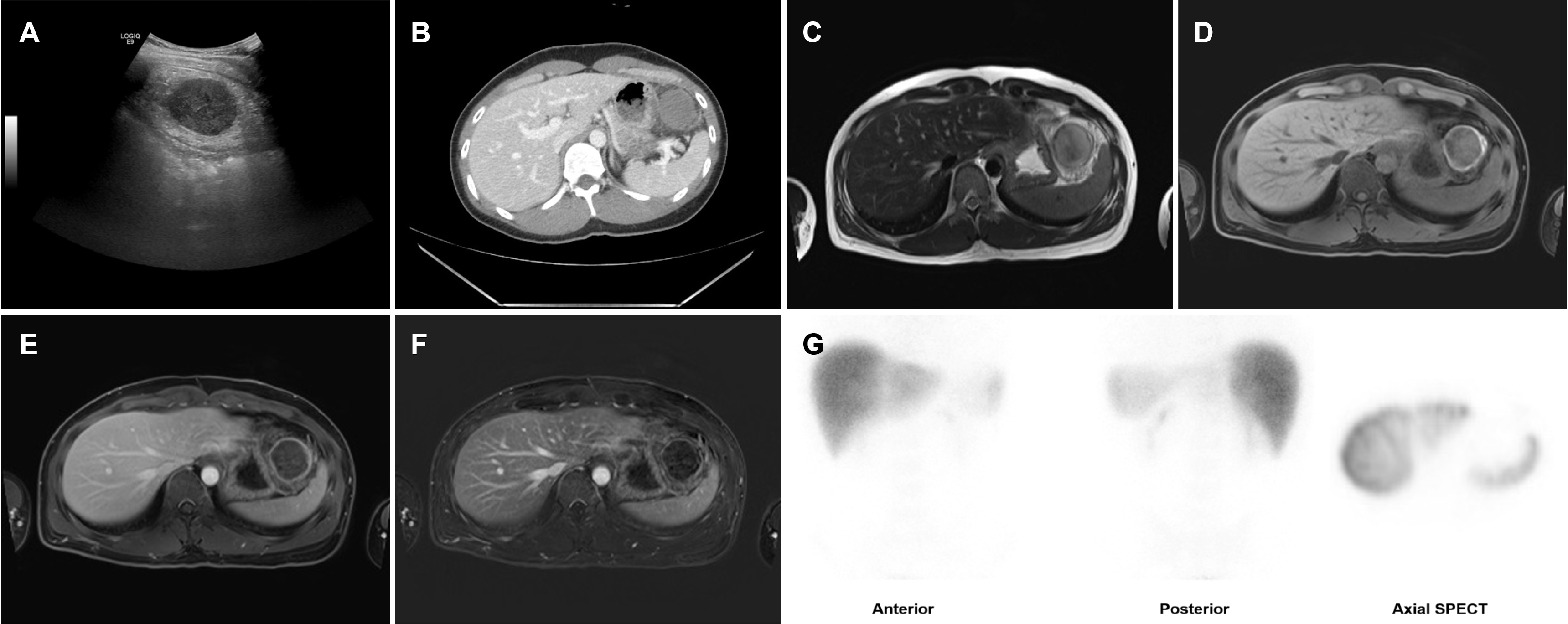
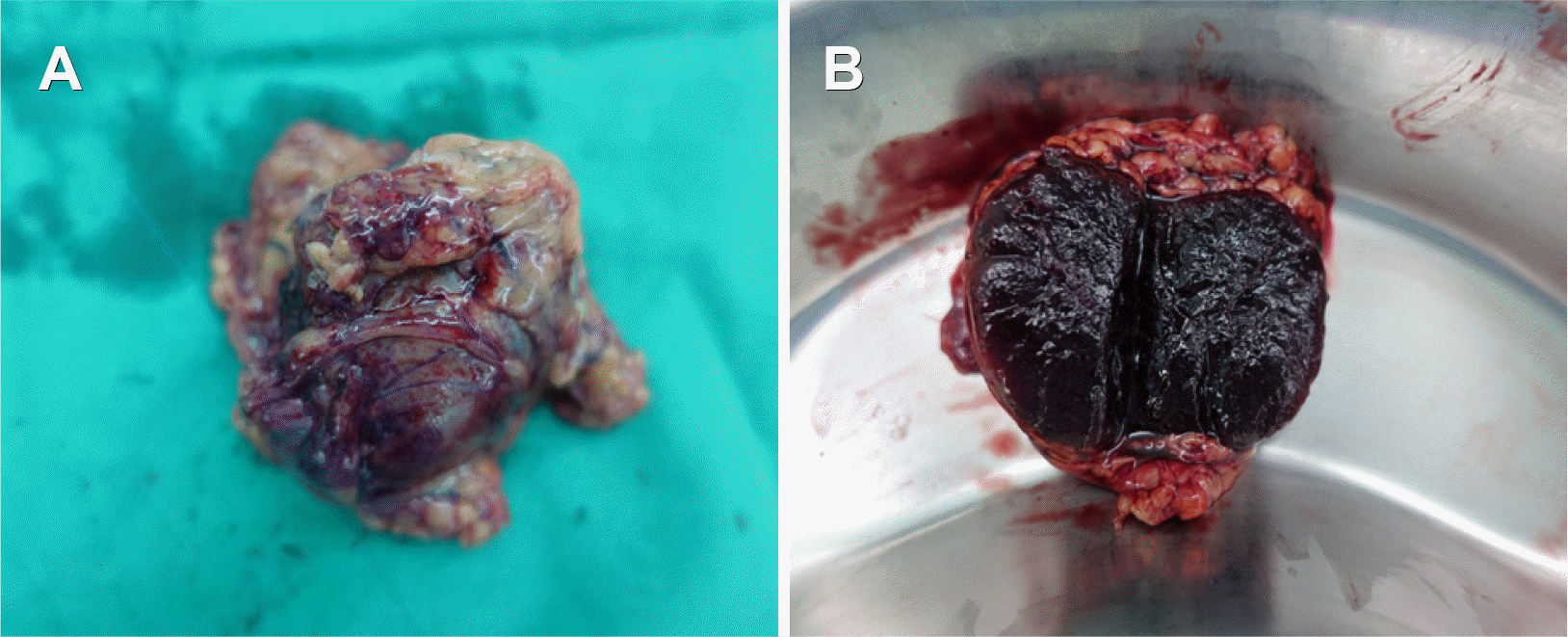
A 30-year-old male was referred to the Ulsan University Hospital with pain in the left upper quadrant that had begun 7 days earlier. The pain was dull and constant and had no apparent precipitating or alleviating factors. His abdomen was soft and flat, and a physical examination at presentation did not reveal tenderness. He had no other medical or family history, including atrial fibrillation or coagulopathy. Upon admission, his vital signs were as follows: blood pressure, 130/72 mmHg; heart rate, 86 beats per min; respiratory rate, 20 times per min; body temperature, 36.4℃. The initial laboratory tests were as follows: white blood cell count 4,880/mm3 (normal range 4,000-10,000), hemoglobin 13.6 g/dL (14.0-18.0), platelet count 324,000/mm3 (140,000-400,000), serum AST 25 IU/L (15-40), ALT 26 IU/L (0-41), amylase 75 IU/L (20-104), lipase 38 IU/L (5.6-51.3), and CRP 4.53 mg/dL (0.0-0.5). Abdominal ultrasound (US) and CT revealed a mass, 4.5 cm in size without enhancement, in the perisplenic area of the left upper abdomen (Fig. 1A, B). The mass was a solitary lesion isolated from adjacent organs, such as the colon, spleen, and pancreas. Abdominal dynamic MRI using a gadolinium-based contrast identified a solid necrotic tumor with subtle peripheral rim enhancement (Fig. 1C-F). Technetium-99m (Tc-99m) phytate scintigraphy and additional single-photon emission CT (SPECT) did not show any significant uptake within the mass (Fig. 1G). The pain subsided during the diagnostic workup, but a decision to remove the mass surgically was made based on the patient’s age and after a discussion with the patient. An intraoperative laparoscopic exam revealed a well-capsulated, 6 cm-sized, round mass between the proximal stomach and spleen. The tissue was purple, implying necrosis of the mass, but there was no evidence of twisted vasculature or vascular torsion (Fig. 2). The laparoscopic excision was successful, and the procedure lasted 85 min with 20 mL blood loss. The patient was discharged from the hospital 5 days after surgery without complications. The histology evaluation of the excised mass showed that the entire accessory splenic parenchyma had undergone diffuse necrotic changes with hilar vascular congestion (Fig. 3A).
Interestingly, the white pulp was spared compared to the red pulp (Fig. 3B). The proximal splenic vessel showed a near-complete luminal occlusion because of an organized thrombus (Fig. 3C, D), which was suspected as being the cause of the current splenic injury. It was unclear if the vessel was an artery or vein because the layers of the vessel were destroyed after the necrotic changes from the infarction. No other histologic abnormalities were observed in the resected specimen. Thus, the patient was diagnosed with an infarcted extrapancreatic accessory spleen. He recovered uneventfully and was discharged on postoperative day 5. A follow-up was conducted for 1 year with a favorable outcome.

DISCUSSION

While one report described an accessory spleen complicated by a hemorrhagic infarct in a 5-year-old boy in Korea,7 to the best of the authors’ knowledge, this is the first report describing an infarcted extrapancreatic accessory spleen in a Korean adult. Several case reports (n=8) on accessory spleens complicated by torsion or infarction have been found, which are summarized in Table 1.7-14 A diagnosis of an accessory spleen is usually made in childhood (median age 12 years, range 5-75 years) and six patients were younger than 10 years of age. This may be because accessory spleens tend to atrophy as children grow.9 The patients usually present with abdominal pain and sometimes with fever or vomiting. Intermittent torsion and detorsion could be responsible for recurrent short episodes of abdominal pain during growth and can be mistaken for dyspepsia or gastroduodenitis. The accessory splenic masses are located predominantly at the splenic hilum but can occur anywhere in the abdominal cavity. A diagnosis is usually made based on the symptoms and radiologic findings.
Total necrosis of the accessory spleen on CT and MR images is usually seen as subtle peripheral/capsular enhancement, no enhancement at all, or a twisted vascular pedicle connecting the accessory spleen to the splenic artery.15,16 On the other hand, the vascular connection or torsion was not definite in the present patient. The differential diagnoses in such cases include mesenteric cyst, enteric duplication cyst, or lymphatic cyst.11 MRI can evaluate the components of the tumor by scintigraphy using Tc-99m-labelled phytate, or heat-damaged red blood cells can be used to detect splenic tissue and is helpful in the diagnosis of a functional accessory spleen.17 Nevertheless, minimally functioning splenic tissue can result in false-negative findings, and small lesions may be missed due to the low image resolution.18 In the present patient, the mass did not show significant radiotracer uptake, probably because it was predominantly necrotic. Although the infarction of an accessory spleen in an adult is unusual, a pre-operative diagnosis could have been made successfully in this case based on symptoms and radiologic findings.
The surgical indication for accessory spleen infarction is when the patient suffers from abdominal pain or complications, such as peritonitis or intestinal obstruction. Careful surveillance may be pursued if the lesion is small and if symptoms do not persist.
In addition to an infarction, an accessory spleen can also be misdiagnosed as pathologic lymphadenopathy or neoplasm. This is particularly true when accessory spleens are located in the pancreatic parenchyma and are mistaken as intrapancreatic mass lesions whose diagnosis and treatment entail invasive and hazardous procedures, such as pancreaticoduodenectomy or distal pancreatectomy, even in the absence of related symptoms. This paper summarizes reported cases of pancreatectomy for intrapancreatic accessory spleens in the Korean population (Table 2) that had been performed to resolve symptoms or to make a diagnosis when a malignancy was suspected.18-22
Currently, EUS-guided fine needle aspiration and fine needle biopsy (FNB) of peripancreatic or intrapancreatic lesions are considered safe and are used for diagnosis23 despite the fear of puncturing the spleen, which is a hypervascular organ.24,25 For example, one report described the safe use of a percutaneous biopsy for accessory splenic necrosis before surgery that resulted in a correct diagnosis.14 An EUS-FNB was not performed in the present case because the patient refused to consent. On the other hand, it would have been helpful to make a diagnosis that may have led to conservative management. Furthermore, major surgery, such as pancreaticoduodenectomy, could also be avoided by EUS-FNB for intrapancreatic accessory spleens. Therefore, EUS-FNB should be considered if it is difficult to differentiate an accessory spleen from malignancies regardless of the imaging modality.
In conclusion, accessory splenic infarction should be considered in patients presenting with left upper quadrant pain and an intra-abdominal mass, even in adults.

Notes

Financial support

None.

Conflict of interest

None.

REFERENCES

1. Morgenstern L, Skandalakis JE. Hiatt JR, Phillips EH, Morgenstern L, editors. 1997. Anatomy and embryology of the spleen. Surgical diseases of the spleen. Springer;Berlin, Heidelberg: p. 15–24. DOI: 10.1007/978-3-642-60574-1_2. PMID: 8996481.
crossref
2. Yildiz AE, Ariyurek MO, Karcaaltincaba M. 2013; Splenic anomalies of shape, size, and location: pictorial essay. ScientificWorldJournal. 2013:321810. DOI: 10.1155/2013/321810. PMID: 23710135. PMCID: PMC3654276.
crossref
3. Varga I, Babala J, Kachlik D. 2018; Anatomic variations of the spleen: current state of terminology, classification, and embryological background. Surg Radiol Anat. 40:21–29. DOI: 10.1007/s00276-017-1893-0. PMID: 28631052.
crossref
4. Mohammadi S, Hedjazi A, Sajjadian M, Ghrobi N, Moghadam MD, Mohammadi M. 2016; Accessory spleen in the splenic hilum: a cadaveric study with clinical significance. Med Arch. 70:389–391. DOI: 10.5455/medarh.2016.70.389-391. PMID: 27994303. PMCID: PMC5136441.
crossref
5. Varga I, Galfiova P, Adamkov M, et al. 2009; Congenital anomalies of the spleen from an embryological point of view. Med Sci Monit. 15:RA269–RA276.
6. Rashid SA. 2014; Accessory spleen: prevalence and multidetector CT appearance. Malays J Med Sci. 21:18–23.
7. Ko HS, Goo HW, Yoon CH. 2004; Unusual infarction of the accessory spleen or polysplenia in two children: case report. J Korean Radiol Soc. 51:555–558. DOI: 10.3348/jkrs.2004.51.5.555.
crossref
8. Babcock TL, Coker DD, Haynes JL, Conklin HB. 1974; Infarction of an accessory spleen causing an acute abdomen. Am J Surg. 127:336–337. DOI: 10.1016/0002-9610(74)90044-0.
crossref
9. Onuigbo WI, Ojukwu JO, Eze WC. 1978; Infarction of accessory spleen. J Pediatr Surg. 13:129–130. DOI: 10.1016/S0022-3468(78)80004-9.
crossref
10. Hems TE, Bellringer JF. 1990; Torsion of an accessory spleen in an elderly patient. Postgrad Med J. 66:838–839. DOI: 10.1136/pgmj.66.780.838. PMID: 2099424. PMCID: PMC2429706.
crossref
11. Sheth H, Chaudhari S, Sinha Y, Prajapati R. 2018; Infarcted accessory spleen masquerading as a mesenteric cyst. BMJ Case Rep. 2018:bcr2018226130. DOI: 10.1136/bcr-2018-226130. PMID: 30115724. PMCID: PMC6101306.
crossref
12. Yousef Y, Cameron BH, Maizlin ZV, Boutross-Tadross O. 2010; Laparoscopic excision of infarcted accessory spleen. J Laparoendosc Adv Surg Tech A. 20:301–303. DOI: 10.1089/lap.2009.0286. PMID: 20059391.
crossref
13. Ishibashi H, Oshio T, Sogami T, Nii A, Mori H, Shimada M. 2012; Torsion of an accessory spleen with situs inversus in a child. J Med Invest. 59:220–223. DOI: 10.2152/jmi.59.220. PMID: 22450011.
crossref
14. Simon DA, Fleishman NR, Choi P, Fraser JD, Fischer RT. 2020; Torsion of an accessory spleen in a child with biliary atresia splenic malformation syndrome. Front Pediatr. 8:220. DOI: 10.3389/fped.2020.00220. PMID: 32432066. PMCID: PMC7212802.
crossref
15. Ozeki M, Asakuma M, Go N, et al. 2015; Torsion of an accessory spleen: a rare case preoperatively diagnosed and cured by single-port surgery. Surg Case Rep. 1:100. DOI: 10.1186/s40792-015-0101-x. PMID: 26943424. PMCID: PMC4596154.
crossref
16. Sadro CT, Lehnert BE. 2015; Torsion of an accessory spleen: case report and review of the literature. Radiol Case Rep. 8:802. DOI: 10.2484/rcr.v8i1.802. PMID: 27330618. PMCID: PMC4900206.
crossref
17. Nishiguchi S, Habu D, Ishizu H, et al. 2001; Accessory spleen in the pelvis diagnosed by Tc-99m phytate scintigraphy. Ann Nucl Med. 15:263–265. DOI: 10.1007/BF02987843. PMID: 11545199.
crossref
18. Kim SH, Lee JM, Han JK, et al. 2008; Intrapancreatic accessory spleen: findings on MR Imaging, CT, US and scintigraphy, and the pathologic analysis. Korean J Radiol. 9:162–174. DOI: 10.3348/kjr.2008.9.2.162. PMID: 18385564. PMCID: PMC2627219.
crossref
19. Chung YH, Park HJ, Park IS, et al. 2009; An epidermoid cyst in an intrapancreatic accessory spleen. Korean J Med. 76:742–745.
20. Im CJ, Kweon JH, Hwang ET, et al. 2009; An intrapancreatic accessory spleen mimicking a tumor of the pancreas. Korean J Med. 77:S326–S331.
21. Kim JH, Chung KH, Oh SY, et al. 2014; Two cases of epidermoid cysts in the intrapancreatic accessory spleen mimicking pancreatic cystic neoplasm. Korean J Pancreas Biliary Tract. 19:52–58. DOI: 10.15279/kpba.2014.19.1.52.
crossref
22. Ko HJ, Shim JR, Lee TB, et al. 2020; Epidermoid cyst in an intrapancreatic accessory spleen in the pancreas head: a case report. BMC Gastroenterol. 20:392. DOI: 10.1186/s12876-020-01540-4. PMID: 33218300. PMCID: PMC7678289.
crossref
23. Barawi M, Bekal P, Gress F. 2000; Accessory spleen: a potential cause of misdiagnosis at EUS. Gastrointest Endosc. 52:769–772. DOI: 10.1067/mge.2000.108666. PMID: 11115915.
crossref
24. Renno A, Hill M, Abdel-Aziz Y, Meawad H, Lenhard A, Nawras A. 2020; Diagnosis of intrapancreatic accessory spleen by endoscopic ultrasound-guided fine-needle aspiration mimicking a pancreatic neoplasm: a case report and review of literature. Clin J Gastroenterol. 13:287–297. DOI: 10.1007/s12328-019-01045-y. PMID: 31549337.
crossref
25. Fritscher-Ravens A, Mylonaki M, Pantes A, Topalidis T, Thonke F, Swain P. 2003; Endoscopic ultrasound-guided biopsy for the diagnosis of focal lesions of the spleen. Am J Gastroenterol. 98:1022–1027. DOI: 10.1111/j.1572-0241.2003.07399.x. PMID: 12809823.
crossref

Fig. 1
(A) Ultrasound image of the accessory spleen shows a hypoechoic mass with a well-defined margin. (B) Contrast-enhanced axial computed tomography image shows a well-defined, hypo-enhancing mass, measuring approx. 4.5 cm, in the left upper quadrant of the abdomen. (C) On axial T2-weighted image, the mass appears as a solid lesion with moderate-to-high signal intensity. On contrast-enhanced MR image obtained in the (D) pre-contrast phase, (E) portal phase, and (F) subtraction image of portal phase, the tumor shows relatively high signal intensity in the pre-contrast image and no enhancement except for a subtle and thin peripheral enhancement, which is indicative of coagulative necrosis due to ischemia. (G) Technetium-99m phytate scintigraphy and single-photon emission computed tomography (SPECT) images do not show radiotracer uptake within the abdominal mass.
kjg-78-3-183-f1.tif
Fig. 2
(A, B) Surgical specimen after laparoscopic surgery.
kjg-78-3-183-f2.tif
Fig. 3
(A-D) Microscopic pathology of the surgical specimen. (A) The accessory splenic parenchyma shows decreased hematoxylin staining, and the hilar vessels show marked congestion (H&E, ×12.5). (B) The parenchyma shows diffuse necrotic changes in the red pulp (empty arrowhead) with relative sparing of the white pulp (arrowhead) (H&E, ×40). (C) The proximal splenic vessel shows luminal occlusion (arrow) (H&E, ×12.5). (D) The vessel was occluded by an organized thrombus (empty arrow) (H&E, ×40).
kjg-78-3-183-f3.tif
Table 1
Previous Reports of Accessory Spleen Torsion and Infarction, Including This Case
Study Age Sex Symptoms Complications Size (cm) Location Treatment
Babcock et al. (1974)8 5 F Diffuse abdominal pain Infarction 4.5×2×1 Right lower quadrant Accessory splenectomy
Onuigbo et al. (1978)9 5 F Abdominal pain, vomiting Infarction 3 Right upper abdomen Accessory splenectomy
9 M Right lower abdominal pain Infarction (-) Right lower abdomen Accessory splenectomy
Hems and Bellringer (1990)10 75 F Poorly localised central abdominal pain, vomiting, constipation Torsion (-) Upper border of the pancreas Accessory splenectomy
Ko et al. (2004)7 5 M Left abdominal pain, fever Haemorrhagic infarction 3.5 Splenic hilum Accessory splenectomy
Sheth et al. (2018)11 20 M Left lower quadrant pain Infarction 7 Mesentery of the terminal ileum Accessory splenectomy
Yousef et al. (2010)12 12 M Left upper quadrant pain Infarction 3.5×2.5×2 Left upper quadrant adhering to anterior abdominal wall Accessory splenectomy
Ishibashi et al (2012)13 3 F Right flank pain, fever Torsion and infarction 7 Greater omentum Accessory splenectomy
Simon et al (2020)14 5 F Right upper quadrant pain, vomiting, fever, malaise Torsion and infarction (-) Right upper abdomen Accessory splenectomy
This case 30 M Left upper quadrant pain Infarction 6 Splenic hilum Accessory splenectomy

F, female; M, male.

Table 2
Korean Reports of Intrapancreatic Accessory Spleen
Study Age Sex Symptoms Complications Size (cm) Treatment
Kim et al. (2008)18 28 F Right epigastric pain Infarction 3×2.5×2.2 Distal pancreatectomy
Chung et al. (2009)19 37 M (-) (-) 6.0×5.0×2.5 Distal pancreatectomy
Im et al. (2009)20 60 M Right low abdominal pain (-) 1.2×1.2 Distal pancreatectomy
Kim et al (2014)21 47 M (-) (-) 2.3×1.8×1.7 Distal pancreatectomy
53 M Dyspepsia, weight loss (-) 2.5×2.0×2.0 Distal pancreatectomy
Ko et al. (2020)22 68 M Abdominal distension (-) 3.7×3.5×2.6 PPPD

F, female; M, male; PPPD, pylorus preserving pancreaticoduodenectomy.

TOOLS
Similar articles