

 PDF
PDF Citation
 Print
Print


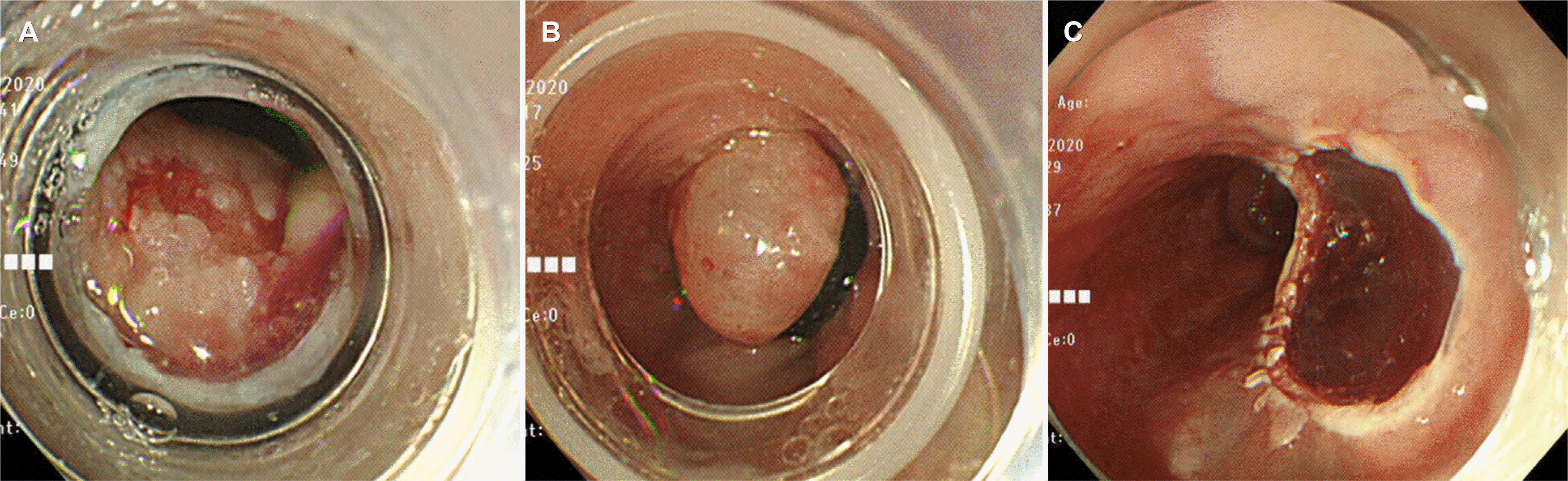
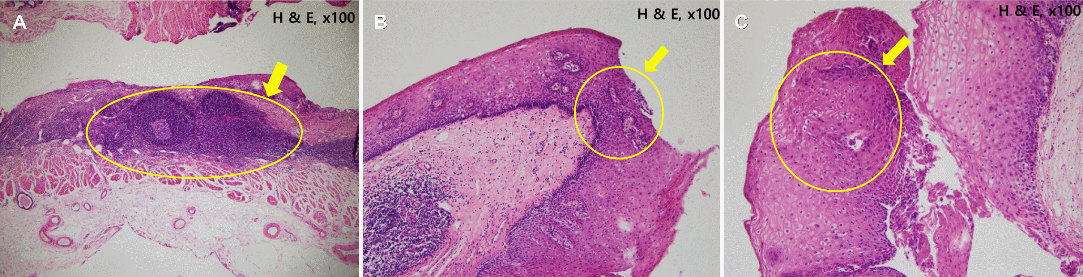
증례: 62세 남자 환자가 식도에 병변이 발견되어 내원하였다. 환자는 건강검진을 목적으로 시행한 식도위십이지장내시경(esophagogastroduodenoscopy) 검사에서 하부식도에 함몰형의 점막병변이 관찰되어 내원하였다. 병변은 상절치(upper incisor)로부터 35 cm 부위 식도에서 관찰되었으며 약 6 mm 크기였다(Fig. 1). 조직 검사 결과 비정형 세포와 국소적인 중등도의 이형성(atypical cells and focal moderate dysplasia)으로 진단되었다. 내원 당시 활력 징후는 혈압 150/90 mmHg, 맥박수 80회/분, 체온 37.3℃로 측정되었다. 혈액 검사 결과 특이 이상 소견은 없었다. 내원 후 시행한 내시경 검사 결과 주변 정상 식도점막과 경계가 잘 구별되는 함몰형의병변이 관찰되었고 루골용액(Lugol’s solution)에 염색이 되지 않았다(Fig. 2). 재시행한 병리 검사 결과도 중등도의 이형성으로 진단되었다(Fig. 3). 초음파 내시경(EUS) 검사 결과 점막층(mucosal layer)에 국한된 병변임을 확인하였다(Fig. 4).밴드결찰술을 이용한 내시경 점막절제술(endoscopic mucosal resection with ligation)을 시행하였다(Fig. 5). 최종 병리 검사에서 완전절제를 확인하였으며, 이형성 외에 편평태선(lichen planus)이 관찰되었다(Fig. 6A). 림프구가 점막하에띠를 이루듯이 관찰되었고 이형성은 국소적으로 분포하였다(Fig. 6B, C). 편평태선과 관련된 구강점막, 생식기 또는 피부의 특이 병변은 발견되지 않았다.
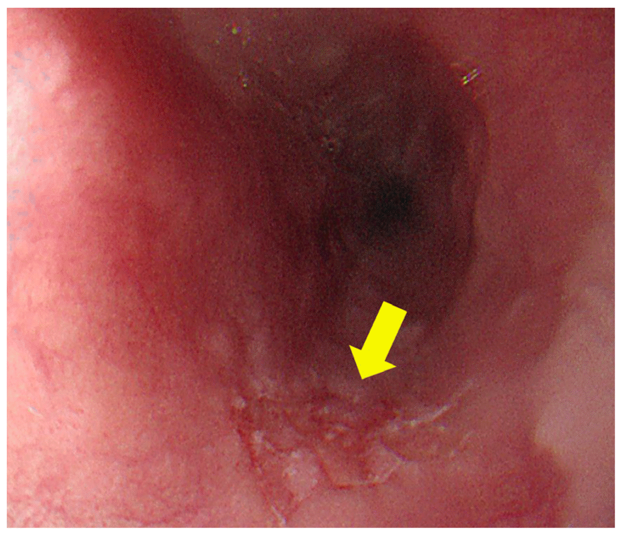 | Fig. 1Initial esophagogastroduodenoscopy findings. Slightly depressed mucosal lesion on the lower esophagus (arrow).
|
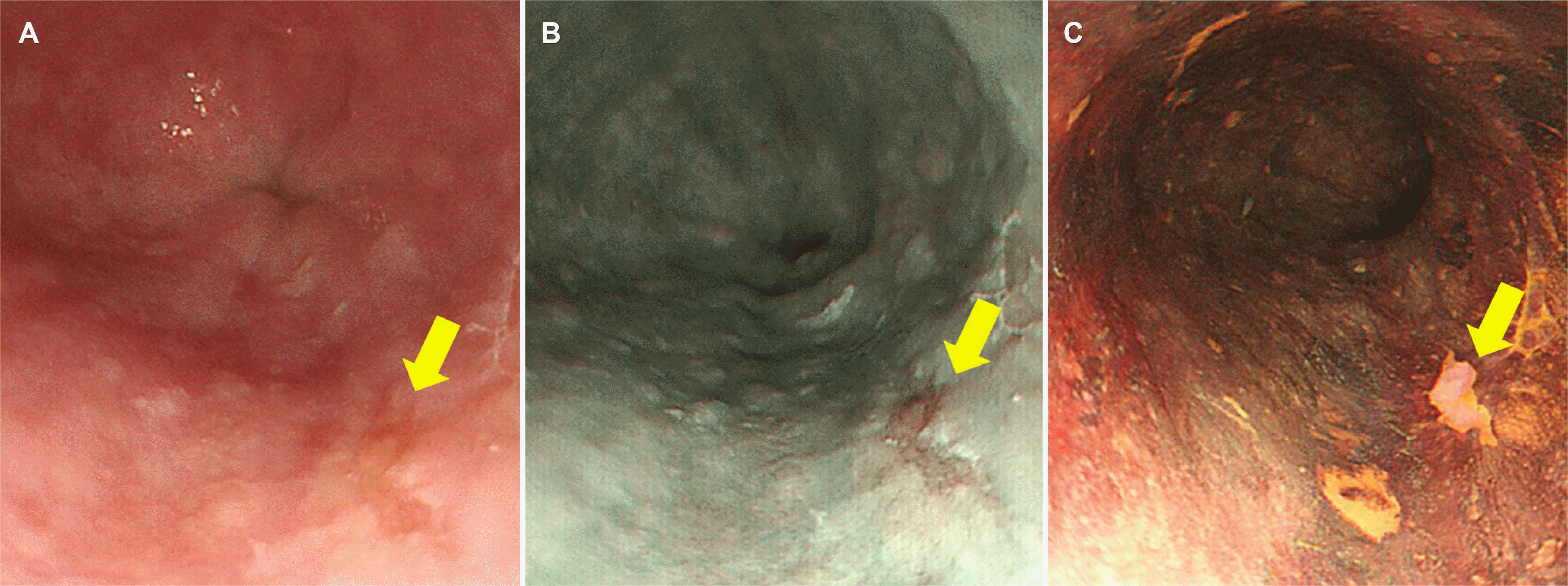 | Fig. 2Second esophagogastroduodenoscopy findings. (A) Detected lesion on white light imaging endoscopy (arrow). (B) On narrow-band imaging (arrow). (C) On chromoendoscopy image with Lugol’s solution staining (arrow).
|
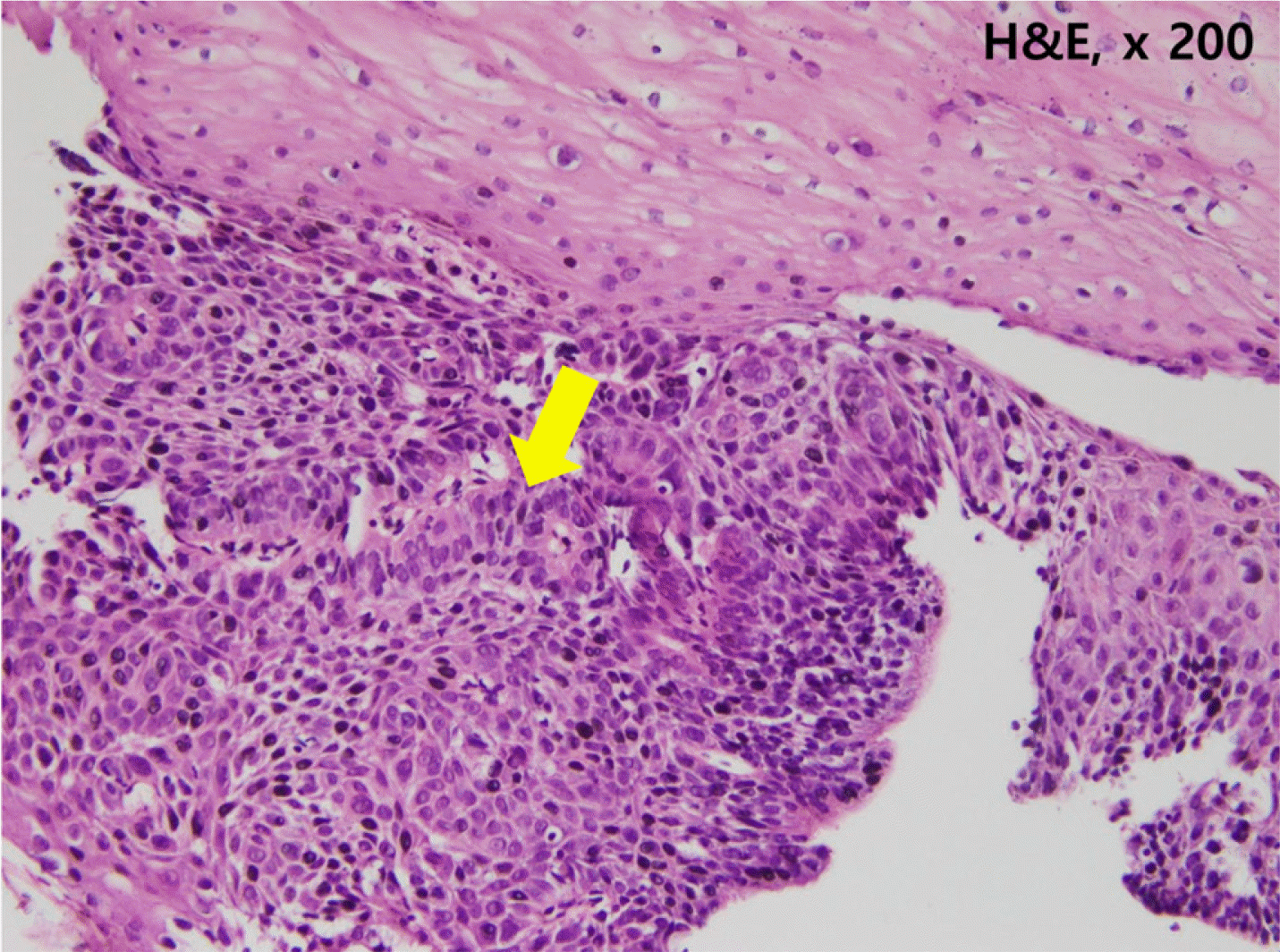 | Fig. 3Histology findings of the biopsied specimen. Focal increased nucleo-cytoplasmic ratio indicating dysplasia (arrow) (H&E, ×200).
|
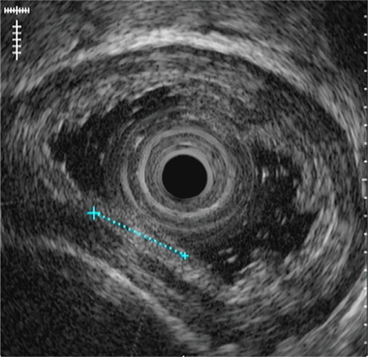 | Fig. 4Endoscopic ultrasound examination. A 6 mm sized isoechoic lesion was confined in the 1st layer of the esophageal wall.
|
진단: 이형성을 동반한 식도 편평태선
본 증례는 피부에 동반된 병변이 없으면서 식도에 발생한 편평태선에 동반된 이형성에 관한 것이다. 편평태선은 주로 피부(사지, 손바닥, 발바닥, 모발, 손톱 등)와 점막(구강, 생식기, 결막, 식도 등)에 발생하는 특발성 염증 질환(idiopathic inflammatory disorder)이다. 유병률은 일반 인구의 0.1-2.3%로 보고되었으며,1 피부 병변 없이 점막병변만 보이는 경우는 전체 환자의 25% 정도로 알려져 있다.2 식도의 편평태선은 드문 것으로 보고되었지만 일부 연구에서는 유병률이 과소평가 되어 있을 가능성을 시사하였다.3-7 최근의 연구에서 추정한식도 편평태선의 유병률은 일반 인구 집단의 0.1%로, 이는호산구성 식도염(eosinophilic esophagitis)의 서양인 유병률인 0.05%보다 높은 수치이다.7 식도의 편평태선 환자 중에서는약 5%에서만 피부병변을 동반하는 것으로 알려져 있다.5 구강이나 생식기에 편평태선이 발생한 경우 식도의 편평태선의 발생과 연관성이 높은 것으로 알려져 있으나, 본 증례에서는 식도외 부위에 편평태선이 발견되지 않았다.7,8
피부병변이 없는 경우 바이러스 감염(human immunodeficiency virus 또는 간염 바이러스)이나 크론병(Crohn’s disease), 비스테로이드소염제(nonsteroidal anti-inflammatory drugs) 또는 nivolumab 등의 면역관문억제제(immune checkpoint inhibitors) 사용, 약제 유발 식도염(pill induced esophagitis), 기타 류마티스 질환 등이 병인으로 추측되고 있다.5,6 중년이나 고령의 여성에서 호발하는 것으로 보고되었고, 위치의 경우 하부식도보다는 중부나 상부식도에서 더 많이 보고되고 있다.5 내시경 육안 소견으로는 백색 결절(white plaque) 또는 점막의 탈락(sloughing), 협착(stricture) 등의 비특이적인 소견이 제시되고 있고, 호산구성 식도염에서 특징적으로 알려져 있는 종축 방향의 골(linear furrow)이나 원모양의 주름(stacked circular rings) 등도 관찰된다.7 병리학적으로 편평상피의 각화증과 과립층의 비대, 기저층의 액화 변성 등의 소견과 특징적인 진피 상부에 띠모양으로 위치하는 림프구의 분포가 잘 알려져 있다.7
식도의 편평태선 환자들이 본 증례와 같이 무증상인 경우도 흔하지만, 소화기 내시경 검사를 시행하는 가장 흔한 이유는 삼킴곤란(dysphagia)이고 장기간 치료받지 않을 경우 식도의 협착(stricture)을 유발할 수 있는 것으로 알려져 있다.5 하지만 명확히 정립된 내시경 및 조직학적 진단 기준이 없기 때문에 비특이적인 식도염(예, sloughing esophagitis, lymphocytic esophagitis, eosinophilic esophagitis, candida esophagitis 등)으로 추정 진단하는 경우가 많을 것으로 추측된다.7 최근의 한 연구에서는 내시경 소견(denudation of the muco-sa, stenosis or stricture, hyperkeratosis, trachealization)과 병리 소견(mucosal detachment, T-lymphocytic infiltrate, intraepithelial apoptosis, dyskeratosis, fibrinogen deposits along the basement membrane)의 항목들을 구조화 하여 식도 편평태선의 진단 기준을 제시하기도 하였다.7 하지만 진단이 적절히 이루어진다고 하여도 추천되는 치료 또한 명확하지않다. Budesonide나 fluticasone과 같은 국소 스테로이드(topical corticosteroids), 면역억제제(immunosuppressants), retinoids, rituximab 등이 시도되었고, 협착을 동반한 식도의편평태선의 경우 내시경 풍선확장술이나 corticosteroids 주입법이 추천되었다.7,9
편평태선은 CD8+ T 림프구가 기저층의 각질형성세포(basal keratinocyte), 조절 T세포(regulatory T lymphocyte) 및 보조 T세포(helper T lymphocyte)에 세포독성(cytotoxic activity)을 보이면서 발생하는 만성 염증과정이다.10 식도의 과각화증(hyperkeratosis)이 전암병변이기 때문에 편평세포암(squamous cell carcinoma) 발생 위험이 증가하는 것으로 알려져 있고 그 발생률은 6.1%로 보고되었다.11,12
본 증례의 경우 내시경 검사를 통해 식도의 미란성 병변으로 발견되었고 조직 검사에서 중등도의 이형성이 확인되어 내시경 절제술로 제거하였지만, 최종 병리진단에는 식도의 편평태선이 확인된 증례였다. 전암 병변으로 암 발생의 위험성이 있음에도 불구하고, 전형적인 육안 소견의 특징 및 환자의 증상이 없는 경우가 흔하기 때문에 주의가 필요하다. 또한 기타 비특이적인 식도염과 감별 진단이 필요하기 때문에 삼킴곤란을 호소하거나 원인이 불분명한 상부 또는 중부 식도의 협착이 있는 경우 질환을 의심해 볼 수 있다.
Go to : 
 XML Download