Journal List > Korean J Gastroenterol > v.78(3) > 1147555

Dahale, Puri, Sachdeva, Agarwal, Kumar, Dalal, and Saxena: Reappraisal of the Role of Ascitic Fluid Adenosine Deaminase for the Diagnosis of Peritoneal Tuberculosis in Cirrhosis

Abstract

Backgrounds/Aims

Although peritoneal tuberculosis (TB) is one of the important differential diagnoses among cirrhotic patients with ascites, a peritoneal biopsy is not always available. High ascitic fluid adenosine deaminase (ADA) has been indicative of peritoneal TB. On the other hand, studies to assess its diagnostic utility based on the confirmation of peritoneal biopsy in cirrhotic patients are scarce.

Methods

Patients with new-onset ascites were enrolled prospectively from a tertiary hospital. Peritoneal biopsy was applied according to clinical judgment when required. Based on pathology diagnosis of the peritoneum, the diagnostic efficacy of ascitic fluid ADA for peritoneal TB was evaluated in total and cirrhotic patients, respectively.

Results

Among 286 patients enrolled, 78 were diagnosed with peritoneal TB. One hundred and thirty-two patients had cirrhosis, and 30 of those were diagnosed with peritoneal TB. The mean ADA was 72.2 U/L and 22.7 U/L in the peritoneal and non-peritoneal TB group, respectively, among the total study population, and 64.0 U/L and 19.1 U/L in the peritoneal and non-peritoneal TB group, respectively, among the subgroup with cirrhosis. The area under the curve for ADA to diagnose peritoneal TB was 0.96 in the total study population with a cutoff value of 41.1 U/L, and 0.93 in cirrhotic patients with a cutoff value of 39.9 U/L.

Conclusions

The ascitic fluid ADA measurements showed high diagnostic performance for peritoneal tuberculosis in patients with ascites regardless of cirrhosis at a similar cutoff value.

INTRODUCTION

Peritoneal tuberculosis (TB) is not uncommon in India, unlike the West, and is a great clinical masquerader.1,2 Cirrhosis is a significant risk factor associated with peritoneal TB, and the co-occurrence of both is not unusual in clinical practice.3-5 Recent reports suggest that more peritoneal TB patients have underlying cirrhosis than older studies.6-8 The gold standard for a diagnosis of peritoneal TB is a demonstration of acid-fast bacilli (AFB) staining and the growth of Mycobacterium Tuberculosis on the culture of ascitic fluid (AF) or a peritoneal biopsy sample.9 Unfortunately, a demonstration of AFB in the ascitic fluid has a sensitivity of only 3%, which can increase only up to 34% by culture. Nevertheless, culture also is a labor-intensive and time-consuming process.9 The diagnostic tool of choice for peritoneal TB is diagnostic laparoscopy with peritoneal biopsy. The laparoscopy findings themselves are highly suggestive and can be taken as synonymous with tuberculosis. More importantly, it provides tissue that can either show histopathological findings or mycobacterial analysis definitive of tuberculosis.9-12 On the other hand, laparoscopy is an invasive procedure, not routinely available everywhere, and is also associated with a small risk of morbidity and mortality, which may be even higher in patients with cirrhosis.13-16 A delay in the diagnosis of peritoneal TB increases morbidity and mortality. Therefore, there is a need for a rapid and accurate test.6 The adenosine deaminase (ADA) level in the ascitic fluid is a good alternative for peritoneoscopy with negligible risk. In meta-analyses, ADA showed excellent sensitivity (93-100%) and specificity (95-97%) in peritoneal TB.17-19 Despite this, its utility in patients with cirrhosis for diagnosing peritoneal TB is still debatable. There are only a few reports with a small sample size and have yielded contradictory results, emphasizing further exploration of the utility of ADA in these patients.20-24
Considering its easy availability, cost-effectiveness, and rapid results, this study evaluated the diagnostic performance of ascitic fluid ADA for peritoneal TB, which was confirmed by peritoneal biopsy, especially in patients with cirrhosis.

SUBJECTS AND METHODS

1. Patients

This study was a prospective observational case-control study that was conducted at the Department of Gastro- enterology, G.B. Pant Hospital, New Delhi, from October 2013 to December 2015. Prior approval for the study was obtained from the institutional ethics committee (IRB No. 11/IEC/MAMC/2011).
The study was designed to find the accuracy of ascitic fluid ADA for the diagnosis of peritoneal TB against the pathological diagnosis based on peritoneal biopsy. Consecutive patients ≥18 years in age presenting with new onset of ascites were evaluated. After a complete diagnostic evaluation, the patients were categorized as peritoneal TB and non-peritoneal TB group with or without cirrhosis. Cirrhosis was diagnosed based on the clinical, radiological, or pathological findings. Patients with coexistent tuberculosis at any other body site or possible mixed ascites from two etiologies were excluded.

2. Evaluation for the cause of ascites

All patients with ascites were evaluated for the etiology as per standard protocol. A complete blood count, liver function tests, kidney function tests, chest X-ray, abdominal ultrasonography, and ascitic fluid analysis, including cell count, albumin, protein, ADA, Gram, and AFB stain, was done in all subjects. The upper gastrointestinal endoscopy, colonoscopy, and computer-enhanced tomography of the abdomen and chest were performed on a case-to-case basis.

3. Measurement of ADA

ADA analysis was performed on freshly collected AF samples within 2 hours of collection of fluid. Ascitic Fluid ADA was determined by commercially available kits. We used BioSystems S.A. Costa Brava, 30.08030 Barcelona (Spain) (Quality System certified according to EN ISO 13485 and EN ISO 9001 standards) kit which uses Slaats Method for ADA determination.

4. Confirmation of peritoneal TB

A peritoneal biopsy was performed on patients in whom peritoneal TB was suspected after a clinical evaluation and baseline investigations, and in other cases, wherever required clinically. Most of these cases were suspected of malignant ascites and miscellaneous categories of rare etiologies with negative routine evaluations. Peritoneal TB was suspected in patients with a low serum ascites albumin gradient (SAAG), high protein, and clinical signs, such as fever, loss of appetite, loss of weight, the recent development of ascites. For peritoneal TB in cirrhotics, the same criteria were applied except for the SAAG and total protein. A peritoneal biopsy was conducted primarily laparoscopically. This was also done under ultrasound guidance in few patients wherever feasible. The visual findings during laparoscopy were noted, and peritoneal biopsy samples were sent for histopathological analysis. The definitive diagnosis of peritoneal TB was considered if a peritoneal tissue biopsy showed any of the following25: 1) positive AFB stain, 2) caseating granuloma, and 3) Multiple large noncaseating epithelioid granulomas with Langhans giant cell with a high clinical suspicion with a response to antituberculosis drugs. All other patients who did not present the pathological findings of TB in peritoneal biopsy or who did not show the clinical findings of TB and skip peritoneal biopsy were referred to as the non-peritoneal TB group.

5. Statistical Analysis

The continuous data were expressed as the mean with standard deviation or 95% CI. The groups were compared using an independent Student t-test in the case of continuous data, while categorical data were compared using a Chi-Square test. The sensitivity, specificity, diagnostic accuracy, and area under the receiver operating characteristic curve (AUROC) were calculated for ADA for diagnosing peritoneal TB confirmed by peritoneal biopsy. Statistical Products and Services Solution (SPSS) software version 23.0 (SPSS, Chicago, IL, USA) was used for statistical analysis. For all statistical tests, a two-sided p-value <0.05 was considered significant.

RESULTS

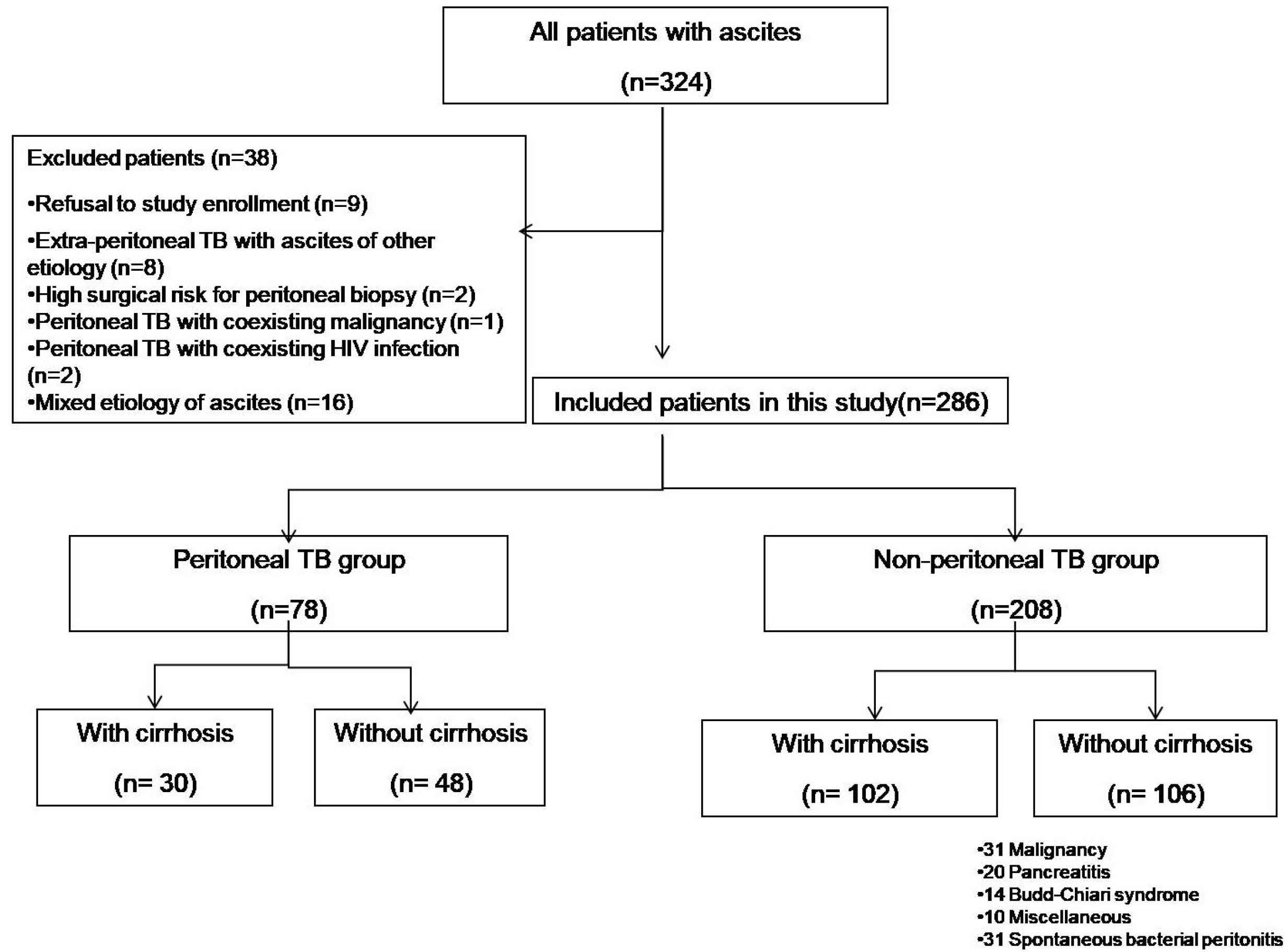
Four hundred and thirty-one patients were presented with ascites for an evaluation during the study period. Of these, 107 cases were lost due to incomplete or no workup for ascites evaluation. The remaining 324 patients were evaluated further for ascites, of which 38 patients were excluded (Fig. 1). Among the remaining 286 patients with ascites, 78 (27%) were diagnosed with peritoneal TB. Thirty out of 78 (38%) in the peritoneal TB group and 102 out of 208 (49%) in the non-peritoneal TB group had cirrhosis.

1. Baseline characteristics of total patients

The patients in the peritoneal TB group were significantly younger than the non-peritoneal TB group (33.3 years vs. 43.7 years). They presented with more frequent symptoms, such as anorexia, weight loss, fever, and abdominal pain, than the non-peritoneal TB group. Approximately 19.23% of the peritoneal TB group presented with lymphadenopathy, but none did in the non-peritoneal TB group. The patients among the peritoneal TB group showed chest X-ray abnormality in 41% and a positive Tuberculin test in 67.94%. The patients in the non-peritoneal TB group showed a lower platelet count and poorer liver functions, including AST, ALT, ALP, bilirubin, PT-INR, and albumin level, compared to the peritoneal TB group. The cell count of ascites was more lymphocyte dominant in the peritoneal TB group, and SAAG was significantly lower in the peritoneal TB group than in the non-peritoneal TB group (0.8 vs. 1.5) (Table 1).

2. Baseline characteristics in the subgroup of cirrhosis

The cirrhotic patients in the peritoneal TB group were relatively younger than the non-peritoneal TB cirrhotic group (35.8 vs. 45.3 years). The patients from the peritoneal TB cirrhotic subgroup had relatively better liver functions, i.e., less bilirubin, PT-INR, high albumin, and low model for end stage liver disease score than the non-peritoneal TB cirrhotic patients. Ascitic fluid analysis of peritoneal TB cirrhotics revealed higher total protein, albumin, ADA, and total leukocyte count, and a low SAAG (1.00 vs. 1.81) (Table 2).

3. ADA level according to the presence of peritoneal TB in total patients

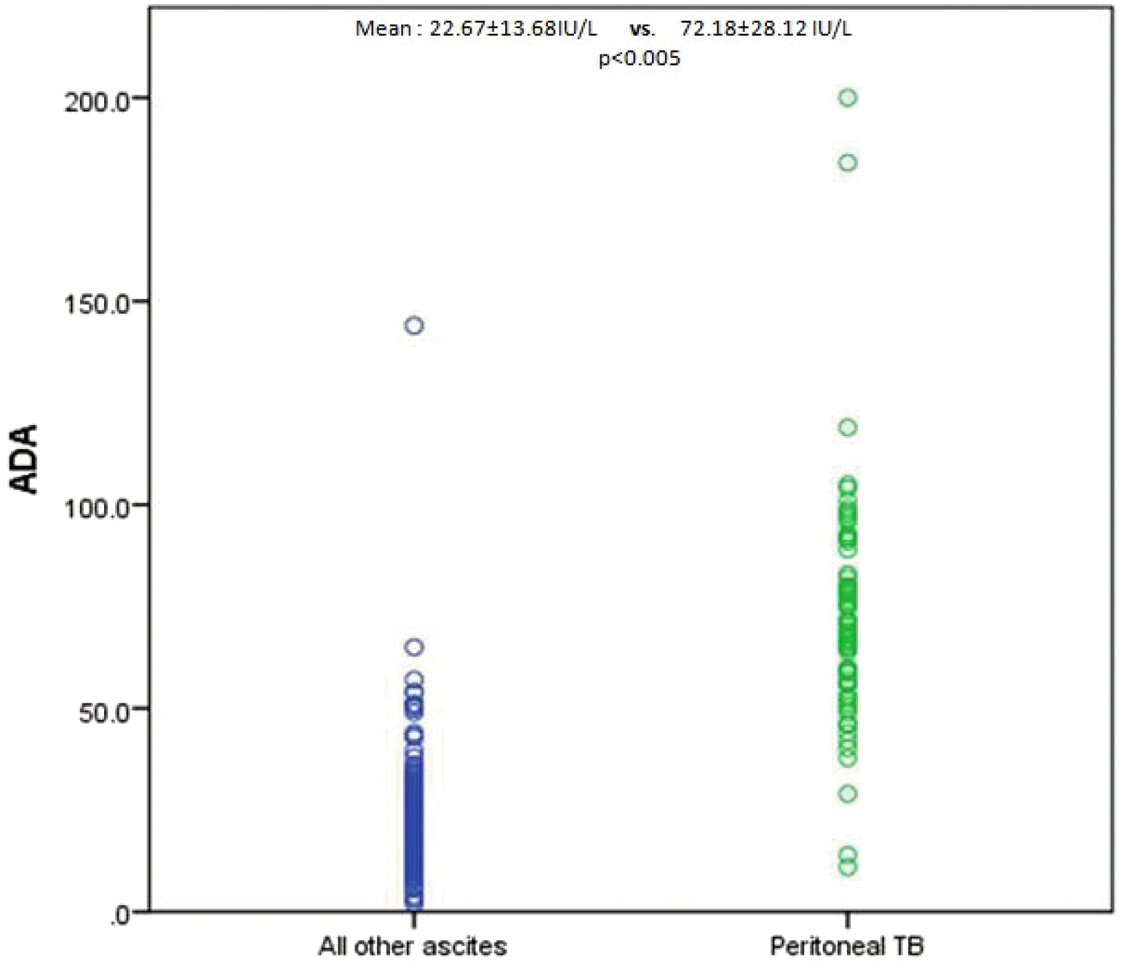
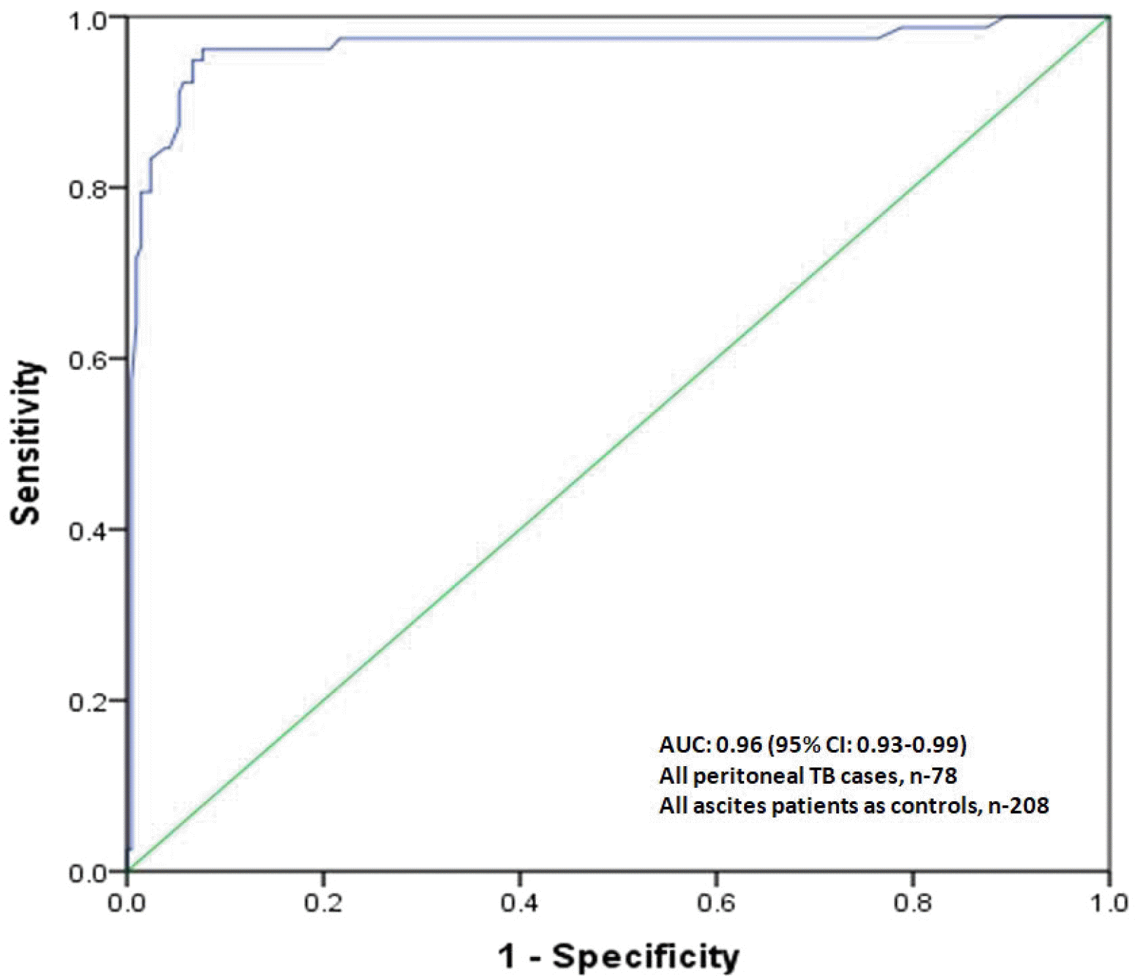
The ADA levels in the ascitic fluid of peritoneal TB patients were significantly higher than the non-peritoneal TB patients (72.2±28.1 U/L vs. 22.7±13.7 U/L, p<0.005) (Fig. 2). The AUROC for ascitic fluid ADA was 0.96 for a diagnosis of peritoneal TB in the total patients with ascites (95% CI 0.93-0.99, p<0.005) (Fig. 3). The sensitivity, specificity, and diagnostic accuracy of the ascitic fluid ADA level for peritoneal TB was 95% (95% CI 87-98%), 93% (95% CI 88-96%), and 93% (95% CI 90-96%), respectively, and the optimal cutoff value was 41.1 U/L.

4. ADA level according to the presence of peritoneal TB in the subgroup with cirrhosis

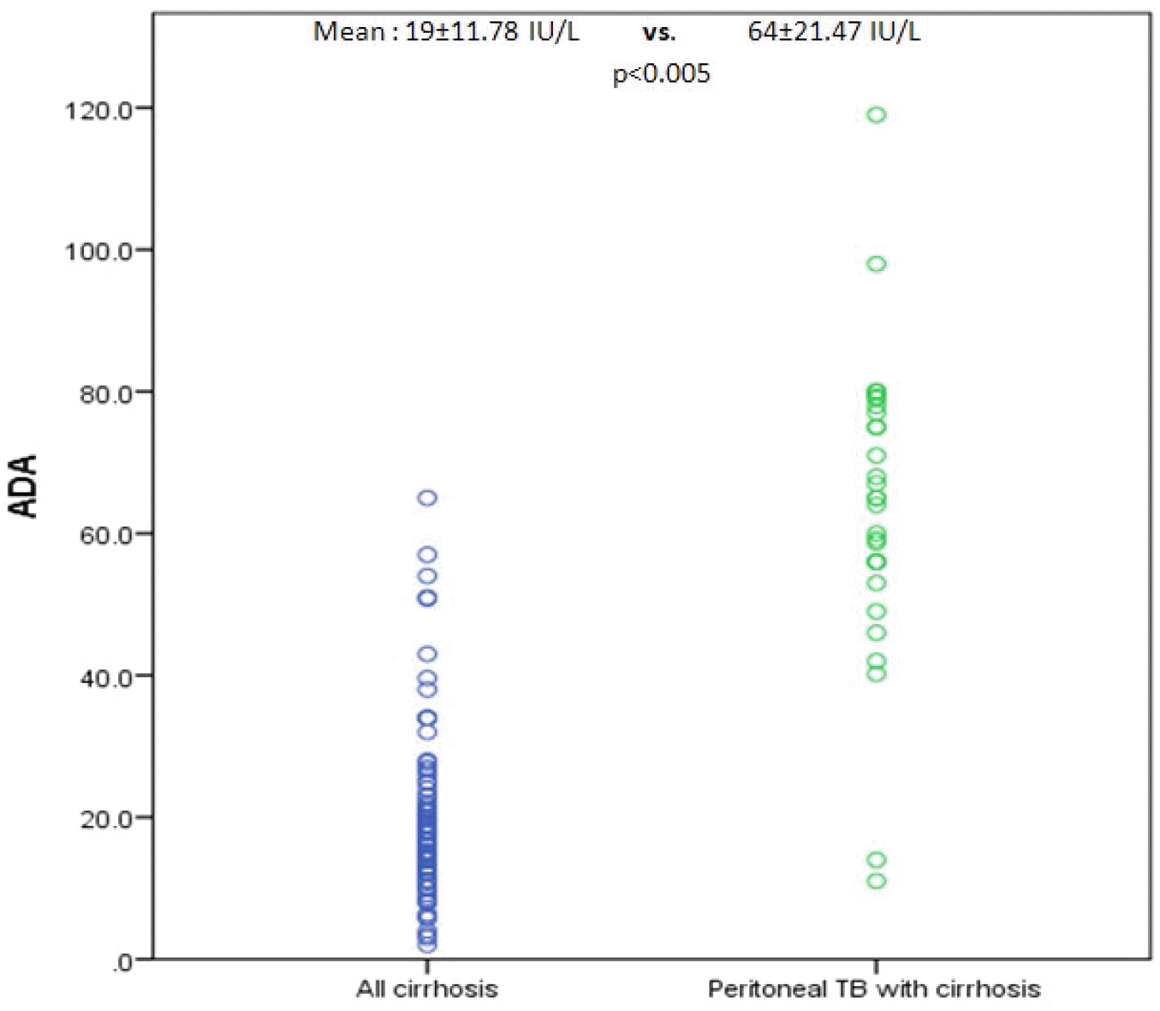
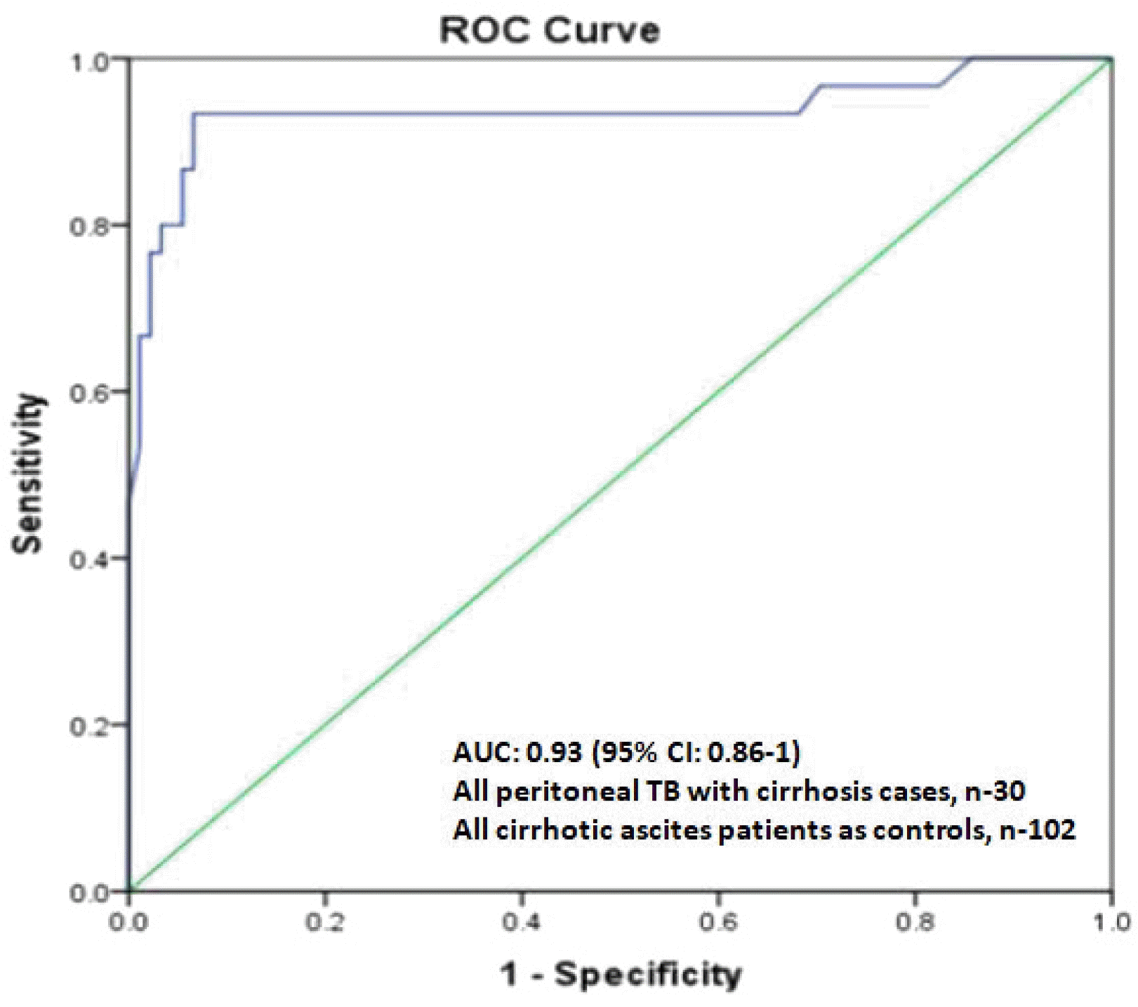
The ADA levels in the ascitic fluid of peritoneal TB patients with cirrhosis were significantly higher than in the non-peritoneal TB cirrhotic patients (64.0±21.5 U/L vs. 19.0±11.8 U/L, p<0.005) (Fig. 4). The AUROC for ascitic fluid ADA was 0.93 for a diagnosis of peritoneal TB in the total patients with cirrhotic ascites (95% CI 0.86-1, p<0.005) (Fig. 5). The sensitivity, specificity, and diagnostic accuracy of the ascitic fluid ADA level for peritoneal TB in the cirrhotics was 93% (95% CI 87-98%), 94% (95% CI 88-96%), and 92% (95% CI 90-96%), respectively, and the optimal cutoff value was 39.9 U/L.

5. Clinical outcome of peritoneal TB

Four patients (8.5%) among peritoneal TB, three with cirrhosis, and one without cirrhosis had an adverse event related to peritoneoscopy. All patients in the peritoneal TB group received antituberculosis therapy according to the national guidelines with the required modification for cirrhosis.26 Four patients died in the patients with cirrhosis on the follow-up within three months versus none in the patients without cirrhosis (13% vs. 0%, p=0.009).

DISCUSSION

Peritoneal TB had very high mortality of approximately 50% in the past, but improvements in diagnostic techniques and anti-TB medication decreased the mortality with the survival rate of almost 91%.8,27,28 One of the important risk factors for peritoneal TB was cirrhosis as in the present study, in which 38% of the peritoneal TB patients had underlying cirrhosis. The clinical presentation was diverse, and in patients with cirrhosis, it may not be suspected initially, leading to a delay in the diagnosis and consequently the start of treatment.9
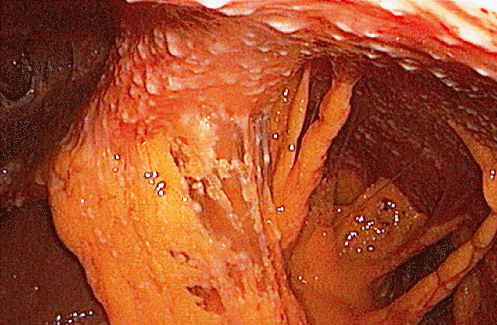
Laparoscopy is the diagnostic tool of choice because it provides a direct examination of the peritoneum, and one can take biopsies for histopathology and microbiological examination. On the other hand, it was associated with morbidity and mortality, even higher in cirrhotic patients.13-16 It is also not practical for patients with severe underlying comorbidities to undergo laparoscopy. Therefore, patients with cirrhosis will often be poor candidates for laparoscopy. Ultrasound is an excellent non-invasive method for an evaluation of the intra-peritoneal cavity, and one can also take peritoneal biopsies.29,30 On the other hand, ultrasound-guided biopsies require expertise and are not feasible for all patients. We were able to do laparoscopy in 47 patients while ultrasound guided biopsies were performed only on 31 patients (39.74%) (Table 3; Fig. 6).
The AFB staining of ascitic fluid has very poor sensitivity, and cultures also take a very long time.9 This is quite a time-consuming process, even if a laparoscopic approach is followed. Good, less invasive, easy to perform tests with good sensitivity and specificity are needed because patients with a delayed diagnosis have higher mortality.6
ADA has been studied in body fluids, including ascitic fluid for diagnosing TB, and has been shown to have high sensitivity and specificity. ADA is an essential enzyme in the purine salvage pathway; it plays an important role in T cell differentiation. Thus far, three meta-analyses have been published dealing with the role of ADA in peritoneal TB diagnosis.17-19 Unfortunately, ADA for diagnosis of peritoneal TB in patients with cirrhosis has been studied.20-24 Hillenbrand et al. found that the ADA is inferior in cirrhosis as a diagnostic method for peritoneal TB. In this study, the sensitivity of ADA was only 30% for diagnosing peritoneal TB in patients with cirrhosis.23 They speculated that this could be because ADA correlates positively with the ascitic fluid total protein, and cirrhotic patients generally have low ascitic fluid total protein. The major drawbacks of this study were the small sample size (n=10), the method of ADA analysis used was different, and the ADA estimation was done on stored samples.23 On the other hand, recent studies revealed high sensitivity (94-100%) and high specificity (92-93%) of ADA in diagnosing peritoneal TB in patients with cirrhosis.20-22,24 On the other hand, these studies also had a small sample size (all four studies altogether had 29 patients) with a small or no control group.
In the present study, ADA overall had very high sensitivity and specificity at a cutoff of 41.1 IU/L. Furthermore, ADA had sensitivity, specificity, and accuracy of 93%, 94%, and 92%, respectively, for diagnosing peritoneal TB in cirrhotic patients at a slightly lower cutoff of 39.9 IU/L compared to non-cirrhotic patients. Therefore, the ADA levels in the cirrhotic peritoneal TB group were much higher than the cirrhotic non-peritoneal group and all other types of ascites patients in the control group.
In the present study, ascitic fluid ADA correlated well with the ascitic fluid total protein (r2 =0.62, p=<0.005). The ADA levels in the cirrhotic peritoneal TB group were much higher despite having a low ascitic fluid total protein than the non-cirrhotic peritoneal TB group. In addition, high total protein does not mean high ADA levels because it can be observed in patients with pancreatic ascites and malignant ascites, where the ascitic fluid total protein was high in all but ADA. Therefore, some other factors act for the increase in ADA in the ascitic fluid in the presence of peritoneal TB.
The ADA has false positivity in 6.9-9.4% ascitic fluids, as depicted in published studies.20,23 In the present study, 14 patients (6.7%) in the control group had an ADA value ≥40 IU/L, including ascitic patients with SBP, pancreatic ascites, malignant ascites, and lymphatic obstruction with ascites. In the SBP patients, post-treatment ascitic fluid ADA was repeated at four weeks and was below the cutoff. To deduce any firm opinion from this observation is difficult, but a repeat ADA measurement after treating the obvious clinical problem when the clinical scenario does not fit might help clinicians. In this study, only four patients in the peritoneal TB groups had ADA <40 IU/L, two patients were already on anti-TB medication, and while the other two patients had neutrophilic dominant ascites (>80% neutrophils in ascitic fluid). Anti-TB medication and low lymphocyte count in the ascitic fluid may have resulted in a lower ADA value in these patients.
The present study is the largest single-center study in terms of the number of patients and the patients with cirrhosis and peritoneal TB. Moreover, a large control with a heterogeneous population makes this study clinically robust. The gold standard in the present study was histopathology on peritoneal tissue, which is highly reliable and accurate (Table 4).
This study had a few limitations. First, the peritoneal biopsy was not applied as the gold standard diagnostic tool for peritoneal TB to all patients with ascites. Instead, the suspected group was selected for peritoneal TB, and the peritoneal biopsy was performed. Therefore, there could be false-negative cases among the non-peritoneal TB group. To the best of the authors’ knowledge, there was no overt peritoneal TB case at least within one month after the initial assessment. In addition, the diagnostic approach might avoid the ethical issues associated with a non-selective biopsy on many non-peritoneal TB patients, as well as the potential complications without clinical benefit. Second, the patients with ascites with mixed causes were excluded from making the diagnostic meaning of ADA clear-cut. Therefore, it is unclear if the ascitic ADA level is useful in these patients. Third, this result is based on the data from a single center in a country. Therefore, care should be taken to apply the findings to other populations.
In conclusion, when a patient visits a clinic for ascites, ascitic ADA measurements in the cases of suspicion of tuberculosis might show excellent diagnostic accuracy for peritoneal TB in non-cirrhotic and cirrhotic patients. Further studies will be needed to delineate the meaning of ADA level in the patients with ascites of mixed causes.
Notes

Financial support

None.

Conflict of interest

None.

REFERENCES

1. Khuroo MS, Khuroo NS. Madkour MM, editor. 2004. Abdominal tuberculosis. Tuberculosis. 1st ed. Springer;Berlin: p. 659–667. DOI: 10.1007/978-3-642-18937-1_37.
crossref
2. Lingenfelser T, Zak J, Marks IN, Steyn E, Halkett J, Price SK. 1993; Abdominal tuberculosis: still a potentially lethal disease. Am J Gastroenterol. 88:744–750.
3. Sanai FM. 2006; The liver in tuberculous peritonitis. Saudi J Gastroenterol. 12:93–94. DOI: 10.4103/1319-3767.27854. PMID: 19858594.
crossref
4. Guirat A, Koubaa M, Mzali R, et al. 2011; Peritoneal tuberculosis. Clin Res Hepatol Gastroenterol. 35:60–69. DOI: 10.1016/j.gcb.2010.07.023. PMID: 21215540.
crossref
5. Muneef MA, Memish Z, Mahmoud SA, Sadoon SA, Bannatyne R, Khan Y. 2001; Tuberculosis in the belly: a review of forty-six cases involving the gastrointestinal tract and peritoneum. Scand J Gastroenterol. 36:528–532. DOI: 10.1080/003655201750153412. PMID: 11346208.
crossref
6. Chow KM, Chow VC, Hung LC, Wong SM, Szeto CC. 2002; Tuberculous peritonitis-associated mortality is high among patients waiting for the results of mycobacterial cultures of ascitic fluid samples. Clin Infect Dis. 35:409–413. DOI: 10.1086/341898. PMID: 12145724.
crossref
7. Yeh HF, Chiu TF, Chen JC, Ng CJ. 2012; Tuberculous peritonitis: analysis of 211 cases in Taiwan. Dig Liver Dis. 44:111–117. DOI: 10.1016/j.dld.2011.08.020. PMID: 21944948.
crossref
8. Manohar A, Simjee AE, Haffejee AA, Pettengell KE. 1990; Symptoms and investigative findings in 145 patients with tuberculous peritonitis diagnosed by peritoneoscopy and biopsy over a five year period. Gut. 31:1130–1132. DOI: 10.1136/gut.31.10.1130. PMID: 2150661. PMCID: PMC1378737.
crossref
9. Sanai FM, Bzeizi KI. 2005; Systematic review: tuberculous peritonitis--presenting features, diagnostic strategies and treatment. Aliment Pharmacol Ther. 22:685–700. DOI: 10.1111/j.1365-2036.2005.02645.x. PMID: 16197489.
10. Wolfe JH, Behn AR, Jackson BT. 1979; Tuberculous peritonitis and role of diagnostic laparoscopy. Lancet. 1:852–853. DOI: 10.1016/S0140-6736(79)91266-2.
crossref
11. Rai S, Thomas WM. 2003; Diagnosis of abdominal tuberculosis: the importance of laparoscopy. J R Soc Med. 96:586–588. DOI: 10.1177/014107680309601204. PMID: 14645607. PMCID: PMC539656.
crossref
12. Bhargava DK, Shriniwas , Chopra P, Nijhawan S, Dasarathy S, Kushwaha AK. 1992; Peritoneal tuberculosis: laparoscopic patterns and its diagnostic accuracy. Am J Gastroenterol. 87:109–112.
13. Hong KD, Lee SI, Moon HY. 2011; Comparison between laparoscopy and noninvasive tests for the diagnosis of tuberculous peritonitis. World J Surg. 35:2369–2375. DOI: 10.1007/s00268-011-1224-2. PMID: 21882025.
crossref
14. Abdelaal A, Alfkey R, Abdelaziem S, et al. 2014; Role of laparoscopic peritoneal biopsy in the diagnosis of peritoneal tuberculosis. A seven-year experience. Chirurgia (Bucur). 109:330–334.
15. Barry RE, Brown P, Read AE. 1978; Physician's use of laparoscopy. Br Med J. 2:1276–1278. DOI: 10.1136/bmj.2.6147.1276. PMID: 152141. PMCID: PMC1608531.
crossref
16. Loffer FD, Pent D. 1975; Indications, contraindications and complications of laparoscopy. Obstet Gynecol Surv. 30:407–427. DOI: 10.1097/00006254-197507000-00001. PMID: 124409.
crossref
17. Tao L, Ning HJ, Nie HM, Guo XY, Qin SY, Jiang HX. 2014; Diagnostic value of adenosine deaminase in ascites for tuberculosis ascites: a meta-analysis. Diagn Microbiol Infect Dis. 79:102–107. DOI: 10.1016/j.diagmicrobio.2013.12.010. PMID: 24629577.
crossref
18. Shen YC, Wang T, Chen L, et al. 2013; Diagnostic accuracy of adenosine deaminase for tuberculous peritonitis: a meta-analysis. Arch Med Sci. 9:601–607. DOI: 10.5114/aoms.2013.36904. PMID: 24049517. PMCID: PMC3776182.
19. Riquelme A, Calvo M, Salech F, et al. 2006; Value of adenosine deaminase (ADA) in ascitic fluid for the diagnosis of tuberculous peritonitis: a meta-analysis. J Clin Gastroenterol. 40:705–710. DOI: 10.1097/00004836-200609000-00009. PMID: 16940883.
20. Liao YJ, Wu CY, Lee SW, et al. 2012; Adenosine deaminase activity in tuberculous peritonitis among patients with underlying liver cirrhosis. World J Gastroenterol. 18:5260–5265.
21. Burgess LJ, Swanepoel CG, Taljaard JJ. 2001; The use of adenosine deaminase as a diagnostic tool for peritoneal tuberculosis. Tuberculosis (Edinb). 81:243–248. DOI: 10.1054/tube.2001.0289. PMID: 11466036.
crossref
22. Huang HJ, Yang J, Huang YC, Pan HY, Wang H, Ren ZC. 2014; Diagnostic feature of tuberculous peritonitis in patients with cirrhosis: a matched case-control study. Exp Ther Med. 7:1028–1032. DOI: 10.3892/etm.2014.1538. PMID: 24669272. PMCID: PMC3964927.
crossref
23. Hillebrand DJ, Runyon BA, Yasmineh WG, Rynders GP. 1996; Ascitic fluid adenosine deaminase insensitivity in detecting tuberculous peritonitis in the United States. Hepatology. 24:1408–1412. DOI: 10.1002/hep.510240617. PMID: 8938171.
crossref
24. Aguado JM, Pons F, Casafont F, San Miguel G, Valle R. 1990; Tuberculous peritonitis: a study comparing cirrhotic and noncirrhotic patients. J Clin Gastroenterol. 12:550–554. DOI: 10.1097/00004836-199010000-00013. PMID: 2146314.
25. Logan VS. 1969; Anorectal tuberculosis. Proc R Soc Med. 62:1227–1230. DOI: 10.1177/003591576906201214.
26. WHO. 2009. Treatment of tuberculosis guidelines. 4th ed. WHO;Genava:
27. Ahmad M, Ahmed A. 1999; Tuberculous peritonitis: fatality associated with delayed diagnosis. South Med J. 92:406–408. DOI: 10.1097/00007611-199904000-00010. PMID: 10219360.
crossref
28. Dineeen P, Homan WP, Grafe WR. 1976; Tuberculous peritonitis: 43 years' expereince in diagnosis and treatment. Ann Surg. 184:717–722. DOI: 10.1097/00000658-197612000-00010. PMID: 826225. PMCID: PMC1345414.
29. Chu YJ, Ryeom H, Lee SY, et al. 2018; The diagnostic usefulness of ultrasound-guided peritoneal biopsy for the solitary peritoneal thickening of an unknown cause visualized as only infiltrated fat tissue on a CT scan. J Korean Soc Radiol. 78:225–234. DOI: 10.3348/jksr.2018.78.4.225.
crossref
30. Vardareli E, Kebapci M, Saricam T, Pasaoglu O, Açikalin M. 2004; Tuberculous peritonitis of the wet ascitic type: clinical features and diagnostic value of image-guided peritoneal biopsy. Dig Liver Dis. 36:199–204. DOI: 10.1016/j.dld.2003.10.016. PMID: 15046190.
crossref

Fig. 1
Study flow chart. TB, tuberculosis.
kjg-78-3-168-f1.tif
Fig. 2
Dot plot analysis chart of adenosine deaminase in peritoneal TB patients versus all patients with ascites. ADA, adenosine deaminase; TB, tuberculosis.
kjg-78-3-168-f2.tif
Fig. 3
AUC for adenosine deaminase in all peritoneal TB patients with all patients with other causes of ascites. AUC, area under the curve; TB, tuberculosis.
kjg-78-3-168-f3.tif
Fig. 4
Dot plot analysis chart of adenosine deaminase in peritoneal TB with cirrhosis patients versus all patients with cirrhotic ascites. ADA, adenosine deaminase; TB, tuberculosis.
kjg-78-3-168-f4.tif
Fig. 5
AUC for adenosine deaminase in peritoneal tuberculosis patients with cirrhosis amongst all patients with cirrhotic ascites. AUC, area under the curve; ROC, receiver operating characteristic curve; CI, confidence interval; TB, tuberculosis.
kjg-78-3-168-f5.tif
Fig. 6
Peritoneoscopic view showing tubercles over parietal peritoneum with adhesions.
kjg-78-3-168-f6.tif
Table 1
Baseline Characteristics in the Total Patients with Ascites
Peritoneal TB (n=78) Non-peritoneal TB (n=208) p-value
Clinical parameter
Age (years) 33.3±13.9 43.7±15.2 <0.005
BMI (kg/m2) 20.2±3.5 20.0±2.6 0.11
Male 42 (54.0) 137 (66.0) 0.06
Associated symptoms
Anorexia 75 (96.0) 70 (34.0) <0.005
Weight loss 74 (95.0) 64 (31.0) <0.005
Fever 63 (81.0) 24 (12.0) <0.005
Abdominal distension 72 (92.0) 101 (97.0) 0.11
Abdominal pain 40 (51.0) 76 (37.0) 0.01
Lymphadenopathy 15 (19.0) 0 (0.0) <0.005
Chest X-ray abnormality 32 (41.0) NA NA
Positive tuberculin skin test 53 (68.0) NA NA
Laboratory findings (serum)
Hb (g/dL) 9.9±1.9 9.4±2.3 0.08
WBC (/mm3) 7,350±2,740 9,830±7,647 0.60
PLT (×103/mm3) 2.6±1.3 1.7±1.3 <0.005
AST (U/L) 43.6±32.4 96.7±349.0 0.031
ALT (U/L) 28.0±26.6 61.1±185.0 0.01
ALP (U/L) 149.7±117.0 213±332 0.01
Protein (g/dL) 7.5±0.9 6.3±0.9 <0.005
Albumin (g/dL) 3.0±0.9 2.6±0.6 <0.005
PT-INR 1.3±0.3 1.6±0.8 0.001
BUN (mg/dL) 11.0±5.5 17±15 <0.005
Creatinine (mg/dL) 0.7±0.3 1.1±0.9 <0.005
Laboratory findings (ascites)
WBC (/mm3) 626±811 386±785 0.02
Lymphocyte (%) 78.4±22.0 67.1±32.6 0.001
Protein (g/dL) 4.5±1.6 2.1±1.2 <0.005
Albumin (g/dL) 2.1±0.9 1.0±0.7 <0.005
SAAG 0.8±0.4 1.5±0.6 <0.005
ADA (U/L) 72.2±28.1 22.7±13.7 <0.005

Values are presented number (%) or mean±standard deviation.

TB, tuberculosis; BMI, body mass index; Hb, hemoglobin; WBC, white blood cell; PLT, platelets; AST, aspartate aminotransferase; ALT, alanine aminotransferase; ALP, alkaline phosphatase; PT, prothrombin time; INR, international normalized ratio; BUN, blood urea nitrogen; SAAG, serum ascites albumin gradient; ADA, adenosine deaminase.

Table 2
Baseline Characteristics in the Subgroup with Cirrhosis
Peritoneal TB with cirrhosis (n=30) Cirrhosis without peritoneal TB (n=102) p-value
Clinical parameter
Age (years) 35.8±10.9 45.3±14.0 0.001
BMI (kg/m2) 20.8± 3.8 20.3±3.0 0.11
Male 24 (80.0) 77 (75.0) 0.80
Associated symptoms
Anorexia 29 (97.0) 19 (19.0) <0.005
Weight loss 28 (93.0) 14 (14.0) <0.005
Fever 25 (83.0) 7 (7.0) <0.005
Abdominal distension 30 (100.0) 97 (95.0) 0.26
Abdominal pain 11 (37.0) 12 (12.0) 0.004
Lymphadenopathy 4 (13.0) 0 (0.0) <0.005
Chest X-ray abnormality 16 (53.0) 5 (5.0) <0.005
Positive tuberculin skin test 21(70.0) 3 (3.0) <0.005
Laboratory findings (serum)
Hb (g/dL) 9.4±2.0 9.1±2.3 0.46
WBC (/mm3) 7,700±3,258 8,751±5,822 0.36
PLT (×103/mm3) 2.2±1.1 1.3±0.8 <0.005
AST (U/L) 54±41 142±520 0.35
ALT (U/L) 31.3±21.0 95±275 0.20
ALP (U/L) 169±152 185±187 0.66
Protein (g/dL) 6.8±0.9 6.3±1.0 0.009
Albumin (g/dL) 2.8±0.9 2.5±0.6 0.76
PT-INR 1.4±0.4 1.7±1.0 0.06
BUN (mg/dL) 11.5±7.0 18 ±17 0.03
Creatinine (mg/dL) 0.83±0.84 1.1±1.2 0.24
Laboratory findings (ascites)
WBC (/mm3) 691±621 137±200 <0.005
Lymphocyte (%) 86.0±8.9 82±21 0.295
Protein (g/dL) 3.4±1.5 1.6±1.0 <0.005
Albumin (g/dL) 1.6±0.9 0.55±0.61 <0.005
SAAG 1.0±0.5 1.8±0.5 <0.005
ADA (U/L) 64.0±21.5 19.0±11.8 <0.005

Values are presented number (%) or mean±standard deviation.

TB, tuberculosis; BMI, body mass index; Hb, hemoglobin; WBC, white blood cell; PLT, platelets; AST, aspartate aminotransferase; ALT, alanine aminotransferase; ALP, alkaline phosphatase; PT, prothrombin time; INR, international normalized ratio; BUN, blood urea nitrogen; SAAG, serum ascites albumin gradient; ADA, adenosine deaminase.

Table 3
Laparoscopic Findings in Peritoneal Tuberculosis
Value
Peritoneoscopy findings (n=47)
Tubercles only 20
Tubercles with adhesions (Fig. 6) 19
Tubercles with peritoneal thickening 3
Peritoneal thickening 1
Cocoon 1
Thickening with adhesions 1
Tubercles with cocoon 1
Tubercles plus thickening plus cocoon 1
Table 4
Histopathological Findings in Peritoneal Tuberculosis
With cirrhosis (n=30) Without cirrhosis (n=48) p-value
Histopathological findings in biopsy of cases (n=78)
Caseating-granulomas 16 (53.0) 21 (44.0) 0.20
Non-caseating granulomas 14 (47.0) 25 (52.0)
Nonspecific 0 (0.0) 2 (4.0) 0.10
Positive AFB stain 12 (40.0) 12 (25.0)

Values are presented number (%).

AFB, acid-fast bacilli.

TOOLS
Similar articles