

 PDF
PDF Citation
 Print
Print


INTRODUCTION
Nonalcoholic fatty liver disease (NAFLD) encompasses a spectrum of liver diseases ranging from nonalcoholic fatty liver (NAFL) to nonalcoholic steatohepatitis (NASH), which may progress to cirrhosis and hepatocellular carcinoma. NAFLD has become a public health problem in the United States and worldwide, affecting almost a quarter of the world’s adult population and imposing a considerable economic burden on the health care systems of many countries.1,2 The prevalence of NAFLD is rising in concert with the rising rates of obesity and type 2 diabetes mellitus (T2DM), with an estimated 33.8% of the population meeting the criteria for obesity and 10.6% for T2DM.3 NAFLD entails a systemic derangement of multiple organ systems, the best characterized of which are the coexistence of cardiovascular disease (CVD), T2DM, and other features of metabolic syndrome. CVD is the leading cause of death among those with hepatic steatosis because of the exacerbated hepatic and systemic insulin resistance, predisposing the individual to atherogenic dyslipidemia and causing the release of several proinflammatory and vasoactive mediators that may promote the development of obesity-related cardiometabolic complications.4-7 Previous studies suggested that NAFLD is associated with subclinical echocardiogram findings indicating a diastolic dysfunction, including increased left ventricular wall thickening, lower early diastolic relaxation tissue velocity (e’), low E/A ratio, and a higher estimated left ventricular filling pressures (E/e’).8-10 These studies are limited by the use of imaging findings of hepatic steatosis alone and do not differentiate histologically between NAFL and NASH. The pathogenesis of cardiac dysfunction in NAFLD is related to the release of inflammatory cytokines among those with NASH.11 However, it is unclear if NASH is associated with a worsened diastolic dysfunction compared to NAFL. Previous studies with histological diagnoses have reported conflicting data, with one study showing no significant difference in cardiac dysfunction between biopsy-proven NAFLD groups.12 On the other hand, more recent studies have shown a possible link between biopsy-proven NAFLD and cardiac dysfunction. An increased cardiac dysfunction is observed in more severe liver disease.13-15 This paper presents a retrospective study of patients with biopsy-proven NAFL and NASH with an assessment of the cardiac structure and function using transthoracic echocardiograms to show that the severity of NAFLD affects the level of diastolic dysfunction.
SUBJECTS AND METHODS
The test population included subjects enrolled in an ongoing tissue and serum repository at Brooke Army Medical Center in San Antonio, Texas, USA. The study protocol for the repository was approved by an ethics review board and conducted in accordance with the principles of the Declaration of Helsinki. All subjects gave their informed consent to participate in the repository. This study underwent expedited IRB approval with a waiver of informed consent to use patients already enrolled in this repository (Brooke Army Medical Center Institutional Review Board, IRB No. C.2016.154d). All subjects had previously undergone a liver biopsy because of the clinical suspicion of NASH. NAFLD was defined by the consumption of less than 20 and 30 g of alcohol every day for women and men, respectively, and a liver biopsy showing fatty liver disease.16 Other common liver diseases were excluded by the self-report or previous documentation, including chronic hepatitis B or C, Wilson’s disease, hemochromatosis, autoimmune hepatitis, primary biliary cirrhosis, primary sclerosing cholangitis, or previously documented NAFLD. No cirrhotic patients were identified prospectively, and a history of liver disease would have excluded any patients with any other liver disease at the baseline. Histology was assessed by a single hepatobiliary pathologist using the NASH-Clinical Research Network criteria.17 The presence of steatohepatitis was defined by steatosis, inflammation, and cytologic ballooning. The participants were categorized based on hepatic fibrosis stage; no fibrosis (F0), mild fibrosis (F1-F2), and advanced fibrosis (F3-F4). They were also categorized by inflammatory histology grade as mild (grade 1), moderate (grade 2), or severe (grade 3), as determined by the pathologist review. The fibrosis and grade were determined at the time of the liver biopsy and were obtained from the medical records. The liver biopsies were not re-examined or confirmed as part of this retrospective study. All subjects in the repository from November 2011 to February 2016 were eligible for inclusion in the study. The primary author performed a chart review of each eligible subject to look for subjects who had a transthoracic echocardiogram performed for any indication within 1 year of the liver biopsy date.
The demographics, baseline characteristics (including past medical history), and laboratory data were obtained and cataloged in the NAFLD repository; the data was used in the analysis. Coronary artery disease and tobacco use at the time of the liver biopsy were obtained from a review of the electronic medical records. A review of the Armed Forces Longitudinal Technologies Application (AHLTA, the military outpatient record) was performed for all subjects in the NAFLD repository study to search for a transthoracic echocardiogram for any indication performed within 1 year of the liver biopsy. The echocardiograms were then reviewed independently by a level III echocardiographer. The echocardiograms were assessed for the E/e’ ratio, E/A ratio, left ventricular ejection fraction (LVEF), LV mass index, left atrial (LA) volume index, tricuspid regurgitation (TR) velocity, left ventricular global longitudinal systolic strain, and estimated pulmonary arterial systolic pressure (PASP) according to recommendations from the American Society of Echocardiography and the European Association of Cardiovascular Imaging.18 The echocardiograms were also assessed for diastolic dysfunction. The presence of any abnormal parameter consistent with diastolic dysfunction would indicate the presence of diastolic dysfunction (as "yes, present" or "no, not present").
1. Statistical analysis
Statistical tests were performed to compare the baseline characteristics and laboratory values between NAFL and NASH (Table 1). Transthoracic echocardiogram data were compared as follows: 1). NAFL vs. NASH; 2). Non-advanced NAFLD (NAFL+NASH Stage 1-2) vs. advanced NAFLD (NASH stage 3-4); 3). Low-grade NAFLD (NAFL+NASH grade 1) vs. high-grade NAFLD (NASH grade 2-3) (Table 2). The cardiac function between NAFL and NASH was also compared between patients with and without type 2 diabetes (Table 3). The mean and standard deviations were reported for summary statistics. Owing to the small sample sizes, the p-values reported were from the Mann-Whitney U test. The categorical data are reported as percentages and analyzed using a Chi-Squared or Fisher Exact Test where appropriate. Furthermore, multi-variable logistic regression was performed to determine if the E/e’ ratio and other statistically significant factors from the baseline characteristics were independently associated with NASH. Only the E/e’ ratio and HDL could be included in the model because of the lack of power. This data is presented with the ORs and their corresponding 95% confidence intervals and p-value. Statistical analysis was performed using SPSS software v22.0 (SPSS Inc., Chicago, IL, USA).
RESULTS
Two hundred and sixty-three subjects were eligible for chart review; 43 had transthoracic echocardiograms within 1 year of biopsy, and 33 patients had sufficient data to complete the analysis. The analysis included 17 subjects with NAFL and 16 with NASH.
Of the 17 NAFL subjects, the indications for TTE included four subjects for “screening“, three for “dyspnea”, three for “abnormal electrocardiogram”, two for “edema”, and one each for “chest pain”, “arrhythmia”, “syncope”, “valvular disease”, and “murmur”. Of the 16 NASH subjects, the indications for TTE included four subjects for “dyspnea”, two for “screening”, two for “arrhythmia”, two for “valvular disease”, two for “abnormal clinical finding”, and one each for “chest pain”, “hypertension”, “murmur”, and “syncope”.
Only subjects with stage F0-F3 were included in the study and analysis. Of the 16 NASH subjects, five, four, and seven had stage 1, 2, and 3 fibrosis, respectively. NASH patients with grade 1-3 inflammation were included. Five had grade 1 inflammation, nine had grade 2 inflammation, and two had grade 3 inflammation. The mean age was 48.8±10.8 years, with 61% male and 48% Caucasian. Approximately 36%, 18%, 18%, 85%, and 85% of subjects had T2DM, coronary artery disease, reported tobacco use, hypertension, and hyperlipidemia, respectively. The NASH subjects were more likely to have T2DM, hypertension, and coronary artery disease. The NASH subjects had lower platelets, higher AST, higher ALT, lower HDL, and lower albumin than the NAFL subjects (Table 1).
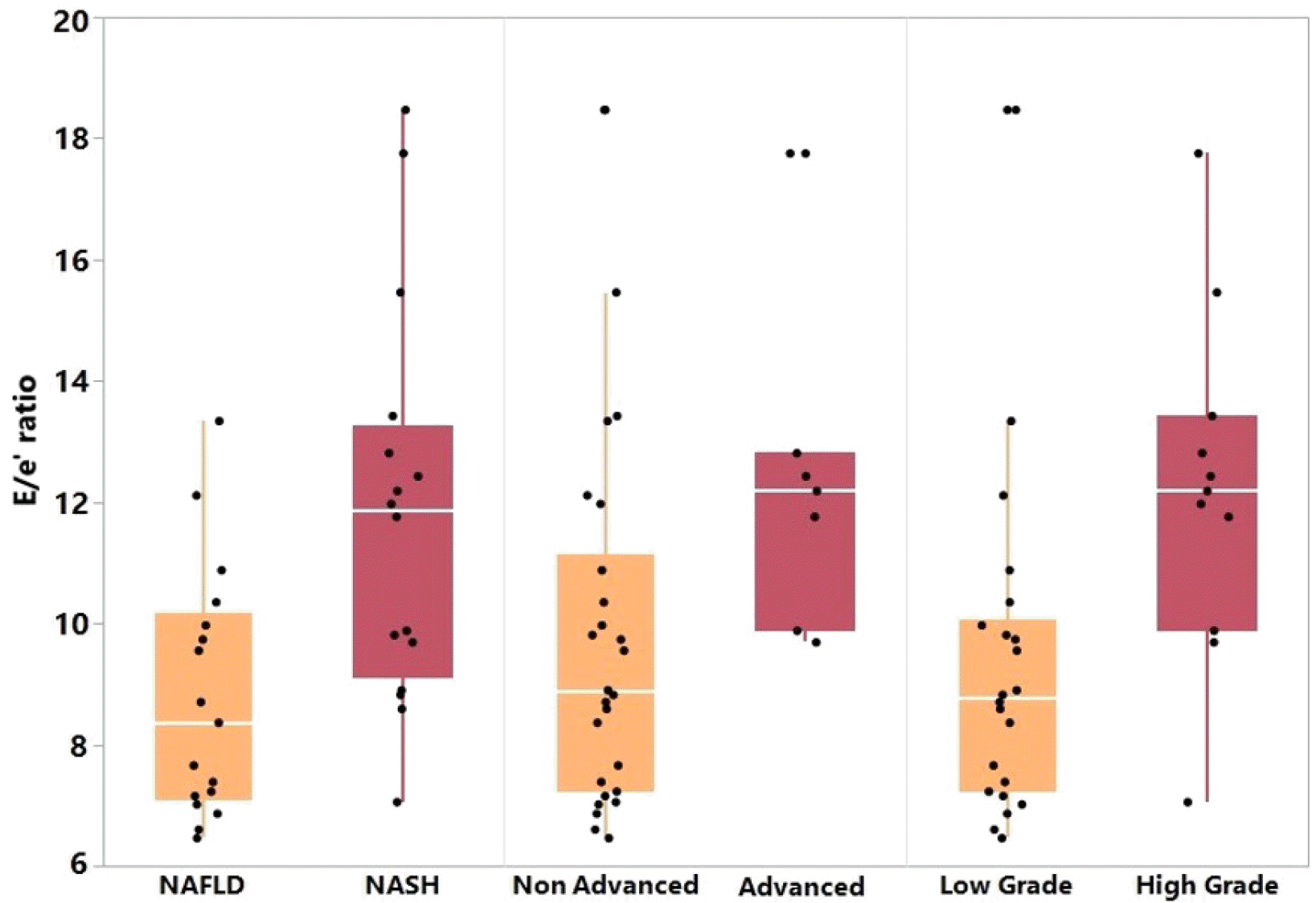
The transthoracic echocardiogram parameters were compared for three different groups: NAFL versus NASH, NAFLD without advanced fibrosis versus advanced stage NASH, and low-grade NAFLD and high-grade NASH. The E/e’ ratio, a marker of left ventricular diastolic dysfunction, was significantly higher in NASH than NAFL (11.8±3.3 vs. 8.8±2.0, p=0.00), advanced stage NASH compared to non-advanced stage NAFLD (12.4±2.7 vs. 9.7±3.0, p=0.02), and high-grade NASH compared to lower-grade NAFLD (12.2±2.9 vs. 9.3±2.7, p=0.00) (Fig. 1). NASH showed a marginally significant association with a diastolic dysfunction compared to NAFL (31% vs. 6%, p=0.05). This trend was maintained with advanced stage NASH compared to NAFLD without advanced fibrosis (43% vs. 12%, p=0.08) and with high-grade NASH compared to low-grade NAFLD (36% vs. 9%, p=0.06). There were no significant differences nor trends towards significance were observed between these three groups in regards to other markers of cardiac dysfunction: E/A ratio, LVEF, LV mass index, LA volume index, left ventricular global longitudinal systolic strain, TR velocity, and estimated PASP.
The measures of diastolic dysfunction were also compared in patients with NAFL versus NASH in those who did and did not have T2DM. The E/e’ ratio was significantly higher in NASH than NAFL in those who did not have T2DM (11.8±3.1 vs. 8.6±2.0, p=0.01), but it was not significantly higher than in those with T2DM (11.8±3.5 vs. 9.7±2.6, p=0.31). The remaining markers of cardiac dysfunction were similar in the NASH and NAFL patients with and without T2DM.
Logistic regression was performed to determine if the E/e’ ratio and other statistically significant baseline characteristics were independently associated with NASH. The baseline characteristics that were possible confounders were assessed, including T2DM, hypertension, coronary artery disease, and HDL. The full model, including T2DM, hypertension, and coronary artery disease, did not converge due to singularity. Therefore, these factors were removed, making the final model contain only the E/e’ ratio and HDL. The ORs for E/e’ and HDL were 1.87 (95% CI 1.13-3.12, p=0.00) and 0.89 (95% CI 0.80-0.98, p=0.00), respectively.
DISCUSSION
Cardiac disease is the leading cause of death of patients with NAFLD, and there are numerous studies suggesting a connection between NAFLD and subclinical cardiac dysfunction. In the present study, a histologic diagnosis of NASH was associated with an increased echocardiographic marker of diastolic dysfunction compared to the less aggressive NAFL. These findings are consistent with previous research showing the relationship between the worsening severity of NAFLD and subclinical cardiac remodeling. Jung et al.19 reported that mild and moderate-to-severe NAFLD had a higher OR of abnormal LV relaxation than the normal group (1.29 and 1.95 respectively) and increased relative wall thickness (1.26 and 1.46 respectively). Petta et al.14 showed that more severe liver fibrosis (F3-F4 fibrosis) was associated with both morphologic and functional abnormalities compared to less severe fibrosis (F0-F2), including an increased posterior wall thickness, increased LV mass/height, increased relative wall thickness, increased left atrial volume, ejection fraction, lower lateral e’, and lower E/A ratio. Furthermore, a meta-analysis by Wijarnpreecha et al.20 compiled 12 studies and 280,645 patients and reported a significant association between patients with NAFLD and diastolic cardiac dysfunction compared to patients without NAFLD (OR 2.02; 95% CI 1.47-2.79). Interestingly, a recent study using biopsy-proven NAFLD showed no increase in the E/e’ ratio at rest according to the fibrosis stage. On the other hand, there was a significant increase in stress E/e’ ratio with increasing fibrosis stage. The peak VO2, ventilatory anaerobic threshold, and exercise time were inversely correlated with the fibrosis stage, and they all decreased with increasing fibrosis stage.21 Although there was no difference in the rest E/e’ ratio, as in the present study, this does add support to an increasing diastolic dysfunction with increasing fibrosis stage similar to the present findings.
These findings show that both an advanced stage of fibrosis and a higher grade of inflammation in NAFLD is associated with an increased marker of diastolic dysfunction. This is the first study to show a positive correlation specifically with the histologic grade of inflammation and stage of fibrosis with early cardiac dysfunction. A systemic inflammation and insulin resistance from the liver fat content likely plays a key role because of the unfavorable metabolism of fatty acids, glucose, and lipoproteins. Patients with NAFLD have increased lipolysis, lipid profiles showing increased LDL and decreased HDL, hyperglycemia with insulin resistance, and secretion of inflammatory markers, including IL-6, TNF-alpha, CRP, and others. These metabolic abnormalities and inflammatory markers have been linked with accelerated atherosclerosis and coronary artery disease.22,23 Insulin resistance can also adversely affect the cardiomyocyte function because insulin signaling has been shown to play a role in myosin gene expression and affect the utilization of fatty acids and glucose in the cardiomyocytes.24
Several studies have investigated the possible association between NAFLD and the risk of CVD, but the independent prognostic role of NAFLD on CVD remains a topic of intense debate. It is unclear if the relationship between NAFLD and incidence of CVD is simply a byproduct of the shared CVD risk factors or if NAFLD actively contributes to the development of CVD. This is a relevant question because the answer could change how patients with NAFLD are screened for cardiac disease and how the treatments for NAFLD may reduce these cardiac events. The focus on identifying early cardiac disease is essential because NAFLD has been associated with several other abnormal cardiac findings, such as increased carotid-artery intima-media thickness, increased arterial stiffness, increased coronary artery calcification, and high-risk coronary plaques.25-27 Furthermore, an increasing fibrosis stage has been correlated with increased cardiac events and increased all-cause and cardiac mortality.28-30 Early changes in TTE could indicate an increased risk of cardiac events in the future.
This study has some limitations, such as the small sample size and the retrospective, single-center design. The small sample size and retrospective design made it difficult to control for possible confounders and show statistical significance between populations. For example, the significance of the E/e’ ratio disappeared when breaking down patients with NAFL and NASH into those with and without T2DM because there were only three patients with NAFL and T2DM. Subjects with NASH had a significant prior medical history and laboratory abnormalities at the baseline, which could be expected based on the understanding of the disease. On the other hand, it was difficult to elucidate if these were confounding. This study also relied on transthoracic echocardiograms being performed for any indication, which may have biased the results in many ways. There were more screening TTEs in the NAFL group (four compared to two), but most patients in the NAFL and NASH groups had TTE performed for symptoms consistent with possible cardiac disease and dysfunction. This may have made the underlying cardiac dysfunction more likely and more difficult to differentiate between the groups than a larger, asymptomatic population of NAFL and NASH. Finally, the small sample size made statistical analysis difficult because the multivariable logistical regression could not be run successfully with all the significant variables, and only E/e’ and HDL were assessed.
In conclusion, histologically proven NASH with advanced fibrosis or higher grade inflammation is associated with an increased E/e’ ratio, a marker of diastolic dysfunction, and trended toward significance in the diastolic dysfunction overall. Overall this study adds to the growing body of evidence that demonstrates an independent correlation between the histological severity of NAFLD and subclinical cardiac dysfunction. More studies with a histological diagnosis of NASH will be needed to clarify these findings.
 XML Download