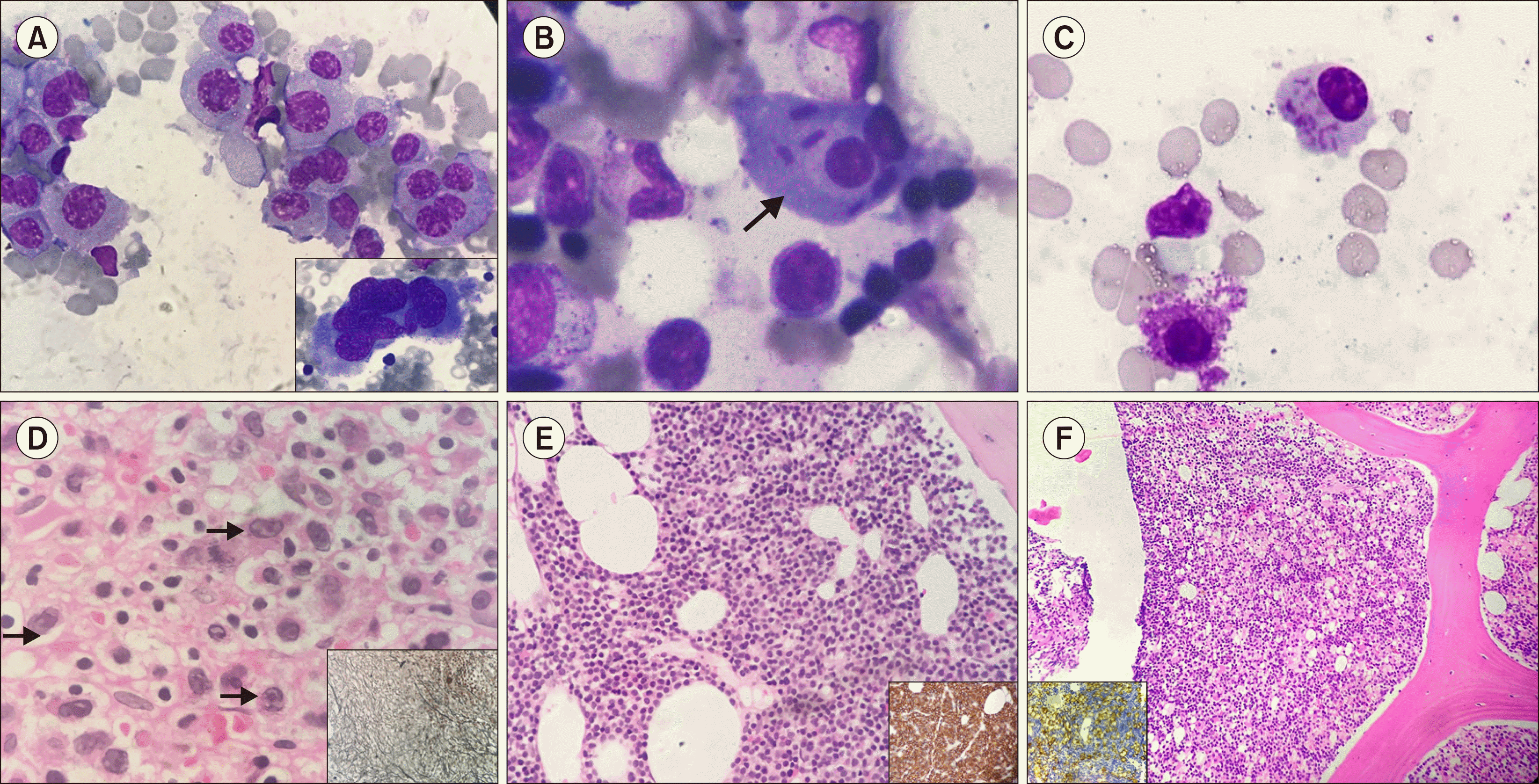
Six challenging morphological variants of plasma cells were identified in various plasma cell neoplasm and confirmed with flow cytometric immunophenotyping or immunohistochemistry. (A) Multinucleated plasma cells, with few resembling a megakaryocyte, in Jenner–Giemsa-stained bone marrow aspirate in an “anaplastic variant” of myeloma (×40). Inset shows a bizarre plasma cell (×100). Classical binucleate plasma cell with thick rod-like inclusion in a smear (B, ×100). A case of MGUS with intracytoplasmic coarse azurophilic Snapper–Schneid granules in plasma cells (C, ×100). In a case with a dry tap, bone marrow biopsy specimen shows large plasma cells with moderate cytoplasm, irregular nuclei with chromatin remodeling and prominent nucleoli (arrow) in a dense fibrotic marrow. Positive staining for CD138 and kappa restriction confirmed a diagnosis of plasmablastic myeloma (D, ×40). Bone marrow biopsy revealed sheet-like arrangement of small cells with scanty cytoplasm that mimic a lymphoma in a “small cell variant” (E, ×20); inset shows CD138-positive staining of all cells with aberrant CD20 positivity and CD45 negativity; FISH showed t(11:14). Bone marrow biopsy specimen showed a histiocytic variant of large plasma cells with abundant pale cytoplasm among classical small lymphoid cells in lymphoplasmacytic lymphoma (×20); inset shows CD138 positivity and lambda restriction (F).


 PDF
PDF Citation
 Print
Print


 XML Download