Journal List > J Bacteriol Virol > v.51(3) > 1147501

Kang, Lee, Jeon, and Jo: Roles of Interleukin-17 and Th17 Responses in COVID-19

Abstract

The ongoing coronavirus disease-2019 (COVID-19) pandemic is caused by severe acute respiratory syndrome coronavirus-2. COVID-19 severity is related to the cytokine storm phenomenon, which is amplified by pro-inflammatory cytokines and chemokines; it may cause extensive pulmonary damage. Among these cytokines, interleukin (IL)-17, produced mainly by T helper 17 (Th17) cells, is responsible for the immunopathological responses present in acute respiratory distress syndrome. This review discusses the roles of IL-17 and Th17 responses in the pathophysiology of COVID-19. Dysregulated Th17-responses, linked to various risk factors, may contribute to pathological inflammation through the amplification of multiple inflammatory cytokines and chemokines, as well as augmentation of neutrophil infiltration in the lungs of severe COVID-19 patients. A more detailed understanding of the roles of Th17 responses, as well as the mechanisms underlying altered IL-17 production and signaling, may improve therapeutic strategies for severe or critically ill COVID-19 patients by targeting the IL-17 pathway.

INTRODUCTION

The outbreak of respiratory coronavirus disease 2019 (COVID-19), caused by the severe acute respiratory syndrome coronavirus-2 (SARS-CoV-2), is the most important public health challenge worldwide (1). SARS-CoV-2 is a positive-stranded, enveloped RNA virus, which can be zoonotically transmitted from animals to humans (2, 3). Bats comprise the most likely animal reservoir of SARS-CoV-2, but the presence of reservoirs in other animals has not been entirely excluded (4, 5, 6). The mortality rate of COVID-19 is approximately 3–4%, but the transmission rate of SARS-CoV-2 among humans is extremely high, presumably because of its air-droplet mode of transmission (7). COVID-19 has a wide range of symptoms, from mild-moderate to severe life-threatening infections; it may cause pneumonia, kidney failure, and death (7). Severe COVID-19 is associated with cytokine storm onset, which involves excessive and uncontrolled secretion of cytokines that may result in fatal sepsis and multi-organ failure (8, 9).
Cytokines and chemokines are protein messengers produced by immune cells, which have important roles in immunological and pathological responses during viral infections (10). Although coordinated cytokine induction is crucial for host immune response, dysregulated production is associated with immunopathology that can cause cytokine storm onset (11, 12, 13). Cytokines associated with COVID-19 include pro- and anti-inflammatory interleukins (ILs), chemokines, and interferons (10, 14). IL-17 is produced by T-helper 17 (Th17) cells; its signaling is associated with immune functions of barrier epithelial tissues and host defense against extracellular bacterial and fungal infections (15, 16, 17, 18). IL-17 includes unique structures composed of IL-17A and IL-17F (17); it is an inducer of antimicrobial proteins, various chemokines, acute-phase response mediators, and inflammatory functions (16, 19). Recent studies have found increased inflammatory responses in COVID-19 patients because of IL-17 overproduction (20, 21). In this review, we discuss the characteristics of IL-17A, its relevance to COVID-19, and the potential for IL-17A-based therapeutics.

OVERVIEW OF IL-17 AND ITS SIGNALING NETWORK

Cytokines, combined with specific receptors, have pleiotropic roles in innate and adaptive immune responses that involve complicated intracellular signal pathways (22). Cytokine dysregulation can cause pathological effects in the host immune system and tissues (23). This section provides a brief overview of IL-17 in terms of biological function and signaling pathways, including JAK and STAT3.

Biological functions of IL-17

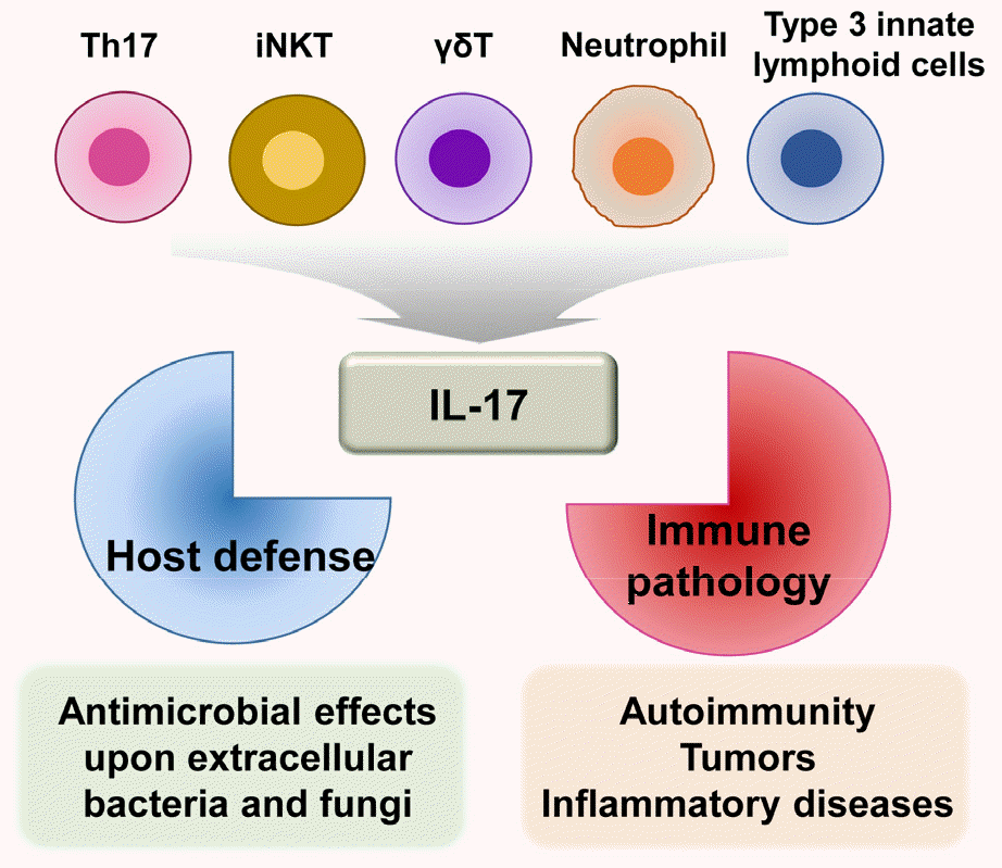
IL-17, produced mainly by Th17 cells (24), has crucial roles in adaptive immunity and inflammatory responses during infection (24). IL-17 is a member of the inflammatory cytokine family that can exert both host-protective and pathological effects (21). The IL-17 family consists of six structurally related members: IL-17A, IL-17B, IL-17C, IL-17D, IL-17E (IL-25), and IL-17F (18). IL-17A is the prototype of the IL-17 cytokine family; it is co-expressed along with IL-17F by Th17 cells (25). IL-17A is also produced by various hematopoietic cells, including CD8+ T cells, invariant natural killer T cells, gamma delta T cells, neutrophils, and type 3 innate lymphoid cells (26, 27). IL-17A is an essential cytokine involved in host defense against extracellular bacterial and fungal infections (15, 17, 18). However, it also contributes to pathologically excessive pro- inflammatory responses related to the development of autoimmunity, tumors, and other inflammatory diseases (15, 28) (Fig. 1).
Fig. 1

IL-17 production and function. IL-17 is mainly produced by Th17 cells and other cell types, including CD8+ T cells, invariant natural killer T cells, gamma delta T cells, neutrophils, and type 3 innate lymphoid cells. IL-17 has a dual function: host defense against extracellular bacterial and fungal infections, as well as pathologic inflammation involved in various diseases (e.g., autoimmune disorders, tumors, and inflammatory diseases).

JBV_2021_v51n3_089_f001.tif
IL-17 binds to receptors to induce an immune response specific to the target cells (15). The IL-17R family consists of five members (IL-17RA, IL-17RB, IL-17RC, IL-17RD, and IL-17RE); IL-17RA is the principal member (25, 27). The IL-17/IL-17R interaction is required for inflammatory responses through various signaling molecules and transcriptional factors, including nuclear factor-κB, Janus kinase (JAK)–STAT, and tumor necrosis factor receptor-associated factor-6 (15, 29). Intracellular signaling pathways involving the JAK–STAT3 interaction are discussed in detail in the following section.

JAK–STAT signaling pathway and STAT3

The JAK–STAT pathway is a representative cytokine signaling pathway that mediates messages from membrane receptors to the nucleus (30, 31). The JAK–STAT signaling pathway is composed of three classes of proteins: tyrosine kinase-related receptors, JAK enzymes, and signal transducer and activator of transcription (STAT) transcription factors (32). JAKs are phosphorylated in the presence of cytokine ligands, thus creating STAT docking sites. STAT molecules are phosphorylated and dimerized by JAK, then transported to the nucleus to regulate expression of cytokine-responsive genes in the nuclear membrane (33).
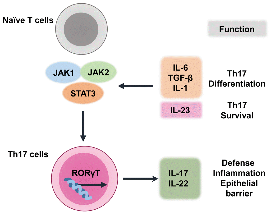
The JAK–STAT signaling pathway is critical for transducing signals of various cytokines and growth factors (e.g., IL-6, granulocyte-macrophage colony-stimulating factor, growth hormone, epidermal growth factor, platelet-derived growth factor, and interferons) in various cell types (30). IL-6, IL-1, and IL-23 activate STAT3, a transcriptional factor required for Th17-cell differentiation (34). Retinoic acid-related orphan receptor (ROR)γt is another transcription factor crucial in Th17-cell development. Th17-cell differentiation is activated by IL-1β, IL-6, and transforming growth factor-β; it is maintained by IL-23 (20, 34). Thus, multiple cytokines and transcriptional factors are involved in IL-17 production by Th17 cells.
STAT3 activates RORγt transcription, which regulates Th17-cell differentiation (20). The STAT3–RORγt interaction is important for Th17-cell differentiation (24). Th17-cell survival and differentiation require IL-23 and IL-21, which are produced by dendritic and T cells, respectively (24). IL-6 regulates Th17 cells through a positive feedback loop that amplifies the expression and activation of IL-17 and STAT3 (35). Th17-cell differentiation is promoted by the binding of IL-6 and transforming growth factor-β to naive CD4+ T cells (36). However, Th17-cell differentiation does not require STAT4 or STAT6, which are transcription factors that regulate Th1 and Th2 reactions, respectively (37). Indeed, STAT3 is essential for Th17-cell differentiation (38). Because STAT3C is an active STAT3 variant, its overexpression increases the number of IL-17-producing lymphocytes. This results in increased RORγt expression in CD4+ T lymphocytes (39). In contrast, STAT3 deficiency in hematopoietic stem cells, especially CD4+ lymphocytes, results in a sharp decrease in the number of Th17 lymphocytes (39, 40). Together, STAT3 and RORγt are crucial for the activation of IL-17-producing Th17 cells. Fig. 2 summarizes the differentiation of Th17 cells and the related cytokine network. Details of IL-17/IL-17R signaling pathways are beyond the scope of this review; they have been discussed in several recent reviews (15, 27, 41). The next section describes the roles of IL-17 and Th17-related risk factors in the pathophysiology of COVID-19.
Fig. 2

Th17-cell differentiation and related cytokine network. In naive T cells that are committed to differentiate into Th17 cells, multiple cytokines (e.g., IL-6, IL-1, transforming growth factor-β, and IL-23) activate STAT3, which has a critical role in Th17-cell differentiation. RORγt is a crucial transcription factor in Th17-cell development, and STAT3 is a key factor in the transcriptional activation of RORγt. Finally, Th17-cell survival and differentiation are assisted by IL-23.

JBV_2021_v51n3_089_f002.tif

ROLE OF IL-17 IN COVID-19

IL-17 and pathological responses in COVID-19 patients

Previous studies have reported upregulation of Th17 cells in COVID-19 patients (42, 43). IL-17 is involved in the pathogenesis of acute respiratory distress syndrome through the enhancement of neutrophil infiltration into the lungs (44, 45, 46). Increased IL-17A signaling and upregulation of Th17 or regulatory T cells (Treg) are positively correlated with acute respiratory distress syndrome severity in Middle East respiratory syndrome-related coronavirus and respiratory syncytial virus infections (47, 48). Similarly, Th17-cell overactivation and exaggerated cytotoxic effects of CD8+ T cells are at least partly responsible for severe immune injury in COVID-19 patients (43). A recent study showed that COVID-19 patients with pneumonia exhibited exhausted T-cell profiles with upregulated Th17 responses (49). IL-17A is associated with lung inflammation and poor clinical outcomes in viral acute respiratory distress syndrome (45). IL-17A signaling amplifies pathological inflammation through the induction of pro-inflammatory cytokines, such as IL-6 and IL-1; thus, it is considered a promising target for adjunctive acute respiratory distress syndrome treatment in COVID-19 patients (44). Despite a known association between IL-17 and acute pulmonary injury in COVID-19 patients, the underlying functional mechanisms are poorly understood.
There is substantial evidence to support an interaction between IL-17 and angiotensin-converting enzyme 2 (ACE2), an entry receptor for SARS-CoV-2 (50, 51, 52, 53). Murine models have demonstrated a relationship between pulmonary human ACE2 expression and COVID-19 development, along with a pro-inflammatory immune response (54). In a murine model of severe bacterial pneumonia, recombinant ACE2 has been shown to inhibit IL-17A-mediated STAT3 activation and decrease pulmonary neutrophil infiltration. This suggests a protective role for ACE2 in bacterial lung infections (55). Although the relationship between ACE2 expression and COVID-19 severity is complex, ACE2 is an important regulator of IL-17A production (53, 56). IL-17A-mediated pulmonary inflammation in COVID-19 patients may lead to an increased number of alveolar neutrophils and organ dysfunction (20); therefore, future research should address pathogenic IL-17A responses and factors related to IL-17A signaling, which can affect the clinical severity of COVID-19. Understanding the mechanism of increased IL-17A production in various tissues during SARS-CoV-2 infection may provide novel strategies for treatment using IL-17-based drugs.

Risk factors and Th17 responses in COVID-19

Risk factors for COVID-19 include age, obesity, sex, cardiovascular diseases, chronic kidney disease, hypertension, and diabetes mellitus (57, 58, 59, 60, 61). These risk factors are known to upregulate Th17 responses and IL-17A signaling (20). It has been suggested that pro-inflammatory responses (e.g., increased IL-17A levels) contribute to lung damage and hypercoagulability in patients with severe COVID-19 (62). Although the effects of these risk factors on IL-17A production and Th17 responses are poorly understood, their relationship supports the possibility for use of IL-17A-targeting modalities in the management of severe COVID-19 patients with high risk of mortality. In this section, we have explored the effects of some risk factors on IL-17 levels and pathological responses in COVID-19.
Age
COVID-19 patients aged ≥ 80 years have a 20-fold greater risk of mortality, compared with such patients aged 50–59 years (63). Although the age-related alterations in Th17 responses are not well understood, there is evidence for upregulated differentiation of IL-17-producing effector cells from naive CD4+ T cells in healthy volunteers aged > 65 years (64). A recent study showed that elderly individuals had increased Th17 responses (including upregulated RORγt expression and IL-17 levels in peripheral blood), compared with middle-aged and young individuals (65). IL-17 treatment of human umbilical vein endothelial cells has been shown to cause elevated endothelin-1 and vascular cell adhesion protein-1 levels. This suggests a relationship between Th17 responses and endothelial cell senescence (65). Another study reported that transcriptional activation of IL-17-, IL-17F-, IL-22-, and IL-17-producing CD4+ T cells significantly increased with age in humans and mice (66). Increased IL-17 and Th17 responses were also reported in aged mice due to increased mRNA expression of human-homologous RORγt (66). Adoptive transfer of naive T cells from aged mice leads to the development of colitis in RAG−/− mice, suggesting a role for Th17 in the pathogenesis of inflammatory disorders (66). Moreover, the balance between Th17 (CD4+ IL23 receptor(R)+) and Treg (CD4+ Foxp3+) cells is dysregulated in old age, resulting in Th17-cell predominance (67). Although these studies suggest that immune senescence is closely related to increased Th17 responses and IL-17 production, the COVID-19-specific roles of age-related alterations in Th17–Treg balance remain poorly understood.
Cardiovascular diseases and hypertension
SARS-CoV-2 spike protein binds to ACE2, an important component of renin–angiotensin and cardiovascular systems, to facilitate viral invasion into host cells (68). Numerous studies have demonstrated a link between COVID-19 and cardiovascular diseases (69, 70, 71, 72). SARS-CoV-2 infection triggers endothelial injury and dysfunction, leading to increased mortality in COVID-19 patients (56, 68, 73). Although the mechanisms underlying myocardial injury are largely unknown, Th17 dysregulation may contribute to COVID-19 outcomes in patients with comorbid cardiovascular diseases and hypertension.
IL-17 is involved in angiotensin II-mediated hypertension and cardiovascular dysfunction (74); it comprises a therapeutic target for cardiovascular diseases. Th17-cell differentiation causes organ damage in hypertensive mice (75), whereas genetically IL-17A-deficient mice have low blood pressures because of Th17–Treg imbalance (74). Patients with hypertension have increased numbers of Th1 and Th17 cells, as well as increased levels of related cytokines (76). Based on these reports, hypertension is presumed to be closely associated with increased Th17 response and Th17–Treg imbalance, thereby contributing to COVID-19 severity.
Obesity
Obesity has been suggested to cause chronic inflammation, thereby leading to Th17-related metabolic and autoimmune diseases (77, 78). An experimental study found greater numbers of IL-17-secreting CD4+ T cells in obese mice than in lean mice (78). The increased Th17 responses associated with obesity are dependent on IL-6 production; they are linked to autoimmune disease susceptibility (78). Altered regulation of intracellular metabolism, including fatty-acid metabolism, might result in obesity-related Th17 responses (79). Studies have shown higher IL-17 and IL-23 levels in obese women than in lean women (80). Based on these findings, IL-17 pathway activation might be amplified in obesity, potentially leading to adverse clinical outcomes in patients with COVID-19 (81). The mechanisms by which obesity induces Th17 inflammatory responses are unclear. Th17–Treg imbalance may cause dysregulated immune homeostasis because lean adipose tissues are rich in Tregs, which control tissue inflammation (82). Notably, acetyl-CoA carboxylase 1 (ACC1) was found as a key regulator of Th17 cell differentiation through regulation of RORγt binding to the Il17a gene (83). Further studies are needed to clarify the molecular mechanisms underlying Th17-cell increase and Treg-cell decrease, as well as the roles of obesity-related metabolic changes.
Diabetes mellitus
Diabetes mellitus and hyperglycemia are considered predictors of poor prognosis in COVID-19 (84, 85). Hyperglycemia is linked to dysregulated innate immunity involving upregulated expression of Toll-like receptors and defective neutrophil function (86). In addition, hyperglycemia leads to protein glycosylation, altered complement function, and phagocytosis inhibition (86). Therapies for COVID-19 may lead to poor glycemic control because of virus-triggered β-cell damage and increased insulin resistance through exaggerated inflammatory cytokine production (87). Thus, a vicious cycle might arise involving diabetes mellitus and COVID-19 (87).
Th17 dysregulation is likely to contribute to complex interactions between COVID-19 and diabetes mellitus. Patients with types 1 and 2 diabetes have increased Th17–Treg imbalances and Th17-cell activation, compared with controls (20, 88, 89, 90). Diabetic patients have lower levels of miR-146a, which regulates Th17-cell differentiation by inhibiting the expression of cytokines such as IL-6 and IL-21 (91). In addition, increased pro-inflammatory cytokine signaling and poor glycemic control in diabetic patients are associated with Th17-response dysregulation (91, 92). These studies suggest that IL-17-targeting therapeutics may be useful in the treatment of diabetic COVID-19 patients.
Sex
Several studies have suggested sex-differences in the clinical outcomes of COVID-19. A recent meta-analysis showed a higher prevalence of COVID-19 in men (93). Smoking and alcohol consumption were also more prevalent in men, and they have been linked to increased Th17-cell frequencies (94, 95). Furthermore, severe COVID-19 was more common in men than in women, although robust analyses are needed to further evaluate these sex differences (96, 97, 98). Male COVID-19 patients had higher plasma inflammatory cytokine levels, such as IL-8 and IL-18, whereas female patients had upregulated T-cell activation. Male COVID-19 patients with severe disease also exhibited a negative correlation between a poor T-cell response and age (97). However, the molecular mechanisms by which sex is associated with COVID-19 severity are unknown.
There is evidence to suggest that estrogen and progesterone have roles in the regulation of Th17 responses (99). Estrogen deficiency increases Th17-cell differentiation and lowers the number of Treg cells. This is partly because of the inhibitory effects of estrogen on RORγt activation, which interfere with Th17-cell differentiation (100, 101). In addition, estradiol treatment impairs antigen presentation and IL-23 production by dendritic cells, thereby inhibiting Th17 immune responses (102). In an experimental viral myocarditis model, Th17-cell responses were significantly greater in male mice than in female mice, indicating sex differences in terms of susceptibility to Coxsackievirus B3 infection (103). Progesterone and estrogen inhibited the production of Th1/Th17 inflammatory cytokines, although they increased the production of anti-inflammatory cytokines in human peripheral blood mononuclear cells from women with recurrent spontaneous miscarriages (104). Further studies are needed to clarify whether men have increased Th17 responses, which may increase COVID-19 susceptibility and severity. Additionally, estradiol and progesterone levels may influence COVID-19-related cytokine storm outcomes via enhanced immune tolerance (105). A greater understanding of the role of sex in COVID-19 may contribute to the development of potential sex-based therapeutic approaches.
Acute and chronic kidney diseases
Several studies have suggested that COVID-19 risks and outcomes may be related to chronic kidney disease and kidney transplantation (106, 107, 108). In addition, COVID-19 may be complicated by acute kidney injury, leading to a greater mortality risk (109, 110). Although the underlying mechanisms are largely unknown, Th17 responses might contribute to COVID-19 outcomes in patients with comorbid chronic kidney diseases. An association between common single nucleotide polymorphisms in IL17RA and a risk of renal disease has been demonstrated (111). The homozygous single nucleotide polymorphism rs4819554 AA in IL17RA increases IL17RA protein expression and Th17 responses, which might contribute to renal damage and impaired kidney function (111). IL-17A is involved in numerous pathological conditions related to the development of acute and chronic renal diseases (112, 113, 114). Chronic kidney disease patients often have Th17–Treg imbalances and increased IL-17A responses (112). Table 1 summarizes potential mechanisms by which several risk factors may alter Th17 responses in COVID-19. Further studies are needed to clarify the implications of renal disease in COVID-19 via altered Th17 responses involving IL-17A hyperactivation.
Table 1.

Potential mechanisms by which the risk factors influence altered Th17 responses in COVID-19

Risk factors of COVID-19 Study model Pathologic outcomes in various disease conditions Reference
Aging Human PBMCs The increased Th17 differentiation from naïve CD4+ T cells in healthy eldery group (64)
Human endothelial cells IL-17 leads to the elevation of ET-1 and VCAM-1 levels (65)
C57BL/6 mice Increased Th17 cytokines and frequncies in aged mice (66)
Cardiovascular disease IL17-/- mice ACE2 enhances aortic T cell infiltration with increased IL-17 and other inflammatory cytokines (74)
C57BL/6 mice ACE2 increases Th17 cells through upregulation of SGK1- FoxO1 pathway (75)
Human PBMCs Increased Th17 cells and IL-17 levels in hypertensive patients (76)
Obesity ACC1-/- mice Human PBMCs ACC1 increases Th17 diffrentiation through RORγt activation (83)
Type 1 diabetes Human PBMCs Elevated Th17 responses positively correlate with CRP (89)
Type 2 diabetes Human PBMCs Increased Th17 responses positively correlate with nephropathy (90)
Alcohol RORγt-/- mice Th17 drives alcohol-associated gut inflammation and the development of ALD (94)
Smoking Human PBMCs Increased CCR6+ Th17 cells and VEGFα secretion (95)
Progesterone deficiency Human PBMCs Progesterone decreases STAT3-IL-6 signaling (99)
Estrogen deficiency BALB/c mice Ovariectomized mice increases RORγt, STAT3, and IL-17 (101)
Male BALB/c mice Upregulated Th17 responses in Coxsackievirus B3-infected male mice increases susceptibility of myocarditis (103)
Kidney disease Human PBMCs Increased Th17 frequencies and IL-17 production from effector memory T cells in end-stage renal disease patients (113)
Human PBMCs Enhanced Th17 diffrentiation and IL-17 levels positively correlate with phosphate levels in chronic hemodialysis patients (114)

ACC1, acetyl-CoA carboxylase 1; ACE2. angiotensin-converting enzyme 2; ALD, alcohol-associated liver disease; COVID-19, coronavirus disease 2019; CRP, C-reactive protein; ET-1, endothelin-1; FoxO1, forkhead box O1; IL, interleukin; PBMC, peripheral blood mononuclear cell; RORγt, retineic-acid-receptor-related orphan receptor gamma T; SGK1, serum/glucocorticoid regulated kinase 1; Th17, T helper 17; VCAM-1, Vascular cell adhesion molecule-1. VEGFα, vascular endothelial growth factor α

IL-17-targeting therapeutics in COVID-19

Despite some side effects, monoclonal antibodies that interact with the IL-17 signaling pathway (e.g., secukinumab, ixekizumab, and brodalumab) have been used in therapeutic trials for several IL-17-related autoimmune diseases, including psoriatic diseases and rheumatic diseases (52, 115, 116). The success of IL-17 antagonists in the treatment of autoimmune diseases has encouraged their use in the treatment of COVID-19. In particular, JAK inhibitors, including baricitinib, ruxolitinib, and fedratinib, showed effective anti-inflammatory responses in COVID-19 patients through suppression of the JAK–STAT pathway (117). A recent experimental study showed that ex vivo treatment of whole-blood samples from COVID-19 patients with baricitinib led to a significant decrease in viral spike-specific cytokine release (118). Another study showed that fedratinib, a Food and Drug Administration-approved Janus kinase 2 (JAK2) inhibitor, suppressed the Th17 pathway; this reduced mortality and ameliorated the cytokine storm that occurs in COVID‐19 patients (42). In addition, ruxolitinib, a JAK1 and JAK2 inhibitor, is currently under investigation (ChiCTR2000029580) for potential therapeutic application involving interference with the inflammatory response in COVID-19 (119). Furthermore, considering the close relationship between IL-17 and IL-6, a combined approach to block these cytokines has been proposed as a potential treatment for COVID-19 (26). However, caution is needed because IL-17 deficiency can contribute to small bowel pathology and cytokine release syndrome through an interferon-γ–STAT1-mediated inflammatory pathway (120). Further studies are necessary to clarify whether IL-17-targeting therapeutics can reduce the severity of COVID-19.

CONCLUSION

IL-17A is a key inflammatory cytokine, mainly produced by Th17 cells, which leads to the activation of various pro- inflammatory cytokines and chemokines. This causes lung injury in COVID-19; it also contributes to other inflammatory diseases. Emerging data suggest a pivotal role for IL-17 in pathological inflammatory responses, including severe COVID-19. In addition, IL-17A has been linked to risk factors for poor COVID-19 outcomes (e.g., immune senescence, cardiovascular diseases, obesity, diabetes mellitus, sex, and kidney injury). However, the underlying molecular mechanisms are largely unknown. Although several studies have indicated that excessive IL-17 production is related to the development of acute respiratory distress syndrome in patients with COVID-19, further studies in larger populations are needed to determine the IL-17 levels in sera, plasma, and bronchoalveolar lavage fluids from patients with different clinical phenotypes. This would clarify the usefulness of IL-17-targeting therapeutics in subgroups of patients with IL-17 hyperactivation. The success of IL-17-targeting therapeutics (monoclonal antibodies and JAK–STAT signaling inhibitors) in several autoimmune diseases encourages their potential use in the treatment of COVID-19. Preclinical and clinical data must be accumulated to identify the factors that determine whether IL-17-targeting therapeutics should be used alone or in combination with other drugs or monoclonal antibodies. These translational studies based on IL-17 and Th17 functions suggest the potential for development of improved COVID-19 treatment strategies.

ACKNOWLEDGMENTS

This work was supported by research fund of Chungnam National University. We are indebted to our medical student colleagues at College of Medicine, Chungnam National University, for discussions and investigations that contributed to this article. We thank Dr. P. Silwal for critical reading of the paper. We apologize to colleagues whose work and publications could not be referenced owing to space constraints.
[DISCLOSURE OF POTENTIAL CONFLICT OF INTEREST]
The authors have no competing financial interests to declare.

References

1. Chang L, Yan Y, Wang L. Coronavirus Disease 2019: Coronaviruses and Blood Safety. Transfus Med Rev 2020;34(2):75-80.DOI: 10.1016/j.tmrv.2020.02.003. PMID: 32107119. PMCID: PMC7135848.
2. Mahdy MAA, Younis W, Ewaida Z. An Overview of SARS-CoV-2 and Animal Infection. Front Vet Sci 2020;7:596391.DOI: 10.3389/fvets.2020.596391. PMID: 33363234. PMCID: PMC7759518.
3. Coronaviridae Study Group of the International Committee on Taxonomy of Viruses. The species Severe acute respiratory syndrome-related coronavirus: classifying 2019-nCoV and naming it SARS-CoV-2. Nat Microbiol 2020;5(4):536-44.DOI: 10.1038/s41564-020-0695-z. PMID: 32123347. PMCID: PMC7095448.
4. Zhou P, Yang XL, Wang XG, Hu B, Zhang L, Zhang W, et al. A pneumonia outbreak associated with a new coronavirus of probable bat origin. Nature 2020;579(7798):270-3.DOI: 10.1038/s41586-020-2012-7. PMID: 32015507. PMCID: PMC7095418.
5. Ji W, Wang W, Zhao X, Zai J, Li X. Cross-species transmission of the newly identified coronavirus 2019-nCoV. J Med Virol 2020;92(4):433-40.DOI: 10.1002/jmv.25682. PMID: 31967321. PMCID: PMC7138088.
6. Cui J, Li F, Shi ZL. Origin and evolution of pathogenic coronaviruses. Nat Rev Microbiol 2019;17(3):181-92.DOI: 10.1038/s41579-018-0118-9. PMID: 30531947. PMCID: PMC7097006.
7. Harrison AG, Lin T, Wang P. Mechanisms of SARS-CoV-2 Transmission and Pathogenesis. Trends Immunol 2020; 41(12):1100-15.DOI: 10.1016/j.it.2020.10.004. PMID: 33132005. PMCID: PMC7556779.
8. Pujari R, Thommana MV, Ruiz Mercedes B, Serwat A. Therapeutic Options for COVID-19: A Review. Cureus 2020;12(9):e10480.DOI: 10.7759/cureus.10480. PMID: 32953365. PMCID: PMC7496561.
9. Behrens EM, Koretzky GA. Review: Cytokine Storm Syndrome: Looking Toward the Precision Medicine Era. Arthritis Rheumatol 2017;69(6):1135-43.DOI: 10.1002/art.40071. PMID: 28217930.
10. Zhang JM, An J. Cytokines, inflammation, and pain. Int Anesthesiol Clin 2007;45(2):27-37.DOI: 10.1097/AIA.0b013e318034194e. PMID: 17426506. PMCID: PMC2785020.
11. Channappanavar R, Fehr AR, Vijay R, Mack M, Zhao J, Meyerholz DK, et al. Dysregulated Type I Interferon and Inflammatory Monocyte-Macrophage Responses Cause Lethal Pneumonia in SARS-CoV-Infected Mice. Cell Host Microbe 2016;19(2):181-93.DOI: 10.1016/j.chom.2016.01.007. PMID: 26867177. PMCID: PMC4752723.
12. Davidson S, Maini MK, Wack A. Disease-promoting effects of type I interferons in viral, bacterial, and coinfections. J Interferon Cytokine Res 2015;35(4):252-64.DOI: 10.1089/jir.2014.0227. PMID: 25714109. PMCID: PMC4389918.
13. Shaw AC, Goldstein DR, Montgomery RR. Age-dependent dysregulation of innate immunity. Nat Rev Immunol 2013;13(12):875-87.DOI: 10.1038/nri3547. PMID: 24157572. PMCID: PMC4096436.
14. Chi Y, Ge Y, Wu B, Zhang W, Wu T, Wen T, et al. Serum Cytokine and Chemokine Profile in Relation to the Severity of Coronavirus Disease 2019 in China. J Infect Dis 2020;222(5):746-54.DOI: 10.1093/infdis/jiaa363. PMID: 32563194. PMCID: PMC7337752.
15. Ge Y, Huang M, Yao YM. Biology of Interleukin-17 and Its Pathophysiological Significance in Sepsis. Front Immunol 2020;11:1558.DOI: 10.3389/fimmu.2020.01558. PMID: 32849528. PMCID: PMC7399097.
16. Gaffen SL, Jain R, Garg AV, Cua DJ. The IL-23-IL-17 immune axis: from mechanisms to therapeutic testing. Nat Rev Immunol 2014;14(9):585-600.DOI: 10.1038/nri3707. PMID: 25145755. PMCID: PMC4281037.
17. Gaffen SL. Recent advances in the IL-17 cytokine family. Curr Opin Immunol 2011;23(5):613-9.DOI: 10.1016/j.coi.2011.07.006. PMID: 21852080. PMCID: PMC3190066.
18. Iwakura Y, Ishigame H, Saijo S, Nakae S. Functional specialization of interleukin-17 family members. Immunity 2011;34(2):149-62.DOI: 10.1016/j.immuni.2011.02.012. PMID: 21349428.
19. Gaffen SL. Structure and signalling in the IL-17 receptor family. Nat Rev Immunol 2009;9(8):556-67.DOI: 10.1038/nri2586. PMID: 19575028. PMCID: PMC2821718.
20. Orlov M, Wander PL, Morrell ED, Mikacenic C, Wurfel MM. A Case for Targeting Th17 Cells and IL-17A in SARS-CoV-2 Infections. J Immunol 2020;205(4):892-8.DOI: 10.4049/jimmunol.2000554. PMID: 32651218. PMCID: PMC7486691.
21. Amatya N, Garg AV, Gaffen SL. IL-17 Signaling: The Yin and the Yang. Trends Immunol 2017;38(5):310-22.DOI: 10.1016/j.it.2017.01.006. PMID: 28254169. PMCID: PMC5411326.
22. O'Shea JJ, Murray PJ. Cytokine signaling modules in inflammatory responses. Immunity 2008;28(4):477-87.DOI: 10.1016/j.immuni.2008.03.002. PMID: 18400190. PMCID: PMC2782488.
23. Tanaka T, Narazaki M, Kishimoto T. Immunotherapeutic implications of IL-6 blockade for cytokine storm. Immunotherapy 2016;8(8):959-70.DOI: 10.2217/imt-2016-0020. PMID: 27381687.
24. Robins E, Zheng M, Ni Q, Liu S, Liang C, Zhang B, et al. Conversion of effector CD4(+) T cells to a CD8(+) MHC II-recognizing lineage. Cell Mol Immunol 2021;18(1):150-61.DOI: 10.1038/s41423-019-0347-5. PMID: 32066854.
25. Brevi A, Cogrossi LL, Grazia G, Masciovecchio D, Impellizzieri D, Lacanfora L, et al. Much More Than IL-17A: Cytokines of the IL-17 Family Between Microbiota and Cancer. Front Immunol 2020;11:565470.DOI: 10.3389/fimmu.2020.565470. PMID: 33244315. PMCID: PMC7683804.
26. Casillo GM, Mansour AA, Raucci F, Saviano A, Mascolo N, Iqbal AJ, et al. Could IL-17 represent a new therapeutic target for the treatment and/or management of COVID-19-related respiratory syndrome? Pharmacol Res 2020;156:104791.DOI: 10.1016/j.phrs.2020.104791. PMID: 32302707. PMCID: PMC7194796.
27. Matsuzaki G, Umemura M. Interleukin-17 family cytokines in protective immunity against infections: role of hematopoietic cell-derived and non-hematopoietic cell-derived interleukin-17s. Microbiol Immunol 2018;62(1):1-13.DOI: 10.1111/1348-0421.12560. PMID: 29205464.
28. Hu Y, Shen F, Crellin NK, Ouyang W. The IL-17 pathway as a major therapeutic target in autoimmune diseases. Ann N Y Acad Sci 2011;1217:60-76.DOI: 10.1111/j.1749-6632.2010.05825.x. PMID: 21155836.
29. Schwandner R, Yamaguchi K, Cao Z. Requirement of tumor necrosis factor receptor-associated factor (TRAF)6 in interleukin 17 signal transduction. J Exp Med 2000;191(7):1233-40.DOI: 10.1084/jem.191.7.1233. PMID: 10748240. PMCID: PMC2193168.
30. O'Shea JJ, Schwartz DM, Villarino AV, Gadina M, McInnes IB, Laurence A. The JAK-STAT pathway: impact on human disease and therapeutic intervention. Annu Rev Med 2015;66:311-28.DOI: 10.1146/annurev-med-051113-024537. PMID: 25587654. PMCID: PMC5634336.
31. O'Shea JJ, Plenge R. JAK and STAT signaling molecules in immunoregulation and immune-mediated disease. Immunity 2012;36(4):542-50.DOI: 10.1016/j.immuni.2012.03.014. PMID: 22520847. PMCID: PMC3499974.
32. Li HX, Zhao W, Shi Y, Li YN, Zhang LS, Zhang HQ, et al. Retinoic acid amide inhibits JAK/STAT pathway in lung cancer which leads to apoptosis. Tumour Biol 2015;36(11):8671-8.DOI: 10.1007/s13277-015-3534-8. PMID: 26044560.
33. Xin P, Xu X, Deng C, Liu S, Wang Y, Zhou X, et al. The role of JAK/STAT signaling pathway and its inhibitors in diseases. Int Immunopharmacol 2020;80:106210.DOI: 10.1016/j.intimp.2020.106210. PMID: 31972425.
34. Chen Z, Laurence A, O'Shea JJ. Signal transduction pathways and transcriptional regulation in the control of Th17 differentiation. Semin Immunol 2007;19(6):400-8.DOI: 10.1016/j.smim.2007.10.015. PMID: 18166487. PMCID: PMC2323678.
35. Camporeale A, Poli V. IL-6, IL-17 and STAT3: a holy trinity in auto-immunity? Front Biosci (Landmark Ed) 2012;17:2306-26.DOI: 10.2741/4054. PMID: 22652781.
36. Tanaka T, Narazaki M, Kishimoto T. IL-6 in inflammation, immunity, and disease. Cold Spring Harb Perspect Biol 2014;6(10):a016295.DOI: 10.1101/cshperspect.a016295. PMID: 25190079. PMCID: PMC4176007.
37. Nishihara M, Ogura H, Ueda N, Tsuruoka M, Kitabayashi C, Tsuji F, et al. IL-6-gp130-STAT3 in T cells directs the development of IL-17+ Th with a minimum effect on that of Treg in the steady state. Int Immunol 2007;19(6):695-702.DOI: 10.1093/intimm/dxm045. PMID: 17493959.
38. Zhou L, Ivanov, II, Spolski R, Min R, Shenderov K, Egawa T, et al. IL-6 programs T(H)-17 cell differentiation by promoting sequential engagement of the IL-21 and IL-23 pathways. Nat Immunol 2007;8(9):967-74.DOI: 10.1038/ni1488. PMID: 17581537.
39. Yang XO, Panopoulos AD, Nurieva R, Chang SH, Wang D, Watowich SS, et al. STAT3 regulates cytokine-mediated generation of inflammatory helper T cells. J Biol Chem 2007;282(13):9358-63.DOI: 10.1074/jbc.C600321200. PMID: 17277312.
40. Harris TJ, Grosso JF, Yen HR, Xin H, Kortylewski M, Albesiano E, et al. Cutting edge: An in vivo requirement for STAT3 signaling in TH17 development and TH17-dependent autoimmunity. J Immunol 2007;179(7):4313-7.DOI: 10.4049/jimmunol.179.7.4313. PMID: 17878325.
41. Krstic J, Obradovic H, Kukolj T, Mojsilovic S, Okic-Dordevic I, Bugarski D, et al. An Overview of Interleukin-17A and Interleukin-17 Receptor A Structure, Interaction and Signaling. Protein Pept Lett 2015;22(7):570-8.DOI: 10.2174/0929866522666150520145554. PMID: 25990083.
42. Wu D, Yang XO. TH17 responses in cytokine storm of COVID-19: An emerging target of JAK2 inhibitor Fedratinib. J Microbiol Immunol Infect 2020;53(3):368-70.DOI: 10.1016/j.jmii.2020.03.005. PMID: 32205092. PMCID: PMC7156211.
43. Xu Z, Shi L, Wang Y, Zhang J, Huang L, Zhang C, et al. Pathological findings of COVID-19 associated with acute respiratory distress syndrome. Lancet Respir Med 2020;8(4):420-2.DOI: 10.1016/S2213-2600(20)30076-X.
44. Wiche Salinas TR, Zheng B, Routy JP, Ancuta P. Targeting the interleukin-17 pathway to prevent acute respiratory distress syndrome associated with SARS-CoV-2 infection. Respirology 2020;25(8):797-9.DOI: 10.1111/resp.13875. PMID: 32557955. PMCID: PMC7323293.
45. Mikacenic C, Hansen EE, Radella F, Gharib SA, Stapleton RD, Wurfel MM. Interleukin-17A Is Associated With Alveolar Inflammation and Poor Outcomes in Acute Respiratory Distress Syndrome. Crit Care Med 2016;44(3):496-502.DOI: 10.1097/CCM.0000000000001409. PMID: 26540401. PMCID: PMC4764422.
46. Muir R, Osbourn M, Dubois AV, Doran E, Small DM, Monahan A, et al. Innate Lymphoid Cells Are the Predominant Source of IL-17A during the Early Pathogenesis of Acute Respiratory Distress Syndrome. Am J Respir Crit Care Med 2016;193(4):407-16.DOI: 10.1164/rccm.201410-1782OC. PMID: 26488187.
47. Mahallawi WH, Khabour OF, Zhang Q, Makhdoum HM, Suliman BA. MERS-CoV infection in humans is associated with a pro-inflammatory Th1 and Th17 cytokine profile. Cytokine 2018;104:8-13.DOI: 10.1016/j.cyto.2018.01.025. PMID: 29414327. PMCID: PMC7129230.
48. Mangodt TC, Van Herck MA, Nullens S, Ramet J, De Dooy JJ, Jorens PG, et al. The role of Th17 and Treg responses in the pathogenesis of RSV infection. Pediatr Res 2015;78(5):483-91.DOI: 10.1038/pr.2015.143. PMID: 26267154.
49. De Biasi S, Meschiari M, Gibellini L, Bellinazzi C, Borella R, Fidanza L, et al. Marked T cell activation, senescence, exhaustion and skewing towards TH17 in patients with COVID-19 pneumonia. Nat Commun 2020;11(1):3434.DOI: 10.1038/s41467-020-17292-4. PMID: 32632085. PMCID: PMC7338513.
50. Peng R, Wu LA, Wang Q, Qi J, Gao GF. Cell entry by SARS-CoV-2. Trends Biochem Sci 2021.DOI: 10.1016/j.tibs.2021.06.001. PMID: 34187722. PMCID: PMC8180548.
51. Gonzalez SM, Siddik AB, Su RC. Regulated Intramembrane Proteolysis of ACE2: A Potential Mechanism Contributing to COVID-19 Pathogenesis? Front Immunol 2021;12:612807.DOI: 10.3389/fimmu.2021.612807. PMID: 34163462. PMCID: PMC8215698.
52. Dubash S, Bridgewood C, McGonagle D, Marzo-Ortega H. The advent of IL-17A blockade in ankylosing spondylitis: secukinumab, ixekizumab and beyond. Expert Rev Clin Immunol 2019;15(2):123-34.DOI: 10.1080/1744666X.2019.1561281. PMID: 30576610.
53. Song J, Zeng M, Wang H, Qin C, Hou HY, Sun ZY, et al. Distinct effects of asthma and COPD comorbidity on disease expression and outcome in patients with COVID-19. Allergy 2021;76(2):483-96.DOI: 10.1111/all.14517. PMID: 32716553.
54. Han K, Blair RV, Iwanaga N, Liu F, Russell-Lodrigue KE, Qin Z, et al. Lung Expression of Human Angiotensin- Converting Enzyme 2 Sensitizes the Mouse to SARS-CoV-2 Infection. Am J Respir Cell Mol Biol 2021;64(1):79-88.DOI: 10.1165/rcmb.2020-0354OC. PMID: 32991819. PMCID: PMC7781002.
55. Sodhi CP, Nguyen J, Yamaguchi Y, Werts AD, Lu P, Ladd MR, et al. A Dynamic Variation of Pulmonary ACE2 Is Required to Modulate Neutrophilic Inflammation in Response to Pseudomonas aeruginosa Lung Infection in Mice. J Immunol 2019;203(11):3000-12.DOI: 10.4049/jimmunol.1900579. PMID: 31645418. PMCID: PMC7458157.
56. Bourgonje AR, Abdulle AE, Timens W, Hillebrands JL, Navis GJ, Gordijn SJ, et al. Angiotensin-converting enzyme 2 (ACE2), SARS-CoV-2 and the pathophysiology of coronavirus disease 2019 (COVID-19). J Pathol 2020;251(3):228-48.DOI: 10.1002/path.5471. PMID: 32418199. PMCID: PMC7276767.
57. Thakur B, Dubey P, Benitez J, Torres JP, Reddy S, Shokar N, et al. A systematic review and meta-analysis of geographic differences in comorbidities and associated severity and mortality among individuals with COVID-19. Sci Rep 2021;11(1):8562.DOI: 10.1038/s41598-021-88130-w. PMID: 33879826. PMCID: PMC8058064.
58. Saeed S, Tadic M, Larsen TH, Grassi G, Mancia G. Coronavirus disease 2019 and cardiovascular complications: focused clinical review. J Hypertens 2021;39(7):1282-92.DOI: 10.1097/HJH.0000000000002819. PMID: 33687179.
59. Gu SX, Tyagi T, Jain K, Gu VW, Lee SH, Hwa JM, et al. Thrombocytopathy and endotheliopathy: crucial contributors to COVID-19 thromboinflammation. Nat Rev Cardiol 2021;18(3):194-209.DOI: 10.1038/s41569-020-00469-1. PMID: 33214651. PMCID: PMC7675396.
60. Carethers JM. Insights into disparities observed with COVID-19. J Intern Med 2021;289(4):463-73.DOI: 10.1111/joim.13199. PMID: 33164230.
61. Figliozzi S, Masci PG, Ahmadi N, Tondi L, Koutli E, Aimo A, et al. Predictors of adverse prognosis in COVID-19: A systematic review and meta-analysis. Eur J Clin Invest 2020;50(10):e13362.DOI: 10.1111/eci.13362. PMID: 32726868.
62. Darif D, Hammi I, Kihel A, El Idrissi Saik I, Guessous F, Akarid K. The pro-inflammatory cytokines in COVID-19 pathogenesis: What goes wrong? Microb Pathog 2021;153:104799.DOI: 10.1016/j.micpath.2021.104799. PMID: 33609650. PMCID: PMC7889464.
63. Williamson EJ, Walker AJ, Bhaskaran K, Bacon S, Bates C, Morton CE, et al. Factors associated with COVID-19-related death using OpenSAFELY. Nature 2020;584(7821):430-6.DOI: 10.1038/s41586-020-2521-4. PMID: 32640463. PMCID: PMC7611074.
64. Lee JS, Lee WW, Kim SH, Kang Y, Lee N, Shin MS, et al. Age-associated alteration in naive and memory Th17 cell response in humans. Clin Immunol 2011;140(1):84-91.DOI: 10.1016/j.clim.2011.03.018. PMID: 21489886. PMCID: PMC3115516.
65. Li Q, Ding S, Wang YM, Xu X, Shen Z, Fu R, et al. Age-associated alteration in Th17 cell response is related to endothelial cell senescence and atherosclerotic cerebral infarction. Am J Transl Res 2017;9(11):5160-8.
66. Ouyang X, Yang Z, Zhang R, Arnaboldi P, Lu G, Li Q, et al. Potentiation of Th17 cytokines in aging process contributes to the development of colitis. Cell Immunol 2011;266(2):208-17.DOI: 10.1016/j.cellimm.2010.10.007. PMID: 21074754. PMCID: PMC3006034.
67. Schmitt V, Rink L, Uciechowski P. The Th17/Treg balance is disturbed during aging. Exp Gerontol 2013;48(12):1379-86.DOI: 10.1016/j.exger.2013.09.003. PMID: 24055797.
68. Amraei R, Rahimi N. COVID-19, Renin-Angiotensin System and Endothelial Dysfunction. Cells 2020;9(7):1652.DOI: 10.3390/cells9071652. PMID: 32660065. PMCID: PMC7407648.
69. Babapoor-Farrokhran S, Gill D, Walker J, Rasekhi RT, Bozorgnia B, Amanullah A. Myocardial injury and COVID-19: Possible mechanisms. Life Sci 2020;253:117723.DOI: 10.1016/j.lfs.2020.117723. PMID: 32360126. PMCID: PMC7194533.
70. Long B, Brady WJ, Koyfman A, Gottlieb M. Cardiovascular complications in COVID-19. Am J Emerg Med 2020;38(7):1504-7.DOI: 10.1016/j.ajem.2020.04.048. PMID: 32317203. PMCID: PMC7165109.
71. Akhmerov A, Marban E. COVID-19 and the Heart. Circ Res 2020;126(10):1443-55.DOI: 10.1161/CIRCRESAHA.120.317055. PMID: 32252591.
72. Clerkin KJ, Fried JA, Raikhelkar J, Sayer G, Griffin JM, Masoumi A, et al. COVID-19 and Cardiovascular Disease. Circulation 2020;141(20):1648-55.DOI: 10.1161/CIRCULATIONAHA.120.046941. PMID: 32200663.
73. Perico L, Benigni A, Casiraghi F, Ng LFP, Renia L, Remuzzi G. Immunity, endothelial injury and complement-induced coagulopathy in COVID-19. Nat Rev Nephrol 2021;17(1):46-64.DOI: 10.1038/s41581-020-00357-4. PMID: 33077917. PMCID: PMC7570423.
74. Madhur MS, Lob HE, McCann LA, Iwakura Y, Blinder Y, Guzik TJ, et al. Interleukin 17 promotes angiotensin II-induced hypertension and vascular dysfunction. Hypertension 2010;55(2):500-7.DOI: 10.1161/HYPERTENSIONAHA.109.145094. PMID: 20038749. PMCID: PMC2819301.
75. Du YN, Tang XF, Xu L, Chen WD, Gao PJ, Han WQ. SGK1-FoxO1 Signaling Pathway Mediates Th17/Treg Imbalance and Target Organ Inflammation in Angiotensin II-Induced Hypertension. Front Physiol 2018;9:1581.DOI: 10.3389/fphys.2018.01581. PMID: 30524295. PMCID: PMC6262360.
76. Ji Q, Cheng G, Ma N, Huang Y, Lin Y, Zhou Q, et al. Circulating Th1, Th2, and Th17 Levels in Hypertensive Patients. Dis Markers 2017;2017:7146290.DOI: 10.1155/2017/7146290. PMID: 28757677. PMCID: PMC5516715.
77. Calcaterra V, Croce S, Vinci F, De Silvestri A, Cordaro E, Regalbuto C, et al. Th17 and Treg Balance in Children With Obesity and Metabolically Altered Status. Front Pediatr 2020;8:591012.DOI: 10.3389/fped.2020.591012. PMID: 33330284. PMCID: PMC7710792.
78. Winer S, Paltser G, Chan Y, Tsui H, Engleman E, Winer D, et al. Obesity predisposes to Th17 bias. Eur J Immunol 2009;39(9):2629-35.DOI: 10.1002/eji.200838893. PMID: 19662632.
79. Endo Y, Yokote K, Nakayama T. The obesity-related pathology and Th17 cells. Cell Mol Life Sci 2017;74(7):1231-45.DOI: 10.1007/s00018-016-2399-3. PMID: 27757507.
80. Sumarac-Dumanovic M, Stevanovic D, Ljubic A, Jorga J, Simic M, Stamenkovic-Pejkovic D, et al. Increased activity of interleukin-23/interleukin-17 proinflammatory axis in obese women. Int J Obes (Lond) 2009;33(1):151-6.DOI: 10.1038/ijo.2008.216. PMID: 18982006.
81. Simonnet A, Chetboun M, Poissy J, Raverdy V, Noulette J, Duhamel A, et al. High Prevalence of Obesity in Severe Acute Respiratory Syndrome Coronavirus-2 (SARS-CoV-2) Requiring Invasive Mechanical Ventilation. Obesity (Silver Spring) 2020;28(7):1195-9.DOI: 10.1002/oby.22831. PMID: 32271993. PMCID: PMC7262326.
82. Panduro M, Benoist C, Mathis D. Tissue Tregs. Annu Rev Immunol 2016;34:609-33.DOI: 10.1146/annurev-immunol-032712-095948. PMID: 27168246. PMCID: PMC4942112.
83. Endo Y, Asou HK, Matsugae N, Hirahara K, Shinoda K, Tumes DJ, et al. Obesity Drives Th17 Cell Differentiation by Inducing the Lipid Metabolic Kinase, ACC1. Cell Rep 2015;12(6):1042-55.DOI: 10.1016/j.celrep.2015.07.014. PMID: 26235623.
84. Vargas-Vazquez A, Bello-Chavolla OY, Ortiz-Brizuela E, Campos-Munoz A, Mehta R, Villanueva-Reza M, et al. Impact of undiagnosed type 2 diabetes and pre-diabetes on severity and mortality for SARS-CoV-2 infection. BMJ Open Diabetes Res Care 2021;9(1):e002026.DOI: 10.1136/bmjdrc-2020-002026. PMID: 33593750. PMCID: PMC7887863.
85. Guan WJ, Liang WH, Zhao Y, Liang HR, Chen ZS, Li YM, et al. Comorbidity and its impact on 1590 patients with COVID-19 in China: a nationwide analysis. Eur Respir J 2020;55(5).DOI: 10.1183/13993003.01227-2020. PMID: 32341104. PMCID: PMC7236831.
86. Jafar N, Edriss H, Nugent K. The Effect of Short-Term Hyperglycemia on the Innate Immune System. Am J Med Sci 2016;351(2):201-11.DOI: 10.1016/j.amjms.2015.11.011. PMID: 26897277.
87. Pal R, Bhadada SK. COVID-19 and diabetes mellitus: An unholy interaction of two pandemics. Diabetes Metab Syndr 2020;14(4):513-7.DOI: 10.1016/j.dsx.2020.04.049. PMID: 32388331. PMCID: PMC7202837.
88. Abdel-Moneim A, Bakery HH, Allam G. The potential pathogenic role of IL-17/Th17 cells in both type 1 and type 2 diabetes mellitus. Biomed Pharmacother 2018;101:287-92.DOI: 10.1016/j.biopha.2018.02.103. PMID: 29499402.
89. Ryba-Stanislawowska M, Skrzypkowska M, Mysliwiec M, Mysliwska J. Loss of the balance between CD4(+)Foxp3(+) regulatory T cells and CD4(+)IL17A(+) Th17 cells in patients with type 1 diabetes. Hum Immunol 2013;74(6):701-7.DOI: 10.1016/j.humimm.2013.01.024. PMID: 23395729.
90. Zhang C, Xiao C, Wang P, Xu W, Zhang A, Li Q, et al. The alteration of Th1/Th2/Th17/Treg paradigm in patients with type 2 diabetes mellitus: Relationship with diabetic nephropathy. Hum Immunol 2014;75(4):289-96.DOI: 10.1016/j.humimm.2014.02.007. PMID: 24530745.
91. Balasubramanyam M, Aravind S, Gokulakrishnan K, Prabu P, Sathishkumar C, Ranjani H, et al. Impaired miR-146a expression links subclinical inflammation and insulin resistance in Type 2 diabetes. Mol Cell Biochem 2011;351(1-2):197-205.DOI: 10.1007/s11010-011-0727-3. PMID: 21249428.
92. Baldeon RL, Weigelt K, de Wit H, Ozcan B, van Oudenaren A, Sempertegui F, et al. Decreased serum level of miR-146a as sign of chronic inflammation in type 2 diabetic patients. PLoS One 2014;9(12):e115209.DOI: 10.1371/journal.pone.0115209. PMID: 25500583. PMCID: PMC4264887.
=93. Abate BB, Kassie AM, Kassaw MW, Aragie TG, Masresha SA. Sex difference in coronavirus disease (COVID-19): a systematic review and meta-analysis. BMJ Open 2020;10(10):e040129.DOI: 10.1136/bmjopen-2020-040129. PMID: 33028563. PMCID: PMC7539579.
94. Chu S, Sun R, Gu X, Chen L, Liu M, Guo H, et al. Inhibition of Sphingosine-1-Phosphate-Induced Th17 Cells Ameliorates Alcohol-Associated Steatohepatitis in Mice. Hepatology 2021;73(3):952-67.DOI: 10.1002/hep.31321. PMID: 32418220.
95. Baskara I, Kerbrat S, Dagouassat M, Nguyen HQ, Guillot-Delost M, Surenaud M, et al. Cigarette smoking induces human CCR6(+)Th17 lymphocytes senescence and VEGF-A secretion. Sci Rep 2020;10(1):6488.DOI: 10.1038/s41598-020-63613-4. PMID: 32300208. PMCID: PMC7162978.
96. Haitao T, Vermunt JV, Abeykoon J, Ghamrawi R, Gunaratne M, Jayachandran M, et al. COVID-19 and Sex Differences: Mechanisms and Biomarkers. Mayo Clin Proc 2020;95(10):2189-203.DOI: 10.1016/j.mayocp.2020.07.024. PMID: 33012349. PMCID: PMC7402208.
97. Takahashi T, Ellingson MK, Wong P, Israelow B, Lucas C, Klein J, et al. Sex differences in immune responses that underlie COVID-19 disease outcomes. Nature 2020;588(7837):315-20.DOI: 10.1038/s41586-020-2700-3. PMID: 32846427. PMCID: PMC7725931.
98. Li X, Xu S, Yu M, Wang K, Tao Y, Zhou Y, et al. Risk factors for severity and mortality in adult COVID-19 inpatients in Wuhan. J Allergy Clin Immunol 2020;146(1):110-8.DOI: 10.1016/j.jaci.2020.04.006. PMID: 32294485. PMCID: PMC7152876.
99. Lee JH, Ulrich B, Cho J, Park J, Kim CH. Progesterone promotes differentiation of human cord blood fetal T cells into T regulatory cells but suppresses their differentiation into Th17 cells. J Immunol 2011;187(4):1778-87.DOI: 10.4049/jimmunol.1003919. PMID: 21768398. PMCID: PMC3155957.
100. Chen RY, Fan YM, Zhang Q, Liu S, Li Q, Ke GL, et al. Estradiol inhibits Th17 cell differentiation through inhibition of RORgammaT transcription by recruiting the ERalpha/REA complex to estrogen response elements of the RORgammaT promoter. J Immunol 2015;194(8):4019-28.DOI: 10.4049/jimmunol.1400806. PMID: 25769926. PMCID: PMC4390502.
101. Tyagi AM, Srivastava K, Mansoori MN, Trivedi R, Chattopadhyay N, Singh D. Estrogen deficiency induces the differentiation of IL-17 secreting Th17 cells: a new candidate in the pathogenesis of osteoporosis. PLoS One 2012;7(9):e44552.DOI: 10.1371/journal.pone.0044552. PMID: 22970248. PMCID: PMC3438183.
102. Relloso M, Aragoneses-Fenoll L, Lasarte S, Bourgeois C, Romera G, Kuchler K, et al. Estradiol impairs the Th17 immune response against Candida albicans. J Leukoc Biol 2012;91(1):159-65.DOI: 10.1189/jlb.1110645. PMID: 21965175.
103. Li Z, Yue Y, Xiong S. Distinct Th17 inductions contribute to the gender bias in CVB3-induced myocarditis. Cardiovasc Pathol 2013;22(5):373-82.DOI: 10.1016/j.carpath.2013.02.004. PMID: 23523188.
104. AbdulHussain G, Azizieh F, Makhseed M, Raghupathy R. Effects of Progesterone, Dydrogesterone and Estrogen on the Production of Th1/Th2/Th17 Cytokines by Lymphocytes from Women with Recurrent Spontaneous Miscarriage. J Reprod Immunol 2020;140:103132.DOI: 10.1016/j.jri.2020.103132. PMID: 32380371.
105. Mauvais-Jarvis F, Klein SL, Levin ER. Estradiol, Progesterone, Immunomodulation, and COVID-19 Outcomes. Endocrinology 2020;161(9):bqaa127.DOI: 10.1210/endocr/bqaa127. PMID: 32730568. PMCID: PMC7438701.
106. Cravedi P, Mothi SS, Azzi Y, Haverly M, Farouk SS, Perez-Saez MJ, et al. COVID-19 and kidney transplantation: Results from the TANGO International Transplant Consortium. Am J Transplant 2020;20(11):3140-8.DOI: 10.1111/ajt.16185. PMID: 32649791. PMCID: PMC7405285.
107. Banerjee D, Popoola J, Shah S, Ster IC, Quan V, Phanish M. COVID-19 infection in kidney transplant recipients. Kidney Int 2020;97(6):1076-82.DOI: 10.1016/j.kint.2020.03.018. PMID: 32354637. PMCID: PMC7142878.
108. Henry BM, Lippi G. Chronic kidney disease is associated with severe coronavirus disease 2019 (COVID-19) infection. Int Urol Nephrol 2020;52(6):1193-4.DOI: 10.1007/s11255-020-02451-9. PMID: 32222883. PMCID: PMC7103107.
109. Hansrivijit P, Qian C, Boonpheng B, Thongprayoon C, Vallabhajosyula S, Cheungpasitporn W, et al. Incidence of acute kidney injury and its association with mortality in patients with COVID-19: a meta-analysis. J Investig Med 2020;68(7):1261-70.DOI: 10.1136/jim-2020-001407. PMID: 32655013. PMCID: PMC7371487.
110. Kunutsor SK, Laukkanen JA. Renal complications in COVID-19: a systematic review and meta-analysis. Ann Med 2020;52(7):345-53.DOI: 10.1080/07853890.2020.1790643. PMID: 32643418. PMCID: PMC7877945.
111. Coto E, Gomez J, Suarez B, Tranche S, Diaz-Corte C, Ortiz A, et al. Association between the IL17RA rs4819554 polymorphism and reduced renal filtration rate in the Spanish RENASTUR cohort. Hum Immunol 2015;76(2-3):75-8.DOI: 10.1016/j.humimm.2015.01.027. PMID: 25636567.
112. Cortvrindt C, Speeckaert R, Moerman A, Delanghe JR, Speeckaert MM. The role of interleukin-17A in the pathogenesis of kidney diseases. Pathology 2017;49(3):247-58.DOI: 10.1016/j.pathol.2017.01.003. PMID: 28291548.
113. Chung BH, Kim KW, Sun IO, Choi SR, Park HS, Jeon EJ, et al. Increased interleukin-17 producing effector memory T cells in the end-stage renal disease patients. Immunol Lett 2012;141(2):181-9.DOI: 10.1016/j.imlet.2011.10.002. PMID: 22004873.
114. Lang CL, Wang MH, Hung KY, Hsu SH, Chiang CK, Lu KC. Correlation of interleukin-17-producing effector memory T cells and CD4+CD25+Foxp3 regulatory T cells with the phosphate levels in chronic hemodialysis patients. Scientific WorldJournal 2014;2014:593170.DOI: 10.1155/2014/593170. PMID: 24558316. PMCID: PMC3914580.
115. Rafael-Vidal C, Perez N, Altabas I, Garcia S, Pego-Reigosa JM. Blocking IL-17: A Promising Strategy in the Treatment of Systemic Rheumatic Diseases. Int J Mol Sci 2020;21(19):7100.DOI: 10.3390/ijms21197100. PMID: 32993066. PMCID: PMC7582977.
116. Hawkes JE, Chan TC, Krueger JG. Psoriasis pathogenesis and the development of novel targeted immune therapies. J Allergy Clin Immunol 2017;140(3):645-53.DOI: 10.1016/j.jaci.2017.07.004. PMID: 28887948. PMCID: PMC5600287.
117. Stebbing J, Phelan A, Griffin I, Tucker C, Oechsle O, Smith D, et al. COVID-19: combining antiviral and anti- inflammatory treatments. Lancet Infect Dis 2020;20(4):400-2.DOI: 10.1016/S1473-3099(20)30132-8.
118. Petrone L, Petruccioli E, Alonzi T, Vanini V, Cuzzi G, Najafi Fard S, et al. In-vitro evaluation of the immunomodulatory effects of Baricitinib: Implication for COVID-19 therapy. J Infect 2021;82(4):58-66.DOI: 10.1016/j.jinf.2021.02.023. PMID: 33639176. PMCID: PMC7904476.
119. Goker Bagca B, Biray Avci C. The potential of JAK/STAT pathway inhibition by ruxolitinib in the treatment of COVID-19. Cytokine Growth Factor Rev 2020;54:51-62.DOI: 10.1016/j.cytogfr.2020.06.013. PMID: 32636055. PMCID: PMC7305753.
120. Kale SD, Mehrkens BN, Stegman MM, Kastelberg B, Carnes H, McNeill RJ, et al. "Small" Intestinal Immunopathology Plays a "Big" Role in Lethal Cytokine Release Syndrome, and Its Modulation by Interferon-gamma, IL-17A, and a Janus Kinase Inhibitor. Front Immunol 2020;11:1311.DOI: 10.3389/fimmu.2020.01311. PMID: 32676080. PMCID: PMC7333770.
TOOLS
Similar articles