

 PDF
PDF Citation
 Print
Print


INTRODUCTION
Osteoporosis is a worldwide disease that is characterized by reduced bone mass and decreased bone strength, resulting in bone fragility and fractures without obvious symptoms or signs, until the occurrence of fracture [1]. The number of patients afflicted with osteoporosis is steadily increasing because of the aging of society that is occurring worldwide. About 200 million people currently suffer from osteoporosis worldwide, and osteoporotic fractures cause approximately 8.9 million fractures a year [2]. At present, osteoporosis is a major public health concern because of its associated healthcare costs. Moreover, fractures caused by osteoporosis are the most important factor in the decrease in the quality of life and survival rates of the aging population [3].
The prevalence of age-related bone loss and osteoporosis is higher in women than in men, and in 25%–30% of aging women, this loss results in major orthopedic problems [2]. The deficiencies in ovarian estrogen hormones such as 17-β-estradiol associated with menopause results in an increased bone turnover rate, causing an imbalance between resorption and formation, and thereby accelerating bone loss [456]. Numerous studies have focused on the effects of isoflavones (ISFs) such as daidzein (DZ) and genistein (GN), which are structurally similar to estrogens, on bone loss in animals [78910]. Various clinical and epidemiological studies in humans have shown that ISFs helped to reduce bone loss and enhanced bone mineral density (BMD) [111213141516]. However, their effectiveness remains controversial. In some studies, soy proteins and ISF did not significantly affect BMD [171819]. The ISF effect on BMD was not statistically significant in a randomized, double-blind, placebo-controlled clinical trial in postmenopausal women [1718]. The consumption of foods containing soy ISF aglycone equivalents for one year did not prevent postmenopausal bone loss and did not significantly affect bone turnover in apparently healthy early postmenopausal white women [19]. Kwak et al. [20] reported that the estrogenic effects of ISFs were variably exerted in individuals according to their metabolic ability to produce a more potent metabolite, equol. Therefore, more effective materials and methods are required to prevent osteoporosis.
The combined intake of ISF and omega-3 oil (OM3) showed an additive effect in preventing bone loss and bone mineral reduction [2122]. OM3, which is mainly composed of unsaturated fatty acids such as eicosapentaenoic acid and docosahexaenoic acid has well-known anti-inflammatory and immunomodulatory effects [23] and increases bone formation [24]. However, double bonds in the fatty acid chains of OM3 are susceptible to oxidation by oxygen and other oxidizing agents. Spoilage by oxidation may result in low molecular weight acids and compounds such as ketones and aldehydes [2526]. These chemicals are the cause of the undesirable unpleasant fishy smell of OM3, which is one of the primary reasons why many people avoid the oil [27].
The current study was performed to assess the inhibitory effect of the combination of lecithin (LCT) instead of OM3 with ISF on bone loss in ovariectomized mice because LCT has no unpleasant odor. LCT, also known as phosphatidylcholine, is a lipid material found in the membranes of all living cells. It is composed of hydrophilic groups, such as phosphoric acid, cholines and esters of glycerol, and lipophilic groups of 2 fatty acids with variable degrees of unsaturation [28]. Therefore, it has an amphiphilic character. When placed in water, it forms micelles or is organized as lipid bilayers with the hydrophobic tails lined up against one another and the hydrophilic head group facing the water on both sides. These unique features make LCT most suitable for use as an excipient for poorly water- and lipid-soluble drugs to enhance their water and lipid solubility in the gastrointestinal (GI) tract [28]. Thereby, it could be effective in increasing the water and lipid solubility of ISFs, which are poorly water- and lipid-soluble, and thus, improve their bioavailability.
In the current study, physicochemical properties, such as ISF solubility and crystallinity, were analyzed to evaluate the effect of the combination of LCT and ISF. Pharmacokinetic (PK) studies were conducted to characterize ISF bioavailability. Animal studies were also carried out to investigate the effect of the ISF and LCT mixture on bone loss compared to ISF uptake alone.
Go to : 
MATERIALS AND METHODS
Materials
Soy ISF containing aglycones (product name: bio-isoflavone; BI) and OM3 (product name: Meganol-ED; OM3) were obtained from SK Bioland and AK&MN Biofarm (Ulsan, Korea), respectively. Soy LCT (SL), oleic acid (OA), tocopherols (TP), safflower oil (SO), and surfactants Tween 20 (T20), 40 (T40), 60 (T60), and 80 (T80) were purchased from Sigma-Aldrich Korea (Seoul, Korea). Perilla oil (PO) was purchased from a Korean grocery store. The analytical standards for DZ and GN for liquid chromatography-mass spectrometry (LC/MS) analyses were purchased from Sigma-Aldrich Korea.
Preparation and characterization of soy ISF-lipid mixtures
The amount of ISFs contained in BI was measured by LC/MS analysis by the Korea Institute of Health Supplements. For the ISF solubility measurements, BI powder was added to different lipids and surfactants, including OM3, OA, TP, SO, SL, T20, T40, T60, and T80, to prepare soy ISF-lipid mixtures, and then the mixtures were stirred in a shaking hybridization oven for one day at room temperature. BI mixture in distilled water (DW) was also prepared as a control. The BI concentration in the water, surfactants, and lipids was 40 mg/mL. To measure and compare ISF solubility in different lipids and surfactants, the mixture was put into an Eppendorf tube and centrifuged at 10,000 rpm for 10 min. The supernatant was diluted with ethanol and filtered through a 0.45-µm membrane filter (Whatman; Sigma-Aldrich, St. Louis, MO, USA) for LC/MS analysis. The LC/MS analysis was carried out using a Waters Acquity H-class liquid chromatograph/SQD2 mass spectrometer (column: Waters BEH C18). Single-ion monitoring mode analysis was used for the ISF aglycones DZ and GN. Calibration curves using methanol solutions of DZ and GN were constructed in the range of 2–200 and 0.2–20 mg/L, respectively, for quantitative analysis. A linear-quadratic model was used to fit the calibration curves. Each ISF solubility data value is expressed as the mean ± standard deviation (sample number, n = 5).
Fourier transform infrared (FT-IR) spectroscopy (Equinox 55; Bruker, Kontich, Belgium) was performed to determine the chemical interaction between soy ISF and LCT. Soy ISF and LCT were assessed by powder X-ray diffraction (XRD) analysis. The XRD pattern was recorded by Rigaku Rotaflex diffractometry (D/Max-2200 Ultima/PC, rotating Cu target, 3 KW X-ray, and set to 40 kV and 40 mA) at an X-ray incident angle of 0.02°. The sample was scanned in the 2θ range 10–60 degrees using a step-scanning mode with an integration time of 10 s at intervals of 0.02°.
For the animal studies, SL was diluted with SO, which had a negligible effect on bone formation [29], because the BI and SL mixture was too viscous to orally administer to small animals, such as mice, using a feeding needle. The change in viscosity of the mixture was measured by Brookfield viscometry (spindle: LV 4 [64]) (n = 3).
PK studies
Male Sprague-Dawley rats, 8-weeks-old and weighing 250–300 g, were fed an ISF-free diet and water until 12 h prior to the experiments. The oral dose of BI suspended in DW (BIP-400) and SL/SO (8:2) (BIL-400) was 400 mg/kg. Six animals were used in each group. Plasma samples were collected at 0.5, 1, 2, 3, 4, 6, 8, 12, 16, and 24 h. Each blood sample was centrifuged at 12,000 rpm for 10 min. The plasma samples (approximately 0.2 mL) were stored at −20°C until analyzed by LC/MS. The concentration of DZ and GN was determined from the calibration curves and expressed as micrograms per milliliter (µg/mL). The non-compartment model of the WinNonlin program (Pharsight, Mountain View, CA, USA) was used to calculate the area under the curve (AUC) values. Each data value is expressed as the mean ± standard deviation (n = 6).
Bone morphometric studies
Eight-week-old female ICR mice were purchased from Dae Han Bio Link (Chungju, Korea). The mice were acclimatized in individual cages in a temperature- and humidity-controlled room at 23 ± 1°C and 60 ± 5% relative humidity under a 12-h light/dark cycle. All animal experiments were approved by the appropriate committees on the care and use of animals in research at Kyungpook National University and were conducted in accordance with the established guidelines for the care and use of laboratory animals (KNU 2019-0063). The acclimatized mice were randomly assigned to sham-operated (Sham) (animal number, n = 8) and surgically ovariectomized groups. BI was not applied to the Sham group. The OVX group was divided into the following 4 groups: OVX applied without BI (OVX) (n = 8), OVX applied with BI powder (BIP-100) (n = 8), OVX applied with a low concentration of BI in SL/SO (8:2) (BIL-60) (n = 8), and OVX applied with a high concentration of BI in SL/SO (8:2) (BIL-100) (n = 8). The BIP-100 mice were orally administered the BI mixture in DW (40 mg/mL) at a dose of 100 mg/kg daily for 4 weeks. The BIL-60 and BIL-100 mice were orally administered the BI mixture in SL/SO (8:2) (40 mg/mL) at doses of 60 and 100 mg/kg daily, respectively. Body weights were measured every week. At the end of the treatment period, the mice were fasted, anesthetized, and euthanized, and body and uterine weights were measured, and then the tibias were collected for bone morphometry.
The bone morphometric parameters of the formaldehyde-fixed tibias were measured using high-resolution micro-computed tomography (µCT, Skyscan 1272; Bruker) with a source voltage of 60 kV, a current of 166 µA, and a resolution of 8 µm. The region of interest for trabecular bone had an offset of -1.936 mm and a height of 1.6 mm. Three-dimensional bone structure images were generated, and the bone volume per total volume (BV/TV), BMD, trabecular separation (Tb. Sp.), and trabecular number (Tb. N.) were evaluated using CTAn software (Bruker). The Student's t-test was used to assess whether the observed differences between groups were statistically significant.
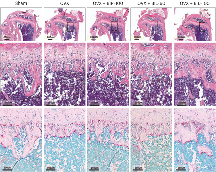
For histomorphometric analysis, fixed tibias were decalcified in 12% ethylenediaminetetraacetic acid and embedded in paraffin. Tissue sections were prepared using a microtome (Leica Biosystems, Nussloch, Germany) for hematoxylin and eosin (H&E) and tartrate-resistant acid phosphatase (TRAP) staining. At least 4 tibias were sectioned for each group, and 3 sections corresponding to the middle sections in each group were used for histomorphometric analysis. The number of osteoclasts per bone perimeter (N. OC/B. Pm) was assessed in the TRAP-stained sections [30]. The Student's t-test was used to assess whether the observed differences between the groups were statistically significant.
Go to :
RESULTS
Characterization of soy ISF-lipid mixtures
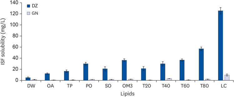
The amount of ISF glycosides daidzin, glycitin, and genistin in BI was 191.61, 89.30, and 20.85 mg/g, respectively, whereas that of the ISF non-glycosides DZ glycitein, and GN was 12.31, 6.00, and 1.77 mg/g, respectively. The solubility of DZ and GN from BI in lipids including OA, TP, PO, SO, OM3, and SL, and Tween surfactants T20, T40, T60, and T80, was measured by LC/MS analysis (Fig. 1). The solubility of DZ and GN in water as controls (DW) was 4.90 ± 1.23 and 1.45 ± 0.24 mg/L, respectively. Their solubility in the experimental lipids was increased compared to water. The solubility of DZ and GN in SL was 125.6 ± 5.78 and 9.7 ± 1.80 mg/L, respectively, which was approximately 25 and 7 times higher, respectively, than in water. The solubility in T80 was the highest among the Tween surfactants.
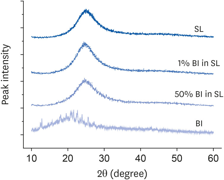
FT-IR analysis was performed to determine the chemical interaction between soy ISF and LCT (no data shown). There were nearly no peak shifts in the FT-IR spectra, and thus, negligible interactions were likely to have occurred between them. Fig. 2 shows the XRD patterns of BI, SL, and the mixtures of 1% and 50% BI in SL. Sharp peaks appeared in the BI pattern, presumably due to the crystalline ISF regions in BI [31]. However, the sharp peaks disappeared in the mixtures because the crystalline regions were considered to dissolve or became amorphous by mixture with SL.
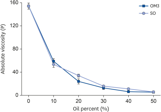
The mixture of BI and SL was too viscous to administer via oral gavage in the animal studies, and thus, SL was diluted with low-viscosity lipids OM3 and SO, as shown in Fig. 3. The absolute viscosity of SL was approximately 154.2 ± 5.2 poise (P). However, it decreased rapidly with increases in the mixing amounts of OM3 and SO. For oral gavage, the mixture of SL and SO, whose weight ratio and viscosity were 8:2 and about 34.0 ± 2.5 P, respectively, was used.
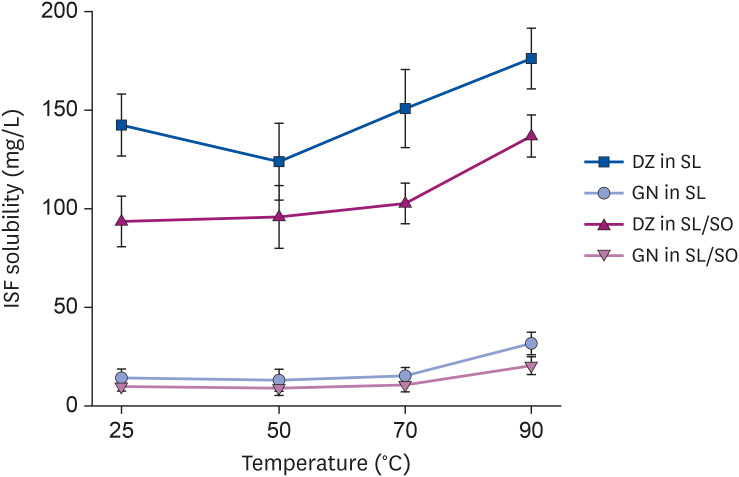
The ISF solubility in SL and SL/SO (8:2) was measured by LC/MS analysis (Fig. 4). The solubility of DZ in SL and SL/SO (8:2) was 142.4 ± 15.7 and 93.5 ± 12.8 mg/L, respectively, while that of GN was 14.2 ± 4.5 and 9.8 ± 2.3 mg/L, respectively. The ISF solubility decreased upon mixing with SO but was higher than those in the other experimental lipids. ISF solubility was slightly increased under elevated temperatures within the experimental range.
PK studies
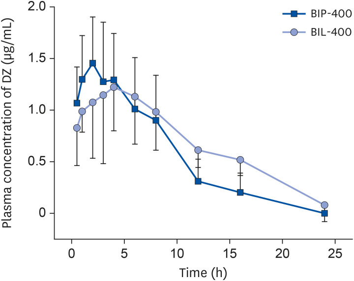
Fig. 5 shows the serum concentration-time profiles of DZ, which were analyzed after oral gavage. The serum concentration of DZ within 4 h after the ingestion of BIP-400 was higher than after the ingestion of BIL-400. Serum concentrations of BIP-400 and BIL-400 were similar for 4–8 h after oral administration. BIL-400 showed higher plasma concentrations than BIP-400 after 8 h. After 24 h, the serum concentration of BIL-400 was 0.08 ± 0.16 mg/L, whereas that of BIP-400 was zero. The AUC values of DZ for BIP-400 and BIL-400 were 13.19 and 16.09 µg·h/mL, respectively. The serum concentration of GN after intake was barely detectable (data not shown).
Bone morphometric studies
Table 1 shows the changes in total body weight during the animal experiments. The OVX mice gained more body weight for 4 weeks after the operation than the Sham and BI-administered OVX groups. There was no significant difference in body weight between the Sham and BI-administered OVX groups. However, the body weight of the BI-administered OVX groups was significantly lower than that of the OVX group at the end of the 4-week experimental period (P < 0.05).
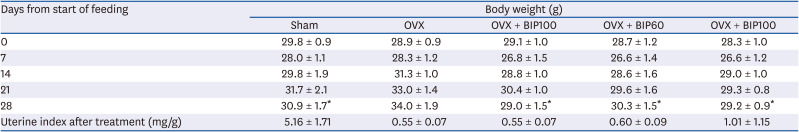
Table 1
Changes in body weight and uterine indices in Sham and OVX mice orally administered BI powder (BIP-100) and mixtures of BI and soy lecithin/safflower oil (BIL-60 and BIL-100)
Each data value is expressed as the mean ± standard deviation (n = 8).
OVX, ovariectomized; BI, bio-isoflavone.
*P < 0.05 compared to the OVX group.
![]()
The uterine index (the ratio of uterine weight to body weight) of the animal groups at the end of the 4-week experimental period is shown in Table 1. The uterine weight had strikingly decreased in OVX mice compared to the Sham group, indicating that the mice were estrogen-deficient. In contrast, there was no significant difference in uterine weight among the OVX groups after the 4-week treatment.
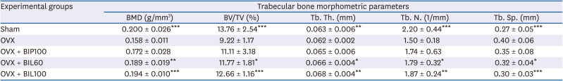
We investigated the effect of soy ISF and LCT on OVX-induced bone loss through µCT at the end of the treatment period. Fig. 6 shows the µCT-scanned images of the proximal tibia. The images show that the OVX mice exhibited a significant reduction in trabecular bone. Table 2 shows the different bone morphometric parameters obtained through μCT analysis. BMD in the OVX group (0.158 ± 0.011 g/mm3) was significantly decreased by 21% compared to the Sham group (0.200 ± 0.026 g/mm3, P < 0.001), whereas the intake of BI powder (OVX + BIP-100: 0.172 ± 0.028 g/mm3) attenuated trabecular bone loss. However, the difference was not significantly different from the OVX group according to the Student's t-test. The BMD from the oral intake of BI and SL mixtures with BI dosages of 60 mg/kg (OVX + BIL-60) and 100 mg/kg (OVX + BIL-100) was 0.189 ± 0.020 and 0.194 ± 0.010 g/mm3, respectively. The BMD of the OVX + BIL-60 and OVX + BIL-100 groups was significantly higher than that of the OVX group (P < 0.01 and P < 0.001, respectively). The BV/TV ratio in the OVX group at 9.21 ± 1.17% was significantly decreased by 33% compared to the Sham group at 13.76 ± 2.54% (P < 0.001), whereas the oral administration of BI powder (OVX + BIP-100: 11.11 ± 3.17%) attenuated trabecular bone loss. The BV/TV ratio of the OVX + BIL-60 and OVX + BIL-100 groups was significantly higher than that of the OVX group (P < 0.05 and P < 0.001, respectively), whereas that of the OVX + BIP-100 group was not significantly higher. Similarly, other bone parameters, such as trabecular thickness (Tb. Th.) and Tb. N., were significantly increased by oral intake of the BI and SL mixtures. However, the Tb. Th. and Tb. N. of the OVX + BIP-100 group were not statistically different from those of OVX according to the Student's t-test. Tb. Sp. was significantly increased due to ovariectomy (P < 0.001) compared to the Sham group. The Tb. Sp. of groups administered mixtures of BI and SL (OVX + BIL-60 and OVX + BIL-100) was significantly decreased (P < 0.05 and P < 0.001, respectively), whereas that of the OVX + BIP-100 group was not statistically different from that of the OVX group.
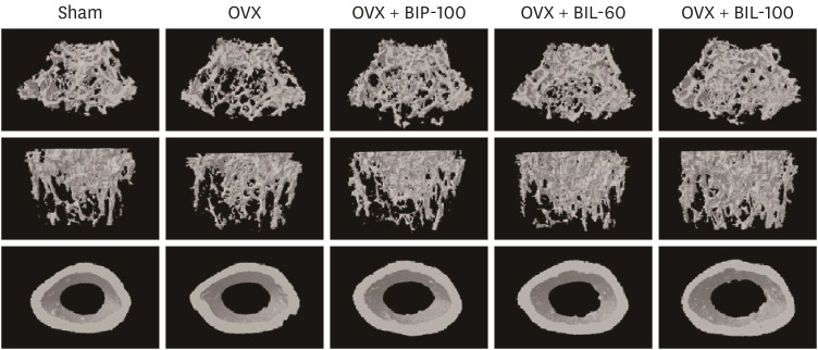 | Fig. 6Micro-computed tomography scanning of tibial trabecular bone of Sham and OVX mice after 4-week treatment with BI powder (100 mg/kg, BIP-100) and mixtures of BI (60 and 100 mg/kg) and soy lecithin/safflower oil (BIL-60 and BIL-100).OVX, ovariectomized; BI, bio-isoflavone.
|
Table 2
Analysis of trabecular bone morphometric parameters in the different groups
Values are the mean ± standard deviation (n = 8).
OVX, ovariectomized; BMD, bone mineral density; BV/TV, bone volume per total volume; Tb. Th., trabecular thickness; Tb. N., trabecular number; Tb. Sp., trabecular separation.
*P < 0.05, **P < 0.01, and ***P < 0.001 compared to the OVX group.
![]()
To further confirm the in vivo results, we analyzed decalcified tibias by H&E and TRAP staining (Fig. 7). The H&E staining figures (upper) show that the purple region of the hematoxylin-stained cells in the OVX group was significantly increased compared with that of the Sham group. The stained cell regions were decreased by BI and SL intake (OVX + BIP-100, OVX + BIL-60, and OVX + BIL-100). As shown in the TRAP-staining figures (lower), the TRAP-stained sections nearly coincided with the hematoxylin-stained sections in the trabecular region, and thus, the cells were considered to be osteoclasts.
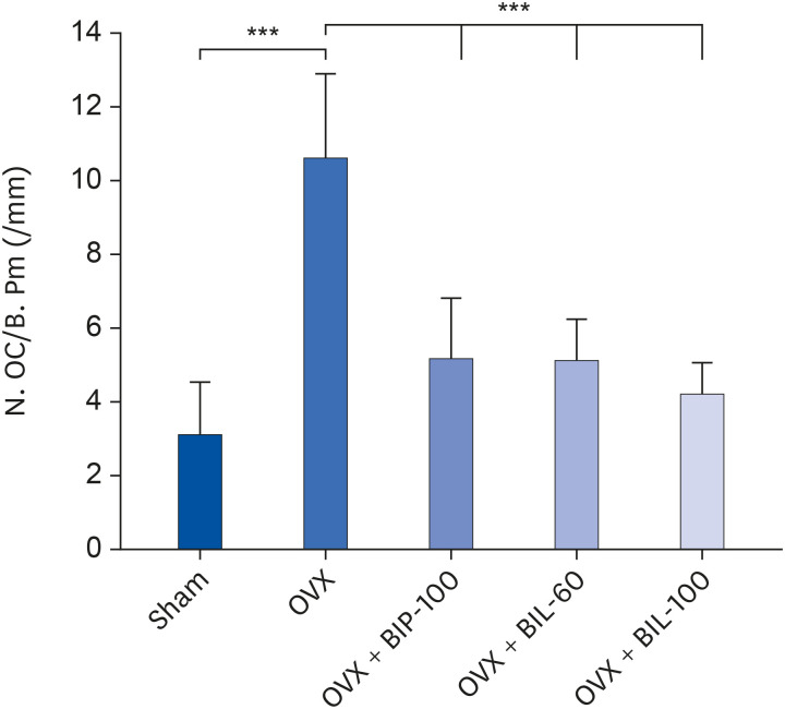
N. OC/B. Pm. was quantified in the TRAP-stained sections (Fig. 8). Consistent with the μCT results, the OVX group exhibited a significant increase in the number of osteoclasts (P < 0.001) compared to the Sham group. BI administration (OVX + BIP-100) decreased the number of TRAP-positive cells compared to the OVX group (P < 0.001). The N. OC/B. Pm. of the groups given mixtures of BI and SL (OVX + BIL-60 and OVX + BIL-100) was also decreased compared to the OVX group. The N. OC/B. Pm. of the OVX+BIL-100 group was significantly different from that of the group administered BI alone (OVX + BIP-100) (P < 0.05).
Go to :
DISCUSSION
For a substance to be absorbed through the digestive tract, it must be soluble in water or oil. Therefore, water or oil solubility is very important for the absorption of substances into the body. The low solubility of ISFs DZ and GN in water indicated that they had poor water solubility (Fig. 1). The low solubility was reported to be due to aromatic rings and fairly strong intermolecular hydrogen bonds among that hydroxyl groups that weakened the solvation process [31]. The poor solubility induced low bioavailability, and thus, their absolute bioavailability in rats after oral administration was reported to be only 6.1% [32]. Although ISF has broad therapeutic effects, its low solubility limits its clinical application. Therefore, research has been conducted to improve water solubility, such as the preparation of a cyclodextrin complex [33]. The solubility of DZ and GN in lipids and surfactants slightly increased compared to those in water (Fig. 1). However, DZ and GN are also poorly soluble in lipids, presumably due to their polar phenolic hydroxyl groups. The solubility of DZ and GN in LCT was the highest among the experimental lipids, presumably because LCT has a phosphatidylcholine group, which is a polar functional group that can interact with polar phenolic hydroxyl groups in DZ and GN [31]. In addition, the dissolution of BI crystalline regions by LCT may improve ISF solubility (Fig. 2).
Controlled-release drug delivery systems (CRDDSs) with the delayed or long-term release of drugs have been studied and used to deliver drugs over long periods, improving drug efficacy, increasing drug safety, and reducing side effects [34]. Phospholipids, such as LCT in CRDDSs enhanced the bioavailability of drugs with low aqueous solubility or low membrane penetration potential, protected sensitive active agents from degradation in the GI tract, and reduced GI side effects [34]. In addition, LCT delayed the release of active ingredients from CRDDSs such as liposomes, solid lipid nanoparticles, and microparticles [28].
According to the PK studies, in the early stage after oral administration when LCT was not fully emulsified, the DZ plasma concentration was lowered by LCT mixing (Fig. 5). This was thought to be because the relatively hydrophobic LCT surrounded the surface of the BI particles, preventing the diffusion of DZ into the digestive tract. This behavior of LCT may be similar to that of LCT in the CRDDSs. In the latter stage after oral administration, the plasma concentration of DZ was increased by LCT mixing. That is, when BI particles were administered orally, the DZ plasma concentration increased and decreased relatively rapidly, whereas, when mixed with LCT, the DZ plasma concentration gradually increased, but remained relatively high for a long time. In addition, the AUC (DZ bioavailability) was increased by mixing with LCT. The DZ bioavailability was considered to be improved due to the increased emulsification and solubility by LCT. However, GN bioavailability could not be analyzed because BI had a low concentration of GN and genistin. Genistin is an ISF glycoside that can be hydrolyzed to GN in the digestive tract.
According to the animal studies, the mean body weight of the OVX group increased during the experimental period, whereas that of OVX + BIP-100 increased less than that of the OVX group. ISF was reported to reduce obesity by decreasing food intake [35]. The mean body weight of the OVX + BIL-60 and OVX + BIL-100 groups was increased significantly less than in the OVX group (Table 1). These findings show that the simultaneous administration of BI and SL could be an effective means to reduce obesity. The oral administration of BI and SL had nearly no effect on the uterine weight of the experimental animals. We suggest that the dose of ISF used in this study did not have adverse effects.
The bone morphometric parameters BMD, BV/TV, Tb. Th., Tb. N., and Tb. Sp. showed that the BI and SL mixtures were more effective at reducing bone loss caused by ovariectomy than BI alone (Table 2). The osteoclast formation in the groups orally administered the mixtures of BI and SL (OVX + BIL-60 and OVX + BIL-100) was significantly reduced compared to the OVX and OVX + BIP-100 groups. These results showed that the simultaneous intake of soy ISF and LCT could be more effective at preventing bone loss than soy ISF intake alone.
In the current study, because SL intake enhanced ISF bioavailability, as shown in the PK studies, the bone loss in the OVX groups with the simultaneous intake of BI and SL was reduced. The ISF bioavailability may be enhanced by the emulsification of LCT and retention of ISF in the micelles, presumably due to the higher ISF solubility in LCT than in water. The colloidal formation of LCT emulsification probably prevented ISF sedimentation in the digestive tract and improved absorption in the body.
Few previous studies have addressed the effect of LCT on preventing bone loss, even though the effect of OM3 on bone metabolism has been investigated in numerous animal models, including mice and rats [2122]. The intake of soy ISF and fish oil, whose main ingredient is OM3, was reported to show ameliorating effects on bone loss, whereas SO intake, whose main ingredients are OA and linoleic acid, was not effective [29]. Therefore, the effect on bone loss may depend upon the chemical structure of the lipids.
In summary, the results of the current study showed that the effect of preventing bone loss and osteoclast formation by the concurrent ingestion of soy ISF and LCT was superior to soy ISF alone because of the improved ISF bioavailability by the emulsification and solvation of LCT. These results raise the possibility that the combination of soy ISF and LCT promoted complete bone formation. However, the effect of LCT on the bioavailability of ISFs DZ and GN could not be accurately studied because the amount of ISFs in BI, especially GN, used as a dietary supplement, was not large. Consequently, further studies using pure ISFs, such as DZ, GN, and equol, are needed to uncover the effect of LCT on ISF bioavailability.
Go to :
 XML Download