INTRODUCTION
External apical root resorption (EARR) is a complication of orthodontic treatment that results in irreversible loss of root structure. During tooth movement, the concentration of forces on the periodontium, particularly on the apical third, can destabilize local homeostasis, resulting in loss of the surface layer of cells that protect the roots of the teeth.
1 Genetic and local factors directly related to orthodontic movement can cause EARR; these include the degree and direction of forces and the treatment duration.
2
The average degree of root resorption in each maxillary anterior tooth is less than 1.5 mm during corrective orthodontic treatment.
3 However, an average of 1.12 mm has also been reported, with a few cases presenting with more than 2.5 mm of resorption.
4 EARR can occur in almost all teeth, although it is more evident in the maxillary and mandibular incisors, with varying degrees of severity.
4
Radiographic monitoring after 6 months following orthodontic treatment initiation is essential for controlling EARR throughout treatment, because resorption can be detected from the initial stages of leveling. The patient’s response to orthodontic movement is indicated during these stages, and this helps the orthodontist to determine the individual limits of each patient.
5
In recent years, the demand for treatment with orthodontic aligners (OAs)
6,7 has increased, considering the superior esthetics, ease of hygiene maintenance, and greater comfort when compared with fixed appliances (FAs).
8 With regard to EARR, the reported results are controversial. While some authors found that patients treated with OA presented a smaller degree of EARR than did those treated with FA,
7,9,10 others observed similar results with the two treatment methods.
6,11
In addition, most studies have been limited by factors such as the lack of a control group, a retrospective study design, and results entirely based on panoramic radiographs. Consequently, differences in EARR between OA treatment and FA treatment remain unclear. Accordingly, the aim of the present randomized controlled clinical trial was to investigate and compare EARR 6 months after treatment initiation between patients treated with OAs and those treated with FAs.
Go to :

MATERIALS AND METHODS
Trial design
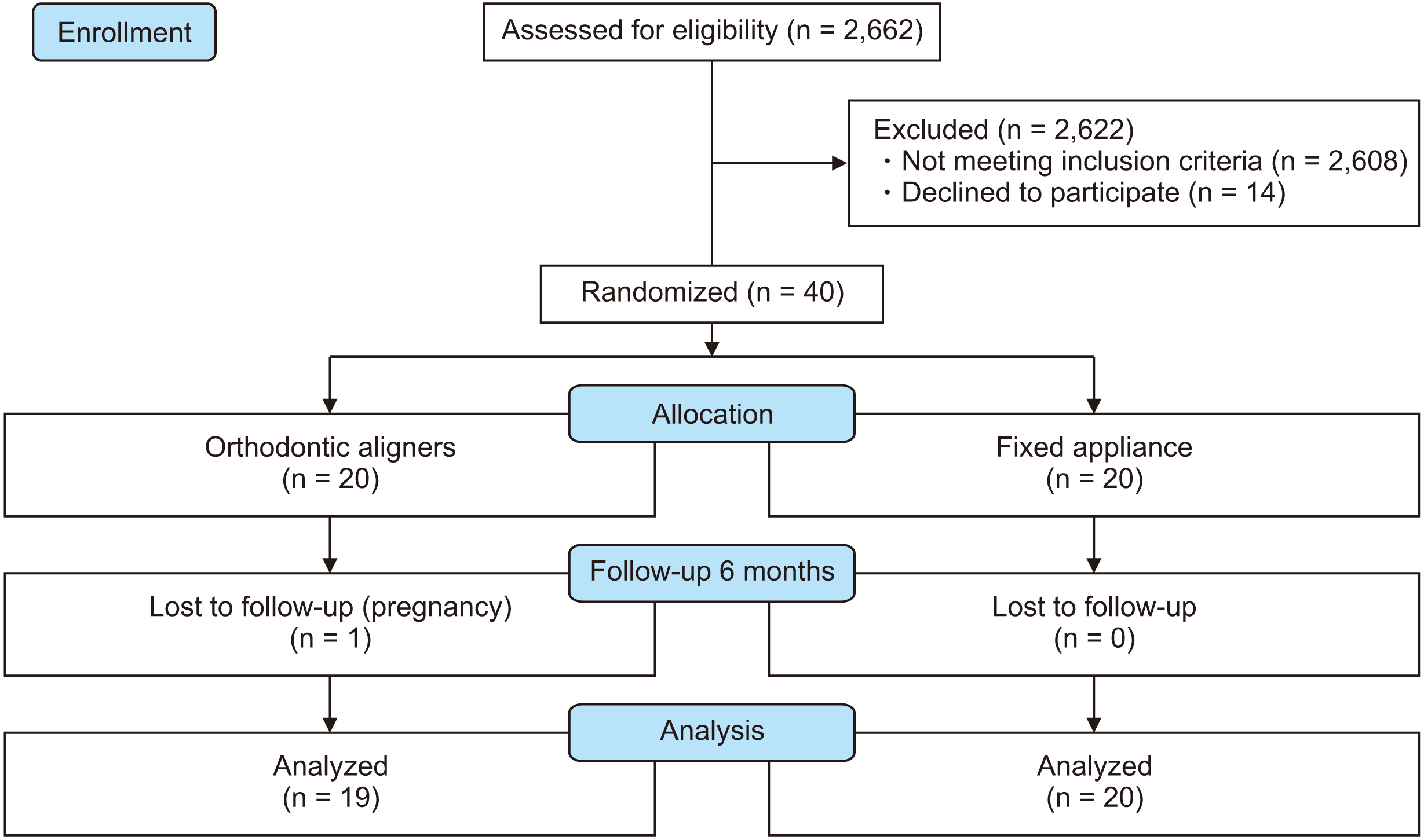
This study was a parallel randomized controlled clinical trial wherein participants were prospectively recruited and randomized into two groups. There were no changes in the methods after commencement of the trial.
Participants, eligibility criteria, and settings
The sample was obtained by the screening of 2,662 individuals on social media or in schools in the city of Londrina, Brazil.
The inclusion criteria were as follows: Angle Class I malocclusion, moderate crowding, facial symmetry, a passive lip seal, and nonextraction treatment. The exclusion criteria were as follows: absence of permanent teeth, anterior and/or posterior open bite and/or crossbite, the need for restorations on the anterior teeth during treatment, a history of trauma to the maxillary incisors, a history of orthodontic treatment, and the presence of previously occurred EARR.
The study was approved by the Ethics Committee of University of North Parana (UNOPAR) (12088219.0. 0000.0108) and registered in the Brazilian Clinical Trials (RBR-9zytwf) registry. Volunteers received treatment in the clinic at UNOPAR and were assisted by orthodontists supervised by a professor of orthodontics with 15 years of experience.
Interventions
For all patients, initial orthodontic assessments included the acquisition of intra- and extraoral photographs, study models, and periapical radiographs (PRs) of the maxillary and mandibular incisors.
Patients were randomized into two groups depending on the treatment method. The OA group included patients treated with OAs (SmartTrack, InvisalignTM; Align Technology, San Jose, CA, USA). Virtual planning was implemented for this group (ClinCheckTM Pro program, version 5.6; Align Technology). The sequence of procedures during treatment, such as the installation of attachments, interproximal reduction, and the application of intermaxillary elastics, among others, followed the virtual plan. The maxillary and mandibular OAs were changed every 10 days, with a recommended daily usage time of 22 hours.
In the FA group, a fixed metallic orthodontic appliance (slot 0.022” × 0.030”, 3M Unitek, Monrovia, CA, USA) was used for treatment. The appliance covered all teeth, and the same sequence of archwires was used (superelastic nitinol: 0.014”, 0.016”, and 0.016” × 0.022”).
In both groups, monitoring procedures were performed monthly. For the present study, the results obtained in the first 6 months were taken into account.
Outcomes (primary and secondary)
Before (T0) and 6 months after (T1) initiation of the orthodontic treatment, PRs of the maxillary and mandibular incisors were obtained for the assessment of root resorption.
For standardization, all radiographs were acquired using the parallel technique with a Cone Indicator Digital Schick CDR positioner (Schick Technologies, Long Island City, NY, USA) at a distance of 40 cm, a Schick Elite CDR intraoral sensor (Schick Technologies), and the CDR DICOM for Windows program (version 5.4). The Dabi Atlante X-ray machine (Dabi Atlante, Ribeirão Preto, Sao Paulo, Brazil) was used (70 kV, 08 mA) with an exposure time of 0.4 seconds for both the maxillary and mandibular incisors.
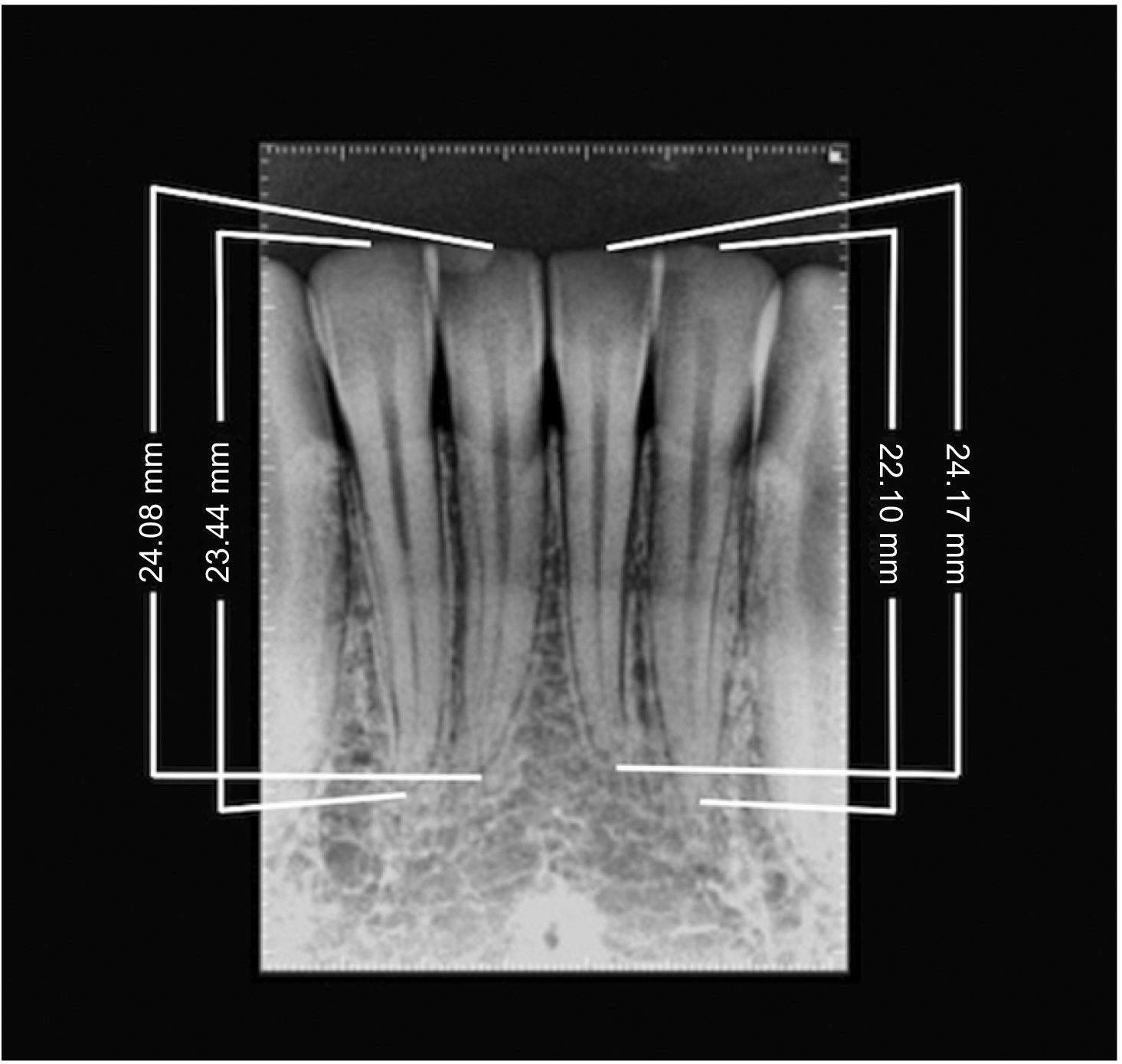
Linear measurement of the degree of EARR was performed (in millimeters) from the root apex to the incisal edge of each incisor (
Figure 1). The same methodology was applied at T0 and T1, and the difference between the two measurements provided the degree of EARR (T1−T0). A single examiner performed all measurements.
 | Figure 1Linear measurements of the tooth length (in millimeters) from the root apex to the incisal edge. 
|
Sample size
The sample size was calculated by taking into account the mean standard deviation (0.51 mm) obtained in a previous study
12 assessing EARR in incisors after orthodontic treatment. Considering a significance level of 5% and a power of 80% for detecting a real mean difference of 0.5 mm in EARR between the two groups, a minimum of 18 patients was required for each group.
Randomization
Simple randomization
13 in a 1:1 ratio was performed by an external researcher using the Excel 2007 program (Microsoft, Redmond, WA, USA). The randomization codes were consecutively inserted in opaque, sealed, numbered envelopes; this ensured concealment of the group allocation.
Blinding
Operator blinding was not possible because patients were still in the active phase of treatment at T1. However, a blind external researcher statistically analyzed the collected data.
Statistical analysis
To check for intra- and interexaminer errors, 30% measurements were repeated, and intraclass correlation coefficients (ICCs) and Bland–Altman analysis for agreement were used for assessment according to the criteria described by Fleiss.
14
The obtained data were checked for normality (Shapiro–Wilk test). Intergroup comparisons were performed using independent t-tests (age, Peer Assessment Rating [PAR] index, and Little’s Irregularity Index) and the chi-squared test (sex). For intragroup comparisons, the dependent t-test was applied. Intergroup comparisons of EARR were performed using covariance analysis using the T0 measures as covariates.
All statistical analyses were performed using IBM SPSS ver. 26 (IBM Corp., Armonk, NY, USA), with a significance level of 5% and a 95% confidence interval.
Go to :

DISCUSSION
EARR observed in teeth subjected to orthodontic treatment is considered iatrogenic, considering the reduction in the tooth length.
15 In the majority of cases, EARR is not severe and remains clinically insignificant. In more severe cases, the orthodontic treatment needs to be modified or even suspended in order to preserve long-term tooth support.
Periapical radiography is the most common method for EARR detection; it can be performed using compact equipment in the dental office itself and is more accessible than other types of diagnostic imaging.
12 Moreover, verification of EARR is more precise on PRs than on panoramic radiographs.
4 Cone beam computed tomography (CBCT) could also be used; however, it is expensive and causes greater radiation exposure. Thus, periapical radiography was the method of choice in the present study.
We measured the lengths of the maxillary and mandibular incisors, which are the most susceptible to EARR,
4 during the first 6 months of treatment because at this time, it is reportedly possible to predict the occurrence of a subsequent increase in resorption.
2
In the intragroup comparisons in the present study, a statistically significant decrease in the length of all teeth was observed. The average degree of EARR (T1−T0) ranged from −0.52 (tooth #32) to −0.88 mm (tooth #41) in the FA group and from −0.52 (tooth #21) to −0.85 mm (tooth #41) in the OA group. In addition, neither group exhibited resorption greater than or equal to 1 mm. The observed EARR represented a linear decrease of 2.88% of the root length, and 97.12% of the original root was intact. These results are similar to those described by Krieger et al.,
16 who assessed 100 patients treated only with OAs. The authors conducted a retrospective study of the degree of EARR in the incisors and first molars using panoramic radiographs and found that all patients exhibited EARR in at least two teeth. In addition, Gay et al.
17 used panoramic radiographs and performed a prospective analysis of the magnitude of EARR in the incisors, canines, first premolars, and first molars of 71 Class I patients treated with OAs. The authors observed results similar to those reported in the literature for patients treated with FAs with light force application.
In our intergroup comparisons, only tooth #21 presented a statistically significant difference. However, the overall differences between groups were not clinically relevant, with the average difference in EARR (T1−T0) ranging from 0.03 mm for tooth #11 and tooth #41 (smallest changes) to 0.35 mm for tooth #21 (greatest change). These results corroborate with those described in other studies,
6,11 where it was observed that FA treatment and OA treatment were associated with the same risk of EARR development when low forces were applied. Barbagallo et al.
11 performed a randomized controlled trial (RCT) with a split-mouth design and assessed the degree of EARR in the premolars using microcomputed tomography. The authors observed that OAs and FAs with low force application resulted in a similar degree of EARR in the premolars, while FA treatment with intense force resulted in the largest amount of EARR. However, in that study, the teeth received orthodontic forces for only 8 weeks and were subsequently extracted for evaluation. Similar results were reported in a retrospective case-control study involving the use of panoramic radiographs.
6
On the other hand, some studies
7,10 have shown that with orthodontic treatment without dental extraction, similar to that in the present study, the incidence and severity of EARR was lower with aligners than with FAs. The authors suggested that orthodontic tooth movements caused by aligners are directed more toward tooth inclination and less toward apical displacement. Eissa et al.
10 performed a pilot study with three treatment groups, namely OA, FA, and self-ligating FA groups, and assessed the degree of EARR in the maxillary incisors using CBCT. According to the authors, EARR represents the biological cost of tooth movement regardless of the technique used. Nevertheless, patients treated with OAs exhibited less EARR than did those treated with FAs. Similarly, Yi et al.
7 observed lesser EARR with OA treatment than with FA treatment. However, the authors retrospectively assessed the degree of EARR in the maxillary and mandibular incisors (in cases without extraction) by evaluating panoramic radiographs, which can distort the tooth size.
4 In a cohort study of 70 patients treated with OAs or FAs, it was concluded that OAs cause lesser EARR than do FAs.
18 Although measurements were performed using CBCT, the study was retrospective, compared EARR in patients with various types of malocclusion, and included cases where teeth were extracted on the recommendation of the orthodontist.
The small differences in EARR between the OA and FA groups in the present study could be related to biomechanical differences between OAs and FAs in terms of the type and location of force application and the moment generated. Torque and intrusion movements are much more frequent in FA treatment; this results in more significant pressure on the apical region, thereby increasing the risk of root resorption.
4 In the present study, considering the time period of 6 months, these potential differences could not be detected. It is also important to note that limited tooth movement along with a short treatment time may not result in visible differences in root resorption between two appliances. This could be another reason that the present results were different from those in some other studies
7,10 showing that OAs cause lesser root resorption than do FAs.
The present study adhered to the Consolidated Standards of Reporting Trials (CONSORT) guidelines, which emphasize on sample calculation, inclusion of a control group, and randomization, which allow for the minimization of selection bias and are thus fundamental components of high-impact RCTs.
19 In accordance with the literature reporting minimal variations in EARR among different techniques and prescriptions,
12,20,21 we did not find significant differences in EARR between patients treated with OAs and those treated with FAs during the initial phase of treatment. Neither group demonstrated resorption of 1 mm or more. Thus, the values obtained may be considered clinically acceptable, as EARR appeared as simple rounding of the apex, which does not compromise long-term stability.
This study also has some limitations. First, the short follow-up time (6 months) is one limitation; however, the patients continue to receive treatment and will be monitored until the complete correction of malocclusion, when fresh assessments will be performed to determine EARR. Moreover, many studies
2,5,22 have pointed out the importance of monitoring during this period in order to control EARR throughout the treatment duration. Compared with those without resorption, patients with detectable root resorption during the first 6 months of active treatment are more likely to experience resorption in the subsequent 6-month period. Second, the present study evaluated root resorption due to treatment with OAs or FAs in the initial stages of treatment in teeth with Angle Class I malocclusion and moderate crowding. Other factors like individual predisposition, force intensity, treatment duration, and degree of orthodontic movement are relevant factors that may influence the degree of EARR.
23
In addition, the biological limits of each patient should be taken into account, and predisposing factors and periodic radiographic monitoring should be observed with the aim of preserving patients and making these techniques absolutely viable. It is also important to emphasize that all patients in the OA group received treatment with the InvisalignTM system, wherein the aligners are fabricated from a single plastic material. The modulus of elasticity differs among materials and could result in differences in the effects of the same movements on the root apex.
Go to :


 PDF
PDF Citation
 Print
Print


 XML Download