

 PDF
PDF Citation
 Print
Print


Song Hee Oh, Sae Rom Lee, Jin-Young Choi, Seong-Hun Kim, Eui-Hwan Hwang, Gerald Nelson
Quantitative cone-beam computed tomography evaluation of hard and soft tissue thickness in the midpalatal suture region to facilitate orthodontic miniimplant placement.
- Korean J Orthod 2021;51:260-269
Go to : 
I read with great interest the article by Oh et al., who evaluated the skeletal and soft tissue thickness in mid-palatal region in search of optimal orthodontic mini-implant placement sites. I would like to congratulate the authors for their fine piece of work. This timely investigation deserves attention from any orthodontic clinicians who practice a palatal approach for placement of the mini-implant as a simple skeletal anchor unit or as a part of other devices such as miniscrew-assisted rapid palatal expander (MARPE) and modified c-palatal plate (MCPP) for various treatment mechanics.
Q1. To classify the patients, the authors used their ‘age’ defined as dental development stages for the patients in 10’s. In my opinion it may have been more advantageous to choose their skeletal age in place of dental age since hand-wrist radiographs are routinely collected in these young patients for diagnostic records and since skeletal maturity information gathered from the hand-wrist films may be more closely related to the palatal bone thickness than the dental development status. Your comments would be appreciated.
Q2. In Table 4 the age group 1, compared to the group 4, displayed significantly thinner hard tissue thickness at 3 mm right and left of the midpalatal suture lines (MR 3 mm and ML 3 mm) in the anterior region. However, it was still thicker than the other midpalatal and lateral suture regions within the group 1. Given that the palatal bone density of the age group 1 is likely lower than the others, would the authors recommend the anterior MR and ML 3 mm as the mini-implant insertion sites of choice in order to take the most advantage of available hard tissue support?
Q3. The results of this investigation were presented in a meticulous manner so that they could serve as a quick reference guide in deciding most opportune mini-implant placement locations in the mid-palatal suture and its immediate neighboring regions. However, some clinicians tend to advocate individual survey of palatal bone density and thickness as well as selection of optimal mini-implants length and position based on cone-beam computed tomography (CBCT). When would you think this more customized approach is necessary?
Q4. In light of the new findings, would the authors describe your preferred mini-implant dimensions and placement sites in the palate for retraction and intrusion.
Questioned by
Seong Ho Han
Division of Orthodontics, Department of Dentistry, St. Vincent’s Hospital, College of Medicine, The Catholic University of Korea, Seoul, Korea
Go to :
A1. First of all, thank you deeply for your considerate comments. We strongly agree with your opinion that the evaluation by age group based on the degree of skeletal maturity with the hand-wrist image was more related to the thickness of the palatine bone. However, hand-wrist radiographs were not taken for all patients in this study. That’s why age-specific patient groups were classified and evaluated based on the age-old development stage suggested by Bjoerk et al.1
A2. Even taking into account that the palatine density in group 1 was likely to be lower than in the other groups, the MR and ML 3 mm regions were thicker than the hard tissue thicknesses at other locations (Midline, MR and ML 1.5 mm). Average 6.16 ± 1.77 mm of hard tissue thickness in this area is quite sufficient to be used as safe and stable place for mini-implant insertion. If we apply static and light force to this mini-implants or use them as indirect anchorages, then there will be no problem in the clinical application (Figure 1). Therefore, we recommend anterior MR and ML 3 mm in group 1 as optimal insertion sites for mini-implant to utilize much hard tissue support as possible.
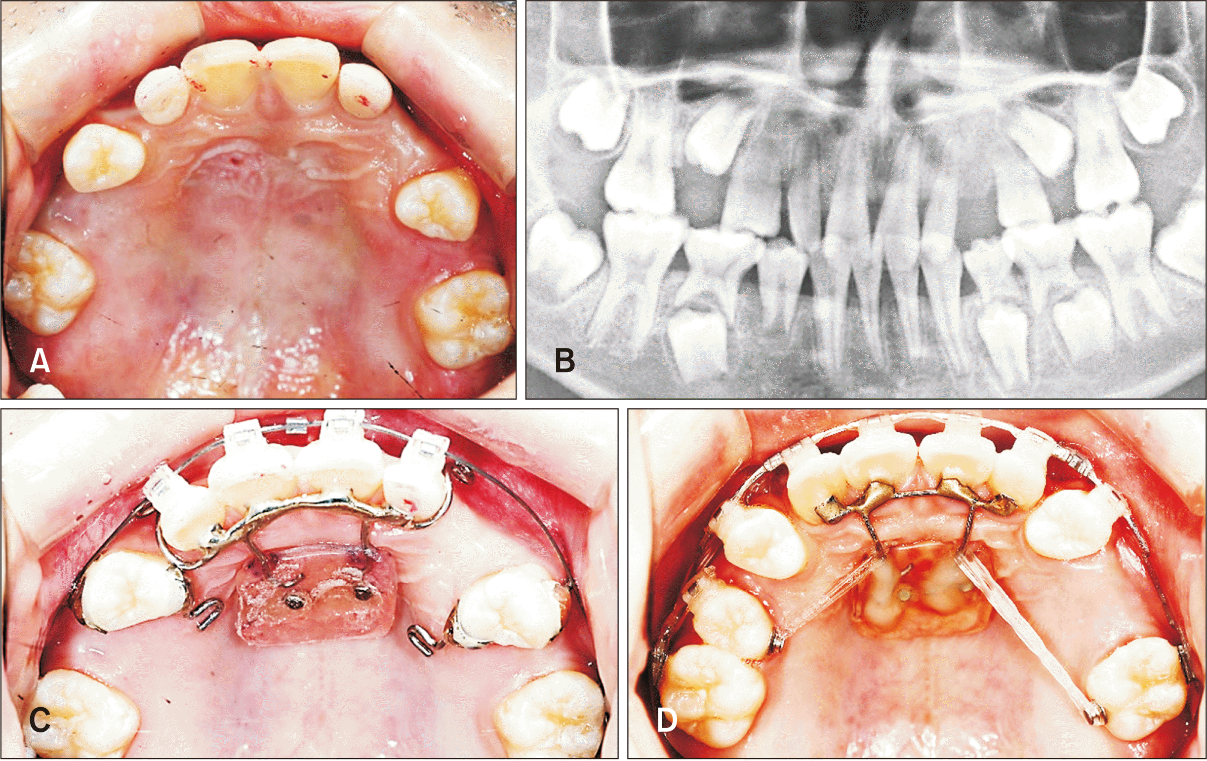 | Figure 1A ten-year-old male patient who requires whole posterior dentition protraction without anterior retraction. A, Pretreatment occlusal photograph. B, Pretreatment panoramic radiograph. C, Two 1.6 × 8 mm self-drilling type miniscrews (Proto type of Bio-Action Screw; Jin Biomed Co., Bucheon, Korea) were placed in the anterior palate area about 3 mm lateral to the midpalatal suture. D, After eight months of treatment occlusal photograph.
|
A3. Of course, we believe that individual survey based on CBCT are ideal for optimal mini-implant length and location selection. However, to take a CBCT for evaluating the individual thickness of hard tissue in this area not only causes unnecessary radiation to the patient, but is also cost-ineffective. The main purpose of this study is to analyze the actual thickness pattern of the midpalatal suture and its adjacent hard and soft tissues according to age and sex, thereby providing anatomical guidelines for clinicians to obtain minimal, safe and effective results when placing mini-implants or palatal miniplates like the previous studies about placement guidelines for upper buccal posteriors and upper lateral palate area.2,3
A4. We strongly recommend to use 1.6 mm in diameter, 8 mm in length mini-implant for midpalatal and lateral palatal area. The same size mini-implant can be used as cross-type c-palatal plate anchoring mini-implant in the posterior midpalatal suture area.4 However, in the case of tooth and bone borne type MARPE or MCPP, clinicians have no choice but to use the predetermined size of mini-implants provided by the manufacturing company.
As demonstrated in the results of this study, both posterior MR and ML 3 mm showed relatively thin hard tissue and thick soft tissue regardless of age and gender. Therefore, in the case of mini-implants for orthodontic anchorage, midpalatal suture or its lateral adjacent regions within 1.5 mm limit are suggested as the recommended insertion locations. Similarly, for effective expansion and stability of MARPE, we recommend to place anchoring mini-implants within the MR and ML1.5 mm.
Replied by
Song Hee Oha and Seong-Hun Kimb
aDepartment of Oral and Maxillofacial Radiology, Graduate School, Kyung Hee University, Seoul, Korea
bDepartment of Orthodontics, Graduate School, Kyung Hee University, Seoul, Korea
Go to :
 XML Download