

 PDF
PDF Citation
 Print
Print


I. Introduction
Congenital abnormalities, aging, and trauma can cause soft tissue deformities. There are several types of injectable materials that are used for facial deformity correction and facial rejuvenation. One of the common and preferred methods is autologous fat transfer which has recently become a popular procedure1. The procedure was first performed in 1893 by a plastic surgeon, Gustav Neuber. He used autologous arm fat to correct a periorbital osteomyelitis scar2. Autologous fat transplantation is a non-invasive method for soft tissue volume loss management3.
Unlike other non-autologous fillers, autologous fat is a long-lasting filler that unites with facial tissues and is well-tolerated4. Also, in some cases, autologous fat transfer is a complementary procedure to a facelift to improve facial contour4,5. Over-treatment, fatty cyst development, fat hypertrophy, fat calcification, and fat embolism are complications of fat injection3. Additionally, the procedure may require “touching up” for better results.
This report reviews the use of autologous fat in maxillofacial reconstruction in 15 patients with malar and periorbital area, nasolabial fold, mandibular angle and body, and perioral area defects. These defects arise in cases such as trauma and congenital deformity that require aesthetic improvement.
Go to : 
II. Materials and Methods
In this study, the data from the patients who were admitted from 2014 to 2016 in Department of Oral and Maxillofacial Surgery, Faculty of Dentistry, Qazvin University of Medical Sciences for trauma defect revision and face augmentation by fat transplants were collected. All patients had been informed about the advantages and possible complications of treatment and signed a formal informed consent for treatment. All patients had the opportunity to ask questions and discuss the advantages and disadvantages of the procedure. This study was approved by the ethical committee of Qazvin University of Medical Sciences with ethical number IR.QUMS.REC.1399.230. There is no conflict with ethical considerations.
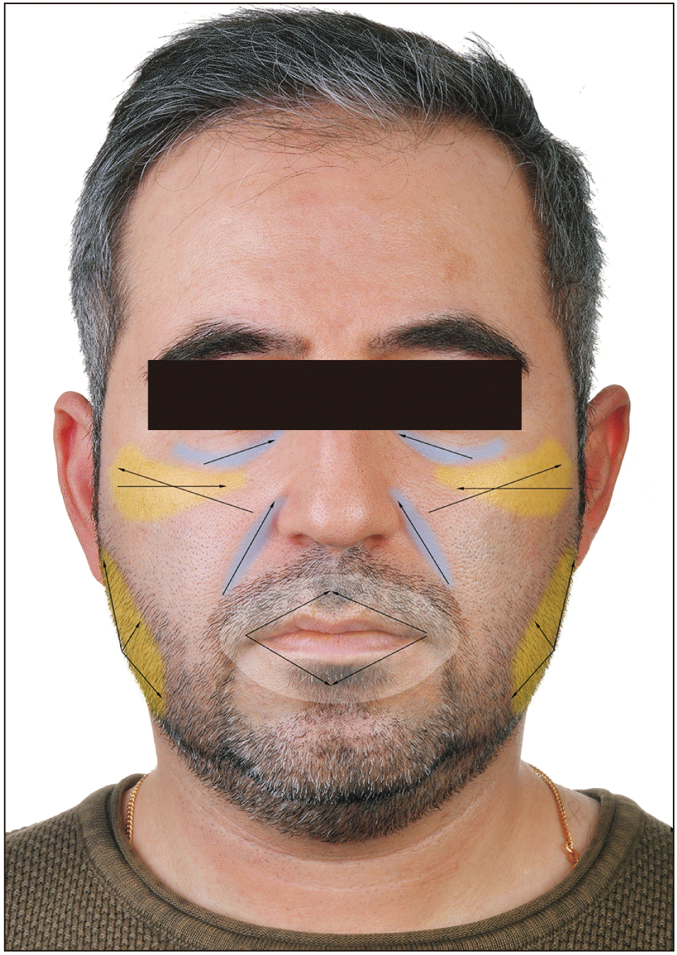
Before harvest, the region was injected by a tumescent solution (0.5% lidocaine+0.0005% epinephrine). After 15 minutes, the fat was harvested by a syringe with low negative pressure (0.75-1.0 atmosphere) and blunt tip cannula (2.1-2.4 mm) through a stab incision on the abdomen or thigh. After processing, harvested fats were injected using 1cc insulin syringes and blunt cannulas (0.7-1.2 mm tip) using a tunneling technique in different planes from deep to surface in fan shape order in the malar and periorbital area, nasolabial fold, mandibular angle and body, and perioral area.(Fig. 1) For malar augmentation, the fat was injected subcutaneously through an incision lateral to the nasolabial fold, as well as via a second incision on the zygomatic arch. The fat was placed near the zygomatic bone, as well as beneath, into, and superficial to the zygomaticus muscles and subcutaneous space. For the periorbital area, fat aliquots were transferred to multiple levels, including subcutaneous, intramuscular, and submuscular. Each nasolabial fold was injected through a lateral incision with feathering on the edges. For the angle and body of the mandible, the jawlines were augmented in the superior, oblique, and anterior directions through an incision on the angle area. The level of injection varies from subcutaneous and supra muscular on the masseter, to slightly deep on the anterior edge of the masseter. For the perioral area, the lip was approached through a lateral incision and a submucosal plan to reach a satisfactory volume.
The indications for fat transplantation in our clinic were: aesthetic (86.7%), asymmetry following trauma (6.7%), and asymmetry following nerve paralysis (6.7%).(Table 1) The autologous fat was used for different maxillofacial region sites: malar and periorbital (80.0%), nasolabial fold (33.3%), mandibular angle and body (13.3%), and perioral area (13.3%).(Table 2)
1. Case 1
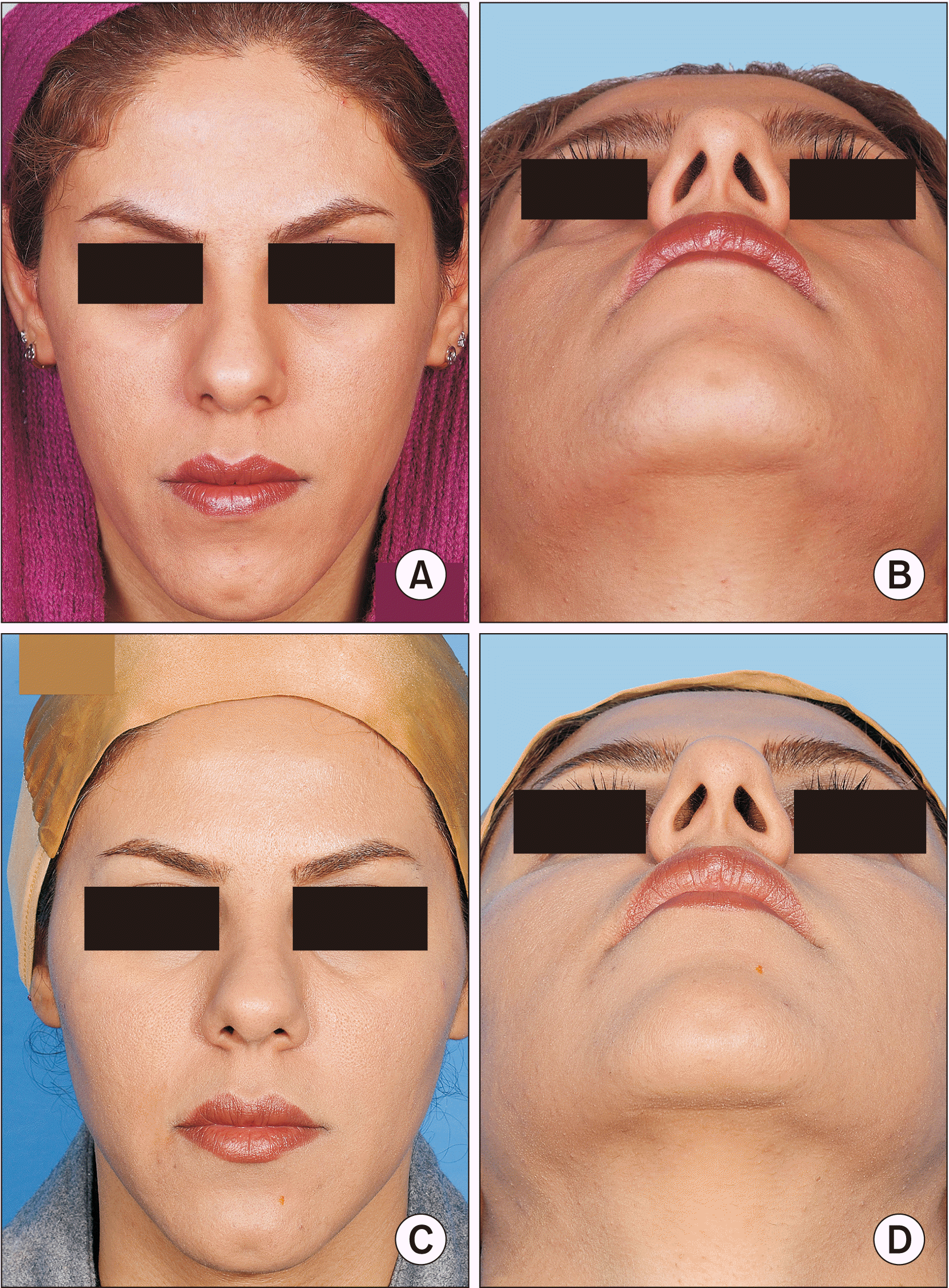
A 26-year-old female patient with a chief complaint of long face, malar flattening, and deep nasolabial fold. Autologous fat harvested from the abdomen was transplanted to the malar area, infraorbital rim, and nasolabial fold.(Fig. 2)
2. Case 2
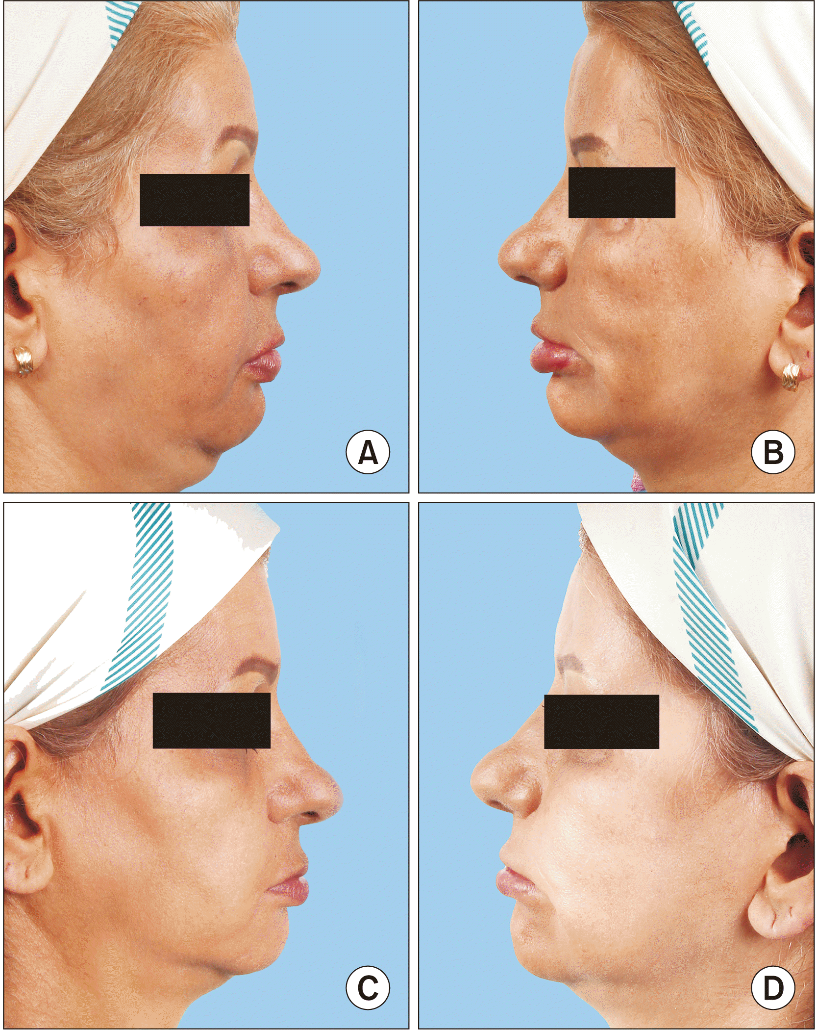
A 45-year-old female patient with a chief complaint of right malar depression, thin upper and lower lips, and inadequate vermilion exposure following facial nerve paralysis. The defects were corrected and formed by fat harvested from the abdomen.(Fig. 3)
3. Case 3
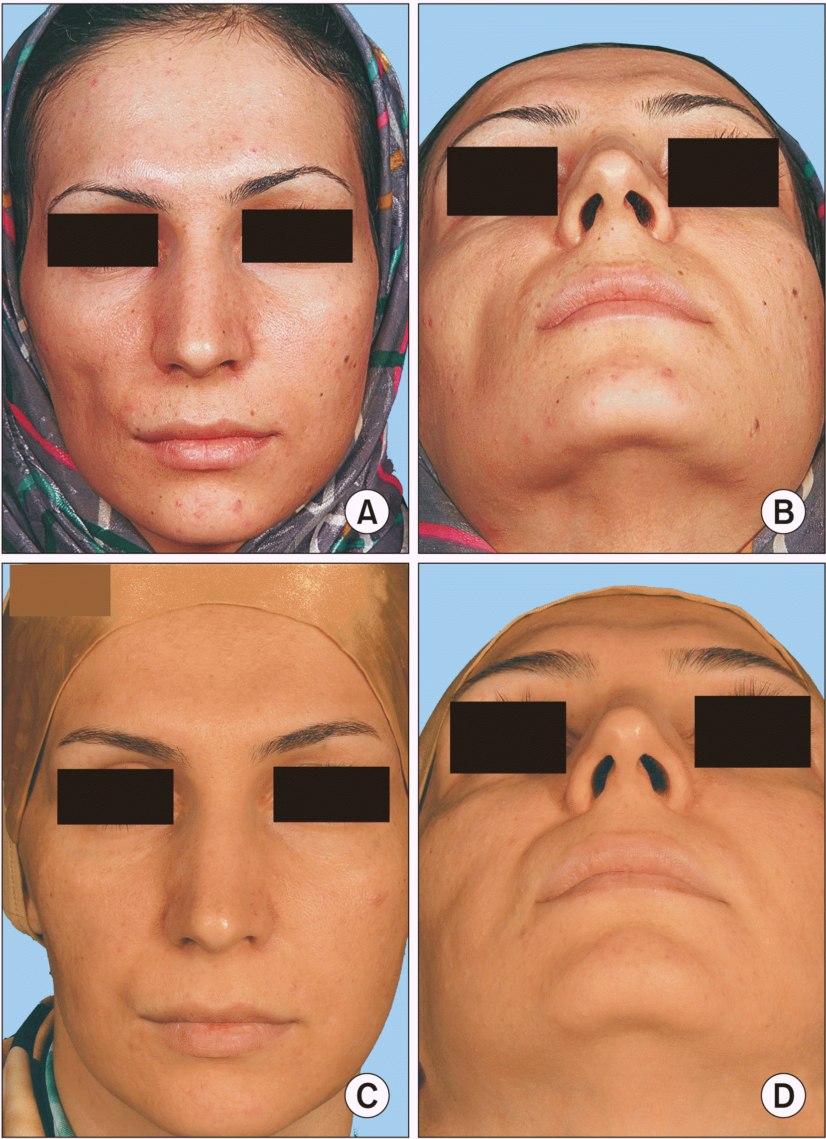
A 35-year-old female patient with depressed right malar area following trauma, a candidate for malar augmentation by fat transferring. The depression was corrected by fat from the abdomen.(Fig. 4)
Go to :
III. Results
A summary of patients are shown in Table 3. The data from 15 patients including 13 female and two male were retrieved. Of all cases, 80.0% had malar flattening, 33.3% had deep nasolabial fold, 13.3% had mandibular asymmetry, and 13.3% of cases also had perioral wrinkles and inadequate vermillion exposure. One patient (6.7%) had soft tissue depression due to surgical scarring, and one patient (6.7%) had soft tissue atrophy due to facial nerve paralysis.
Table 3
Summary of patients
![]()
Of 15 patients, two were not satisfied due to fat graft resorption, and further injections were performed six months after the first injection using preserved fat grafts. One patient continued to be dissatisfied with the final results. There were no other complications related to fat transplants.
Go to :
IV. Discussion
The primary manifestations of aging are subdermal tissue loss, fat atrophy, and vertical displacement of the tissues2,6,7. Reduced skin elasticity and underlying bone loss are two important factors affecting mid-face aging. The first successful free fat transplantation was reported by Neuber in 18932. In 1909, Hollander introduced a technique using a cannula for fat injections. In 1990, Coleman introduced centrifuge use in the fat processing technique8.
To regain volume, morphology, and contour of facial deformities, there are several injectable fillers available in the market such as hyaluronic acid-based fillers, Restylane, calcium hydroxyapatite, and poly L-lactic acid. However, while the results are predictable and satisfactory, these fillers are only appropriate for short-term use9.
Autologous fat is biocompatible, available in large amounts, economically cost-effective, and has long-lasting results in comparison with other fillers. Autologous fat transplantation is one of the best options suggested in tissue augmentation9. An autologous fat graft is a vital tissue, and there are no concerns about antigenicity and possible infection. Additionally, a fat graft has the ability to promote host tissue repair.
However, while being a reliable method for improving facial morphology, autologous fat transfer is a sensitive, challenging technique7,10. Since the fat graft is a vital tissue, the graft has the potential for atrophy and/or hypertrophy; therefore, further injections may be required. According to Xie et al.’s study10, long-term outcome can be expected after one to three injections. This research group reported 73.5% complete satisfaction and 4.5% dissatisfaction among their patients10. In our patients, 93.3% of patients were completely satisfied, 13.3% of patients needed further injections, and one patient (6.7%) was not completely satisfied.
The fat transplant must be performed with minimum tissue trauma6, and fat processing is a sensitive phase that is critical for fat cells’ survival7. Coleman and Katzel8 suggested fat harvesting by using a 10 mL syringe to minimize soft tissue trauma and blunt cannulas for fat transfer to avoid intravascular injection. This method of injection also minimizes resorption, necrosis, cyst formation, and large fat drops (larger than 0.1 mL)8. In a review of 550 articles, 596 patients received fat grafts; the survival rate for autologous fat graft in the breast area was 30%-83%. Based on an animal study, fat graft survival rate after one year follow-up in rabbits was about 14%-14.5%11. Protocols like insulin injection, adipose-derived stem cells, and hyperbaric oxygen were effective in fat cell survival improvement (79.16% after 4 weeks)12. Methods like platelet-rich plasma (69%-77% fat cell survival after 1 year)13,14, progenitor-supplemented adipose tissue (56% fat cell survival after 6 months)15, and mesenchymal stem cells (82% fat cell survival after 1 year)16 were effective in improving fat cell durability. Tanna et al.17 achieved 83% fat durability in soft tissue reconstruction in cases of craniofacial microsomia. Rare, but possible, complications related to autologous fat graft include vision loss, cerebrovascular accident, ophthalmic artery embolism, middle cerebral artery embolism, and even death11.
Go to :
V. Conclusion
Fat transplantation is a safe, reliable, and non-invasive method for facial contour and facial soft tissue defect restoration. Occasionally multiple fat injections are required. Additional methods such as use of mesenchymal stem cells along with fat injection increase the survival rate of transferred fat.
Go to :
 XML Download