

 PDF
PDF Citation
 Print
Print


I. Introduction
Many cultural and societal values emphasize the appearance of individuals, which increases individual interest in appearances. In particular, the main factors that affect facial appearance are asymmetry and mandibular prognathism. A positive correlation among social pressure, physical dissatisfaction related to appearance, and mental health has been reported, particularly in younger generations1. Moreover, a study reported that symmetrical faces are perceived as more attractive2.
Mandibular prognathism is more prevalent in East Asian countries, such as Korea, Japan, and China, than in the West. It is characterized by a long face, a large gonial angle, anterior open bite, incomplete lip closure, and a concave appearance. A face is considered asymmetrical when the dental midline deviation is 2.2 mm or more3, and facial asymmetry is common in the general population and increases toward the mandible2,4-7. However, mandibular prognathism cannot be corrected with conventional orthodontic treatments but requires orthognathic surgery.
Sagittal split ramus osteotomy (SSRO) is the most widely used surgical method for mandibular prognathism. It is advantageous because it allows smooth and rapid bone healing by rigid internal fixation through wide contact between the cancellous bones. However, treatment of asymmetry mandibular prognathism with bilateral SSRO (BSSRO) requires rotation of the distal segment of the mandible. Therefore, the gap between the proximal and distal segments widens. Fixation results in an inwardly displaced proximal segment, causing poor postoperative stability8-11. In addition, intraoral vertical ramus osteotomy (IVRO) is not optimal because it requires a longer healing period than that of SSRO, although the temporomandibular joint (TMJ) is physiologically well positioned with this approach.
In this study, we combined the SSRO and IVRO approaches to achieve a shorter healing period and stable TMJ position in patients with mandibular prognathism. Additionally, we performed lateral corticectomy on the IVRO side to resolve facial asymmetry. The purpose of this study was to evaluate the postoperative anteroposterior stability and improvement of facial asymmetry using a combination of IVRO, SSRO, and lateral corticectomy on the IVRO side in patients with asymmetric mandibular prognathism.
Go to : 
II. Materials and Methods
The patients’ records for admission to the Department of Oral and Maxillofacial Surgery at Dankook University Dental Hospital for treatment of asymmetric mandibular prognathism from July 2009 to October 2018, were reviewed after obtaining approval from the Institutional Review Board of Dankook Dental University Hospital (DKUDH IRB 2020-11-004).
1. Subjects
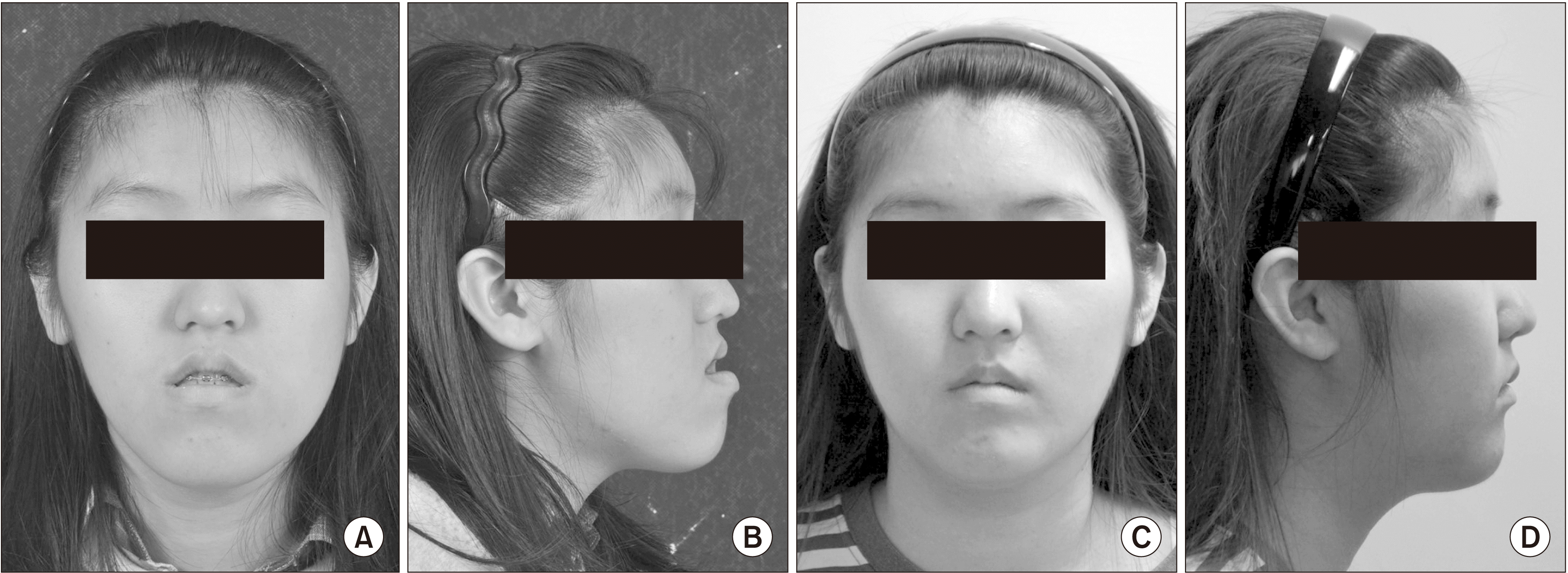
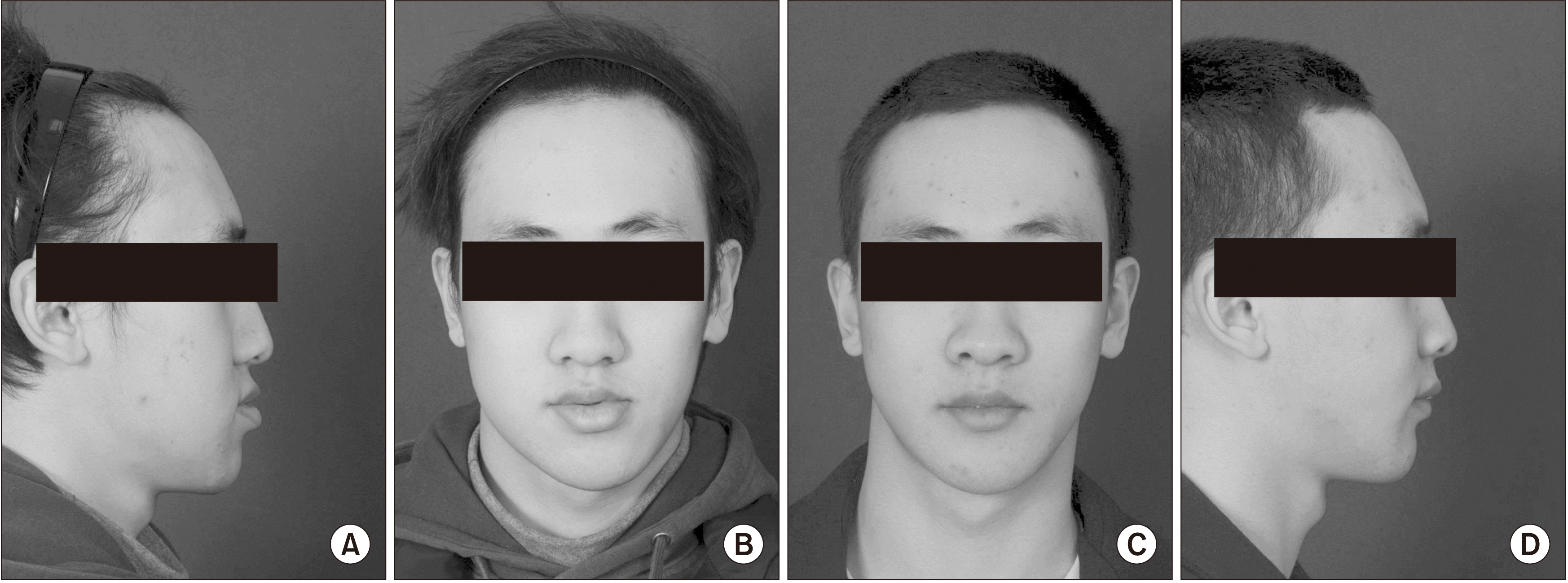
This retrospective study was conducted based on clinical photos, information, and radiographs of 11 patients who visited Dankook University Dental Hospital between July 2009 and October 2018. The patients were diagnosed with mandibular prognathism accompanied by facial asymmetry and underwent surgery at the hospital based on posteroanterior cephalometric radiograph (PA Ceph) and lateral cephalometric radiograph (Lat Ceph). None of the patients had underlying disease. There were six male and five female patients with an average age of 19.5 years (range, 18-23 years). Intermaxillary fixation was performed for approximately 28.1 days (range, 23-37 days), and the average menton deviation was 6.95 mm (range, 2.8-16.6 mm).
2. Surgical methods
An oral and maxillofacial surgeon performed LeFort I osteotomy on the maxilla and SSRO, IVRO, and lateral corticectomy on the IVRO side on the mandible. The surgical plan was based on a three-dimensional computed tomography model. All surgeries were performed using an intraoral approach. For lateral corticectomy, we defined the anterior, upper, and posterior boundaries as the posterior of the mental foramen, the part under the sigmoid notch, and the IVRO osteotomy line, respectively. Holes were created with a bur along the oblique ridge, and the boundaries were cut by connecting these holes with a reciprocating saw. The cortical bone was removed using a chisel in the gap made by this cut.(Fig. 1) Intermaxillary fixation was performed after surgery, followed by postoperative patient management. After discharge, the patients underwent periodic follow-up, during which clinical photographs and radiographs were recorded.(Fig. 2-4)
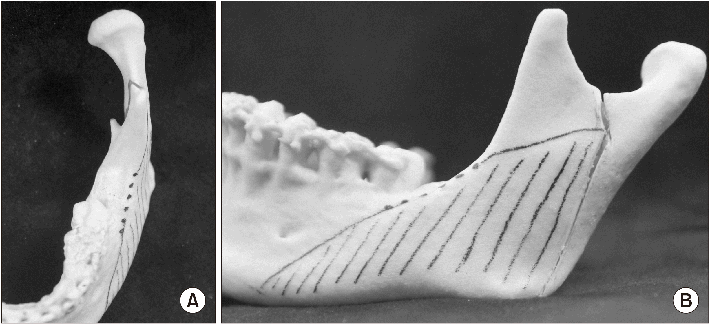 | Fig. 1A. Several holes are made by using bur on the oblique ride line of mandible. B. Adequate cortical bones are removed by lateral corticectomy procedure.
|
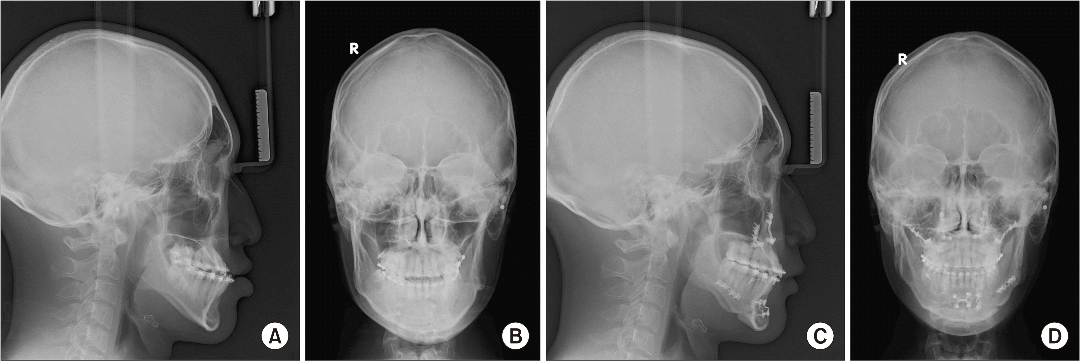 | Fig. 2A. Preoperative posteroanterior (PA) cephalometric. B. Preoperative lateral cephalometric. C. Postoperative PA cephalometric. D. Postoperative lateral cephalometric.
|
3. Study methods
Standardized cephalometric radiographs were used for precise comparison. The Lat Ceph was used to evaluate postoperative anteroposterior stability, and PA Ceph was used to evaluate facial asymmetry. Preoperative Lat Ceph was set to T0, to T1 for postoperation, and to T2 at 12 months. Similarly, preoperation PA Ceph was set to S0, and that at the 12-month follow-up was S1.
The reference points for evaluating anteroposterior stability after surgery were the sella turcica (S), orbitale (Or), porion (Po), and B point. In the Lat Ceph, the Frankfort line was set as the X-axis, and the line passing through the sella turcica perpendicular to the Frankfort line was set as the Y-axis.(Fig. 5) We set the distance from the Y-axis to the B point as the B point distance (Table 1) to evaluate postoperative anteroposterior stability and measured the difference in the distance to the B point (T2-T1) after surgery and at 12-month follow-up. To evaluate the statistical significance, we compared the Lat Cephs from pre-and postoperative (T0, T1), postoperative and 12-month follow-up (T1, T2), and at 12-month follow-up and preoperative (T2, T0) timepoints.
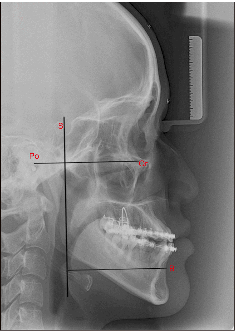 | Fig. 5Reference points and axes on lateral cephalometric radiograph. (S: sella turcica, Or: orbitale, Po: porion, B: B point, X-axis: Frankfort line, Y-axis: The line which is through the sella turcica and perpendicular to the Frankfort line)
|
Table 1
Postoperative anteroposterior stability and facial asymmetry evaluation
![]()
To evaluate the improvement in facial asymmetry, condylion (Co), antegonion (Ag), menton (Me), and mid-sagittal reference (MSR) lines were used (Fig. 6), and five measurements were recorded.(Table 1) Values were measured from the left and right sides, and the small values were divided by larger values to obtain the ratios, noted as Ag angle, Co-Ag, Ag-Me, Co-Me, and MSR-Ag.(Table 2) Each indicator was compared performed preoperatively (S0) and postoperatively (S1). A single observer performed the measurements in duplicate, and the study was conducted using the average values. Reference points were set using Adobe Photoshop 2021 (Adobe, San José, CA, USA), and the length and angle were measured.
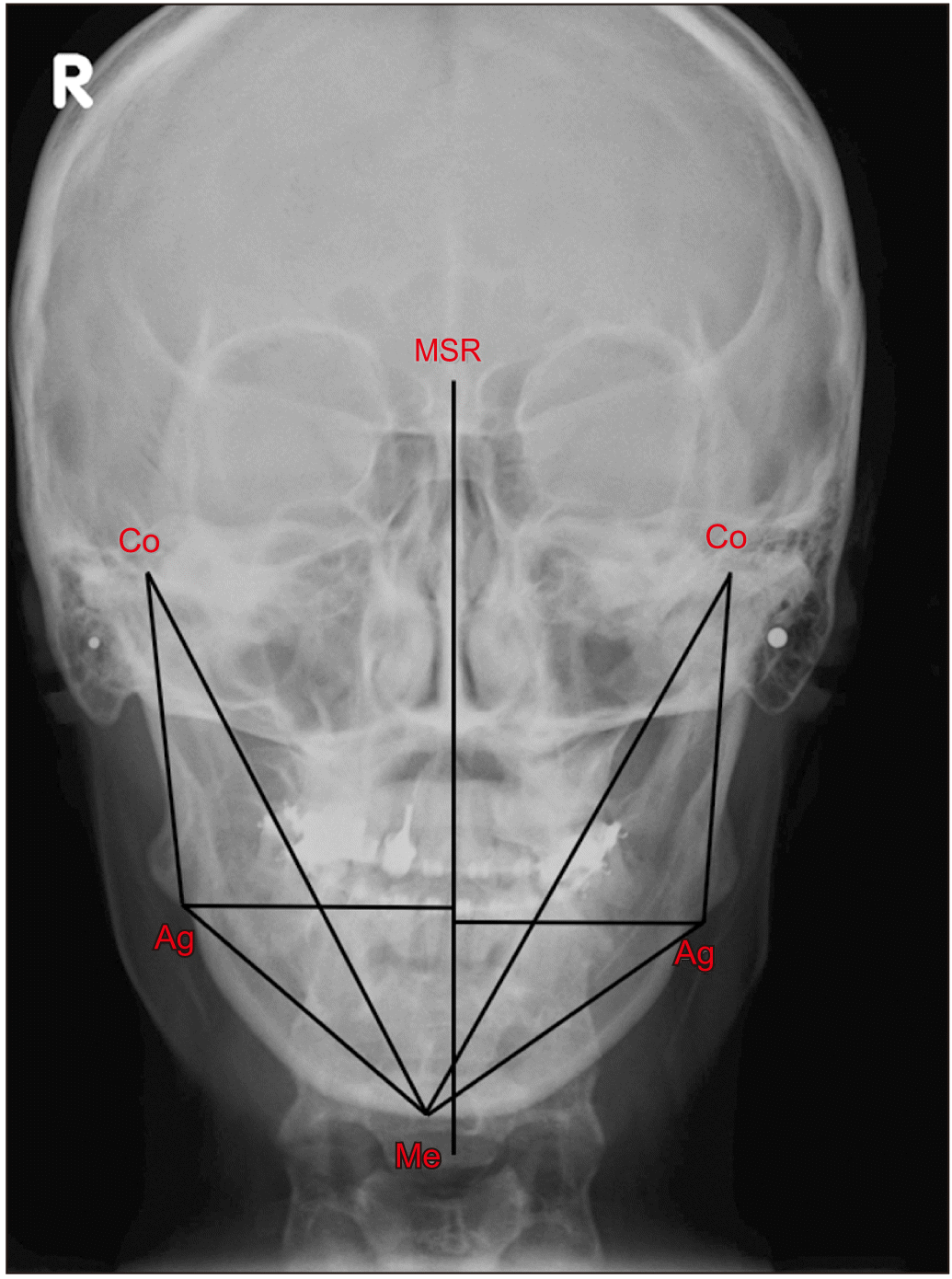 | Fig. 6Reference points and lines on posteroanterior cephalometric radiograph. (Co: condylion, Ag: antegonion, Me: menton, MSR: mid-sagittal reference line)
|
Table 2
Facial asymmetry indicators
![]()
4. Statistics methods
The Wilcoxon signed-rank test was performed using the IBM SPSS Statistics program (ver. 25; IBM, Armonk, NY, USA). The values of (T0, T1), (T1, T2), and (T2, T0) at timepoints were evaluated to assess postoperative anteroposterior stability. For facial asymmetry, S0 and S1 were compared and evaluated for each indicator. A statistically significant difference was noted when the P-value was <0.05.
Go to :
III. Results
1. Postoperative anteroposterior stability
The preoperative (Fig. 2. B) and postoperative (Fig. 2. D) radiographs indicated minimal changes in condylar position following surgery. Table 3 summarizes the average values of the B point distance in T0, T1, and T2 for the 11 cases. The average B point distances for preoperative, postoperative, and at 12 months follow-up were 7.30 cm, 6.56 cm, and 6.67 cm, respectively, with a 0.11 cm mean difference between T2 and T1. B point distances for T0 and T1 were significantly different (P=0.007), whereas those for T1 and T2 were not significantly different (P=0.1). In addition, there was a significant difference between the B point distances of T2 and T0 (P=0.026). Because the Z value is based on positive ranks, T0 was greater than T1 and T2.
2. Facial asymmetry
Table 4 summarizes the values for indicators of facial asymmetry. On comparing the indicators for facial asymmetry before and after surgery, all five indicators demonstrated statistically significant differences between the S0 and S1; the P-values of Ag angle, M-Ag, Co-Ag, Co-Me, and Ag-Me were 0.003, 0.003, 0.008, 0.006, and 0.004, respectively. Because the Z value was based on negative ranks, S1 was greater than S0.
Table 4
Facial asymmetry indicators
| Value | |
|---|---|
| Ag angle (%) | |
| S0 | 91.95±4.08 |
| S1 | 98.44±1.06 |
| MSR-Ag (%) | |
| S0 | 83.22±6.41 |
| S1 | 94.90±4.48 |
| Co-Ag (%) | |
| S0 | 94.37±4.39 |
| S1 | 98.07±1.44 |
| Co-Me (%) | |
| S0 | 93.64±3.71 |
| S1 | 98.22±1.01 |
| Ag-Me (%) | |
| S0 | 87.58±7.85 |
| S1 | 97.32±3.67 |
| (S1, S0) Ag angle | |
| Z | –2.934 |
| P-value | 0.003* |
| (S1, S0) MSR-Ag | |
| Z | –2.934 |
| P-value | 0.003* |
| (S1, S0) Co-Ag | |
| Z | –2.667 |
| P-value | 0.008* |
| (S1, S0) Co-Ag | |
| Z | –2.756 |
| P-value | 0.006* |
| (S1, S0) Ag-Me | |
| Z | –2.845 |
| P-value | 0.004* |
![]()
Go to :
IV. Discussion
The primary problem with orthognathic surgery is postoperative stability. Proffit et al.12 reported that this approach is problematic when the relapse rate is greater than 2 mm. According to Jakobsone et al.13, relapse usually occurs within 6 months of orthognathic surgery. Based on this, we evaluated postoperative stability by measuring the B point distance before and after surgery and at the 12-month follow-up. Lai et al.9 evaluated postoperative relapse as the amount of change in B point distance after a combination surgery of SSRO and IVRO. They reported postoperative anteroposterior stability and no significant difference from that of BSSRO and bilateral IVRO (BIVRO).
In this study, we performed SSRO and IVRO combination surgeries and observed a significant difference in the B point distance between preoperation and postoperation and at the 12-month follow-up and preoperation. However, there was no significant difference in this distance between from postoperation to the 12-month follow-up, with an average difference of 0.11 cm. Therefore, we assessed and observed anteroposterior stability after surgery.
IVRO is useful for addressing the gap problem for facial asymmetry because fixation is not needed. Therefore, postoperative stability was possible despite severe facial asymmetry. According to Beyer and Lindauer3, facial asymmetry occurs when the dental midline deviation is 2.2 mm or greater. We only included patients with a deviation of 2 mm or more in this study, which is consistent with the approach for previous studies8,9,14.
Numerous analytic methods have been suggested for evaluating facial asymmetry, such as Rickett’s method15 and Grummon’s method16. Masuoka et al.5 reported that facial asymmetry becomes more severe from the maxilla to the mandible, especially in the menton, which is where the maximum deviation appears. In addition, menton deviation showed the greatest correlation with subjective judgment of facial asymmetry. Therefore, we focused on mandibular asymmetry and used effective indicators for evaluation, as presented by Grummon and Kappeyne van de Coppello16 and Lee et al.4. According to Hwang et al.17, antegonion and gonion can be used as reference points for evaluating mandibular asymmetry as well as reflection of changes in the soft tissue. Other studies also use these two reference points; however, according to Major et al.18, gonion is inaccurate as a reference point, and antegonion is more accurate than gonion. Therefore, we used antegonion in this study for accurately comparing facial asymmetries.
Surgical procedures performed to treat facial asymmetry include angle shaving, masseter resection, and cortical bone resection. Angle shaving is not an effective approach for resolving asymmetry because it requires bone resection, which increases asymmetry. Additionally, the approach is limited because the frontal profile after surgery is not significantly different from that before surgery19,20. Masseter resection usually is performed with angle shaving; however, it frequently is associated with hematoma and limited mouth-opening capacity as postoperative complications. In contrast, lateral corticectomy reduces the width of the mandible and changes the frontal profile. Therefore, to achieve sufficient frontal profile changes, lateral corticectomy should be performed instead of angle shaving21. According to Deguchi et al.20, because of postoperative masseter muscle atrophy, even lateral corticectomy without resection of the muscle results in a decreased width of the mandible, similar to that with angle shaving and masseter resection. Additionally, Han and Kim22 reported that, even without resecting the masseter muscle, the amount of reduction in the width of the mandible was similar to that of resection. However, nerve damage can occur because it has a bone resection line similar to that of SSRO.
In this study, we performed corticectomy on the IVRO side to resolve facial asymmetry. SSRO was performed at sites that could heal quickly due to contact between the cancellous bones with rigid internal fixation. However, when the evaluation is conducted through model surgery, an appropriate amount of bone is required for fixation on the SSRO side; hence, lateral corticectomy could not be performed sufficiently. On the contrary, because rigid internal fixation was not required on the IVRO side, we performed cortical bone resection to resolve facial asymmetry. Therefore, we efficiently improved the facial asymmetry using lateral corticectomy on the IVRO side.
Previous studies calculated asymmetry as the difference between the left and right measured values of facial characteristics4,6,7. Although the difference in length affects the symmetry, the proportion of the difference(s) in the measured values between the left and right sides with respect to the mandible length can be small when the mandible is large. Therefore, evaluation of asymmetry by comparing ratios is a better method.
Facial asymmetry evaluation revealed a statistically significant difference between the preoperative and postoperative values for the five indicators. Because the Z value is based on negative ranks, postoperative values were higher than preoperative values. This suggests that the left and right sides become symmetrical as the value approaches 100%, and facial asymmetry is improved.
The primary limitation of this study was the small number of cases, which required nonparametric statistics. When we performed nonparametric statistics, we did not calculate the difference between the mean values before and after surgery because only the increase or decrease in values was known. Therefore, compared with BSSRO and BIVRO, it was not possible to identify the most effective procedure for resolving facial asymmetry, and more cases are required for adequate comparison.
Go to :
V. Conclusion
In this study, we achieved postoperative anteroposterior stability and improvement of facial asymmetry in patients with mandibular prognathism. The treatments included LeFort I osteotomy, SSRO, IVRO, and lateral corticectomy on the IVRO side. Based on the results, we determined that there was no significant difference in the B point distances at postoperation and at the 12-month follow-up; there were significant differences in all five indicators related to facial asymmetry before and after surgery, with all showing postoperative increase.
Go to :
 XML Download