

 PDF
PDF Citation
 Print
Print


INTRODUCTION
Acute eosinophilic appendicitis (AEA) is a rare type of appendicitis in that the appendiceal muscularis propria is infiltrated by eosinophils instead of neutrophils observed in acute suppurative appendicitis. Despite this difference, the symptoms of this disease are similar to those of acute suppurative appendicitis.1 Lower gastrointestinal hemorrhage (LGIH) is a very unusual presentation of appendicitis.2 This paper reports a case of an elderly patient with AEA presenting as LGIH and discusses the characteristics of this disease with reference to the literature.
CASE REPORT
The Institutional Review Board of Wonkwang University Hospital (IRB No. WKUH 2021-03-014) approved this study, and informed consent was waived.
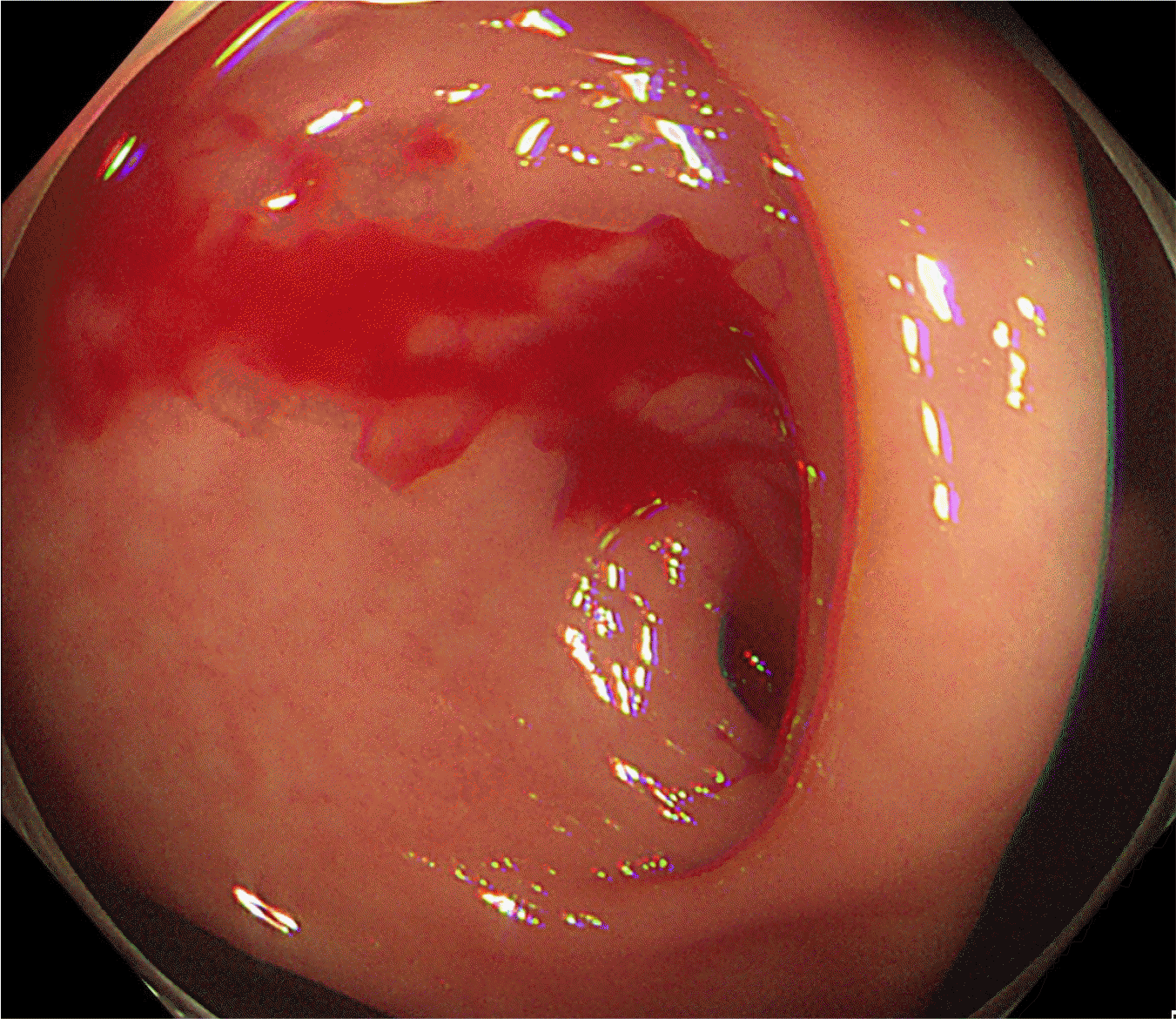
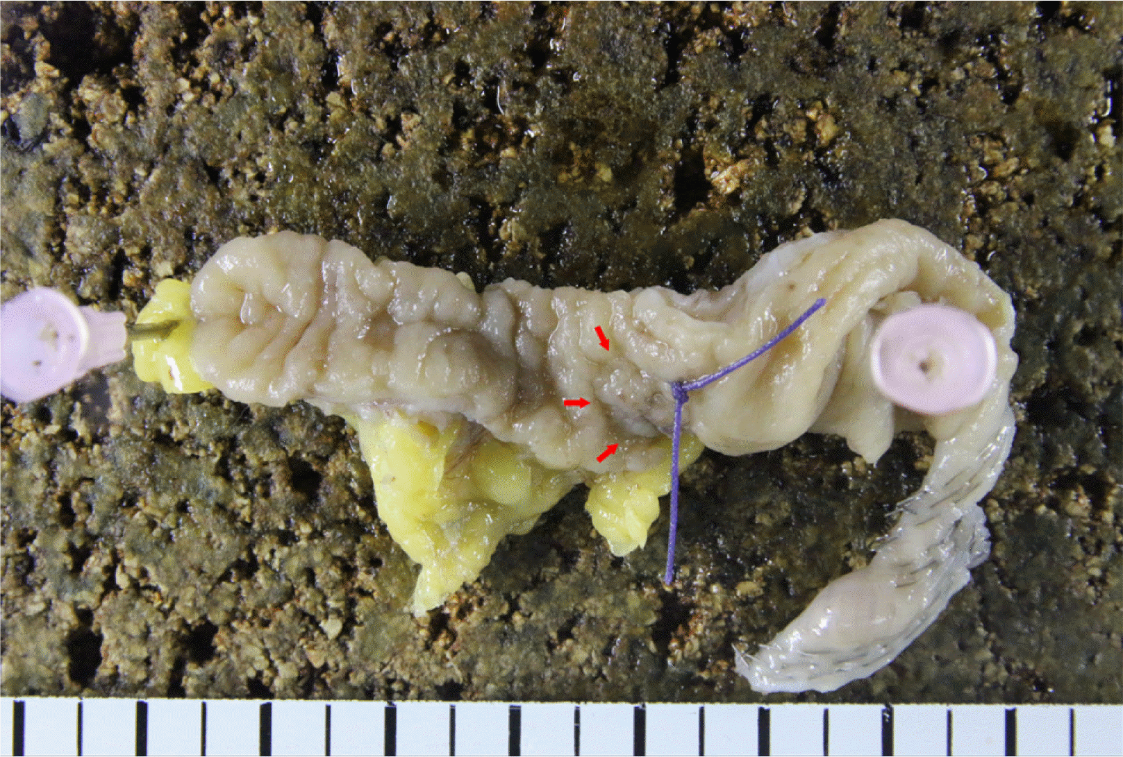
An 82-year-old man visited the emergency department of Wonkwang University Hospital with hematochezia that occurred on the day before his visit. The patient had hypertension and had taken clopidogrel for 2 years because of a history of cerebral infarction. At the time of the visit, his blood pressure, heart rate, and body temperature were 110/80 mmHg, 102 beats/min, and 37.3℃, respectively. Laboratory tests revealed a hemoglobin of 10.9 g/dL, white blood cell count of 6.29×10³/mm3, and CRP of 1.71 mg/L. An 82-year-old man visited the emergency department of Wonkwang University Hospital with hematochezia that occurred on the day before his visit. The patient had hypertension and had taken clopidogrel for 2 years because of a history of cerebral infarction. At the time of the visit, his blood pressure, heart rate, and body temperature were 110/80 mmHg, 102 beats/min, and 37.3℃, respectively. Laboratory tests revealed a hemoglobin of 10.9 g/dL, white blood cell count of 6.29×10³/mm3, and CRP of 1.71 mg/L. The other blood chemistry parameters were within the normal range. The patient underwent colonoscopy under the clinical impression of LGIH. Colonoscopy revealed arterial pulsating hemorrhage in the appendiceal orifice (Fig. 1). Blood clots were observed in the terminal ileum, but there was no evidence of hemorrhage after washing it off. Small bowel hemorrhage was subsequently ruled out. A laparoscopic partial cecectomy was performed without a further evaluation because the patient’s vital signs became unstable. In the gross findings, the appendix size was 6.5 cm×1.0 cm. Ulceration was observed 1.5 cm distal from the appendiceal orifice (Fig. 2). Histologically, marked eosinophilic infiltration was noted with severe central ulceration in the muscularis propria of the appendix and accompanying edema (Fig. 3). Because tissue eosinophilia was confirmed in a biopsy, stool parasite ova exam and parasite antibody ELISA were performed to exclude a parasite infection, but there were no specific abnormalities. Based on these results, the patient was diagnosed with an appendiceal hemorrhage due to AEA. With the absence of postoperative complications, the patient was discharged from the hospital 7 days after surgery.
DISCUSSION
LGIH is defined as hemorrhage below the ligament of Treitz and accounts for approximately 20% of all gastrointestinal tract hemorrhages. In general, appendiceal hemorrhage is a rare cause of LGIH, with only a few cases reported.2-4 The causes of appendiceal hemorrhage have been reported diversely as acute appendicitis, diverticulitis, Crohn’s disease, intussusception, angiodysplasia, aorto-appendiceal fistula, ulcer, erosion, tuberculosis, endometriosis, and gastrointestinal tumors. Appendiceal hemorrhage due to AEA is an even rarer condition; this is the second reported case to the authors’ knowledge.2,5
Eosinophilic infiltration of the appendiceal muscular layer was previously described as “subacute appendicitis”, but no evidence of clinical relevance was provided.6,7 Jona et al.8 described similar findings presenting as acute appendicitis in children as a variant of eosinophilic gastroenteritis. On the other hand, later studies showed that appendices with eosinophilic infiltration in the muscular layer are not associated with atopy or peripheral blood eosinophilia. In most cases, the symptoms are similar to those of acute suppurative appendicitis, and unlike eosinophilic gastroenteritis, no case of steroid treatment has been reported. Thus, there is insufficient evidence to explain it as a variant of eosinophilic gastroenteritis.1,9-11
Aravindan12 suggested that eosinophilic infiltration in the muscularis propria of the appendix was an early event of appendicitis- related type I hypersensitivity to allergens. He further explained that a local allergic reaction could cause edema, and the appendix is vulnerable to ischemia because of its small size, which results in mucosal damage. Neutrophilic infiltration and acute suppurative appendicitis occur if the damaged mucosa is infected with microorganisms.1,12 In a follow-up study in 2010, Aravindan et al.1 used the term “acute eosinophilic appendicitis” to describe this disease entity. They suggested it as a variant of acute appendicitis. The histological feature of AEA is the infiltration of eosinophils in the muscularis propria of the appendix instead of neutrophils and accompanying edema lesions.1 This hypothesis is novel but requires further studies.
In the present case, marked eosinophilic infiltration was observed in the muscular layer of the appendix, which is consistent with AEA. No other causes of tissue eosinophilia, such as a parasitic infection, were found. The patient had no history of atopy or peripheral blood eosinophilia. The histological findings confirmed the ulcer, which was believed to result from the mucosal damage caused by AEA. The ulcer was the presumed cause of this hemorrhage. Because the damaged mucosa had not yet been infected with microorganisms, LGIH presented as the first symptom instead of typical right lower quadrant pain and fever seen with acute suppurative appendicitis. The patient has taken clopidogrel because of cerebral infarction. Only a few cases of appendiceal bleeding have been reported in patients using aspirin, NSAIDs, or ticlopidine. Because of the small number of cases, it is difficult to clarify the association between a specific drug and appendiceal bleeding.2
Various modalities, such as mesenteric arterial angiography, colonoscopy, and multidetector CT (MDCT), are used to diagnose appendiceal hemorrhage. Colonoscopy is a well-established and safe method for diagnosing LGIH. On the other hand, appendiceal hemorrhage may be close to the ileocecal valve, making it difficult to distinguish it from a small bowel hemorrhage. Moreover, in emergency settings, the approach itself may be complex because of the presence of fecal material from the lack of bowel preparation. In these cases, a combination of colonoscopy and MDCT can improve diagnostic accuracy. In addition, MDCT can help identify the underlying cause of appendiceal hemorrhages, such as appendicitis or tumor.2,3,13,14
An appendiceal hemorrhage is treated mainly by surgery, including appendectomy, cecectomy, ileocecectomy, and right hemicolectomy.2,13 Almost all were treated surgically except for two previously reported cases. One of them was treated successfully with endoscopic hemostasis for angiodysplasia, which was confirmed by the absence of other lesions in the appendix. The other was treated with percutaneous transcatheter arterial embolism. Eventually, this patient required surgery because of rebleeding. In many previously reported cases, treatments with endoscopic and embolization approaches were difficult because appendicitis was the cause, or a tumor was present.14,15 Thus, the surgical option appears to be the most feasible method for diagnosing and treating appendiceal hemorrhage.
In conclusion, an unusual case of appendiceal hemorrhage caused by AEA was reported. This disease entity is very rare and requires further study. AEA should be considered in a differential diagnosis of appendiceal hemorrhage. In addition, proper diagnosis is important because appendiceal bleeding can be treated using simple procedures, such as appendectomy, once the location is evaluated using the appropriate modalities.

 XML Download