

 PDF
PDF Citation
 Print
Print


INTRODUCTION
A diverticulum is a sac-like protrusion observed in the bowel wall and can occur anywhere from the esophagus to the colon. A duodenal diverticulum is observed in 2-5% and 7% of patients during upper gastrointestinal tract barium studies and ERCP, respectively.1 In general, it is asymptomatic in most cases but might be associated with complications, such as cholestasis, bleeding, and perforation.2 Duodenal diverticulitis is difficult to diagnose if it is not considered because its clinical symptoms may be similar to acute cholecystitis, acute pancreatitis, or peptic ulcer, which may bring fatal complications in the case of a delayed diagnosis.3 Septic thrombophlebitis or phlephlebitis, mainly superior mesenteric vein thrombosis (SMVT) and portal vein thrombosis (PVT), can be accompanied by diverticulitis, especially in the colon.4,5 On the other hand, cases of duodenal diverticulitis associated with septic thrombophlebitis are very rare, and those with PVT have not been reported.6 This paper reports a patient with duodenal diverticulitis and PVT who underwent endoscopic therapy and showed a rapid clinical improvement.
Go to : 
CASE REPORT
A 44-year-old woman presented to the emergency department of the Dankook University Hospital with severe right upper abdominal pain and nausea for 4 days. She had no specific medical and surgical history. Her vital signs on presentation were as follows: blood pressure, 116/78 mmHg; heart rate, 112 beats/min; respiration rate, 18 breaths/min; and body temperature, 38.2℃. The physical examination revealed tenderness in the right upper abdomen. The initial laboratory data were as follows: white blood cell (WBC) 13,660/mm3; AST 25 U/L; ALT 31 U/L; total-bilirubin 2.49 mg/dL; amylase 33 U/L; lipase 21 U/L; CRP 20.91 mg/dL; procalcitonin 0.99 ng/mL; and D-dimer 1,128 ng/mL. The liver function tests except for bilirubin and coagulation battery were within the normal limits.
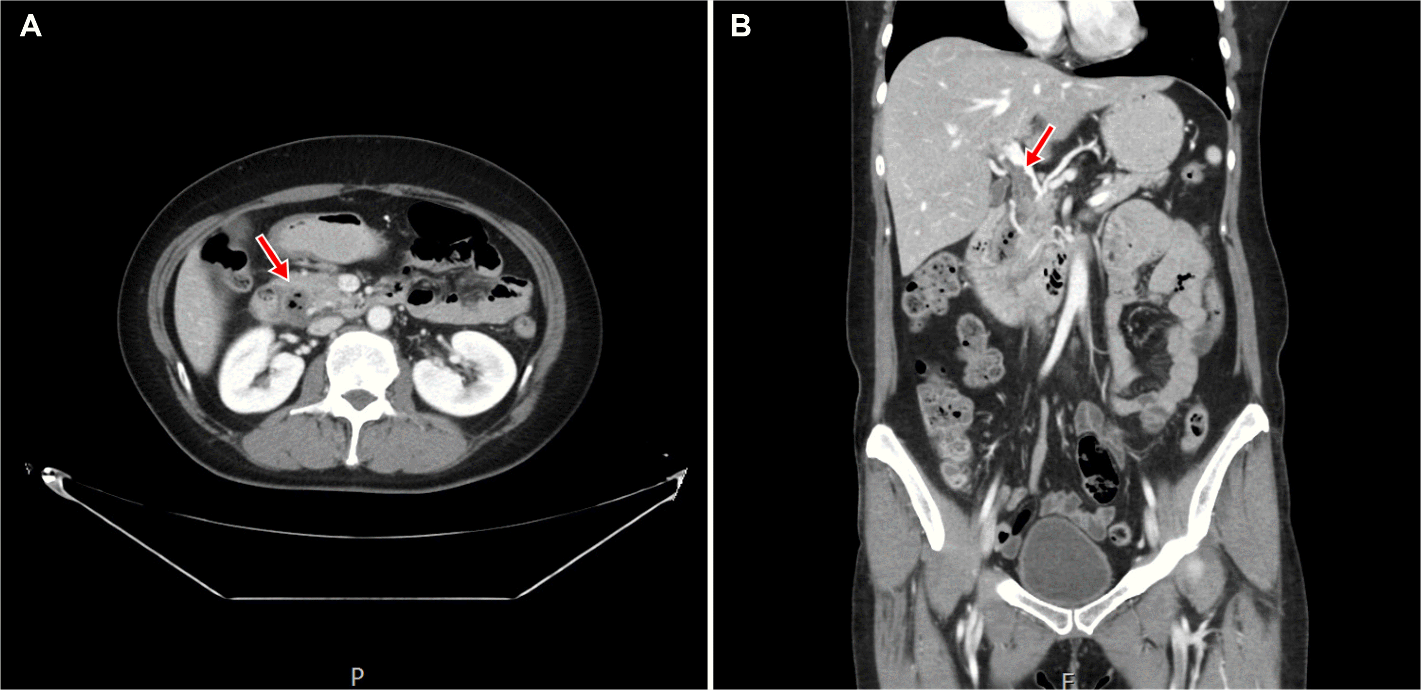
An initial abdominopelvic CT scan revealed a 2.3-cm-sized diverticulum filled with fluid and air in the second portion of the duodenum with linear marginal enhancement, fat stranding, and a small amount of fluid along the periduodenal space (Fig. 1A). In addition, short segmental partial thrombosis of the distal main portal vein (PV) was observed (Fig. 1B). CT and MRCP showed that the common bile duct and main pancreatic duct were preserved, and there was no sign of stone in the common bile duct.
Considering acute duodenal diverticulitis, the patient was fasting, and empirical antibiotics were administrated. However, the abdominal pain and tenderness aggravated; thus, an endoscopic evaluation was considered. Side-viewing duodenoscopy into the duodenal lumen (Fig. 2). Under fluoroscopy, balloon-ocrevealed swollen duodenal mucosa with a narrowed diverticular cluded contrast injection into the diverticulum showed no signs orifice in the 11 o’clock direction of the normal-looking major of contrast leakage into the common bile duct (Fig. 3A). Then, papilla. Purulent fluid was noted from the diverticular orifice endoscopic balloon dilatation of the diverticular orifice was performed after sweeping with an extraction balloon catheter and saline irrigation. Subsequently, pus and food debris gushed out into the duodenal lumen (Fig. 3B-D). Endoscopic placement of a continuous drainage stent was unnecessary because the orifice was sufficiently wide to allow fluid drainage. After the procedure, follow-up laboratory data was normalized rapidly.
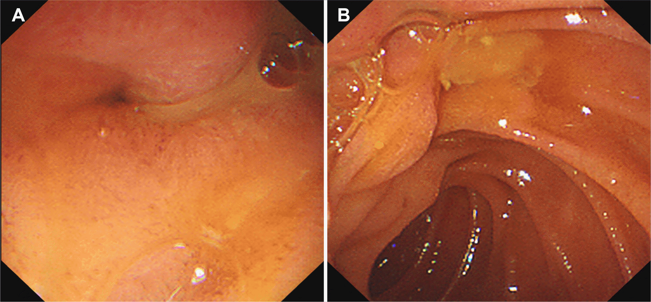 | Fig. 2Endoscopic findings. (A) Swollen diverticular orifice in the 11 o’clock direction of the normal-looking ampulla, and (B) the internal exudate was flowing out.
|
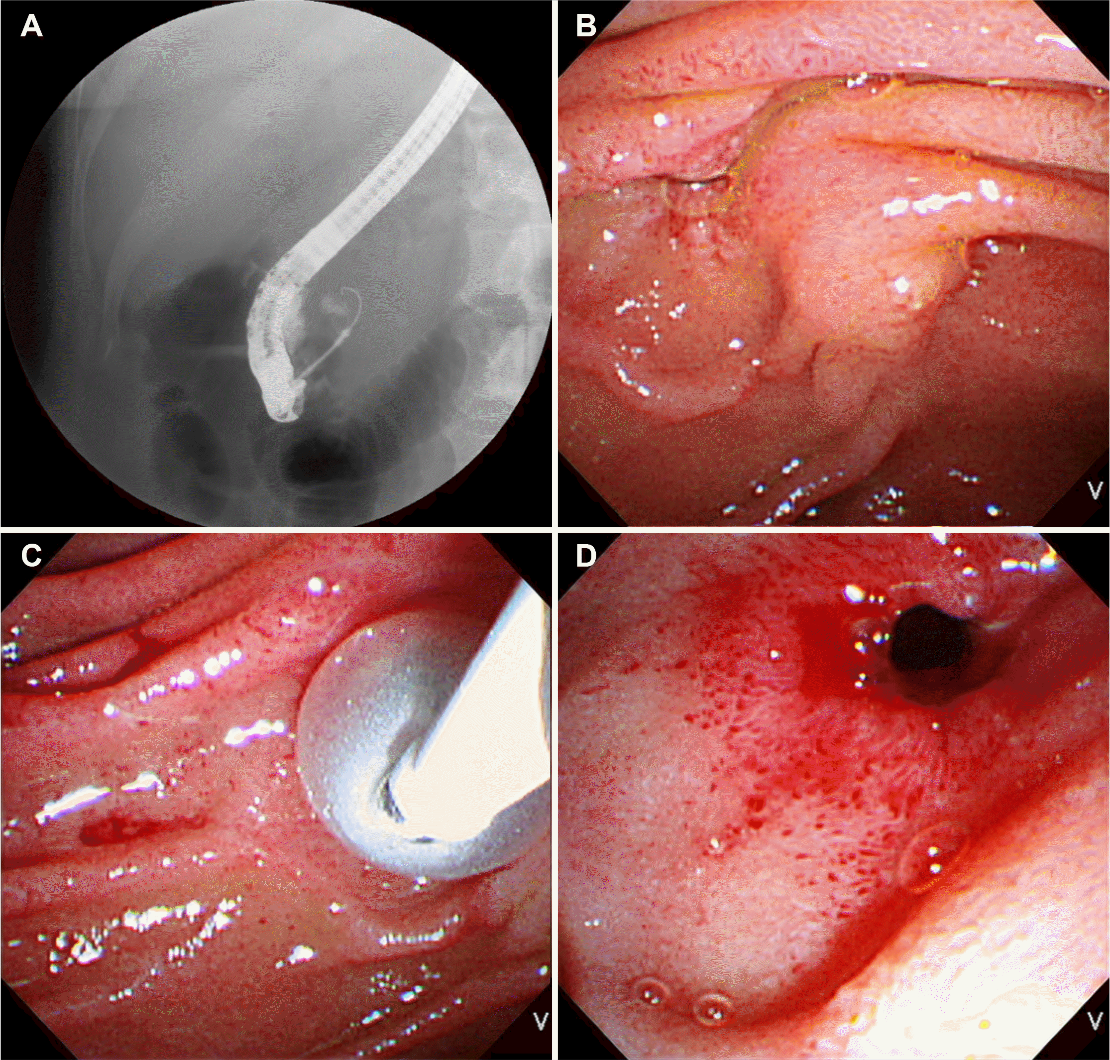 | Fig. 3(A) Fluoroscopic findings. Balloon-occluded contrast injection into the diverticulum showed no sign of contrast leakage into the common bile duct. (B-D) Endoscopic findings. Endoscopic balloon dilatation, sweeping, and irrigation with a normal saline were performed, and the pus and food debris were drained.
|
The patient recovered and was discharged uneventfully with oral antibiotics and warfarin on the 5th-day post-hospitalization. After 1 month, a follow-up CT scan and duodenoscopy revealed a normal-appearing duodenum and complete resolution of the duodenal diverticulitis and PVT (Fig. 4).
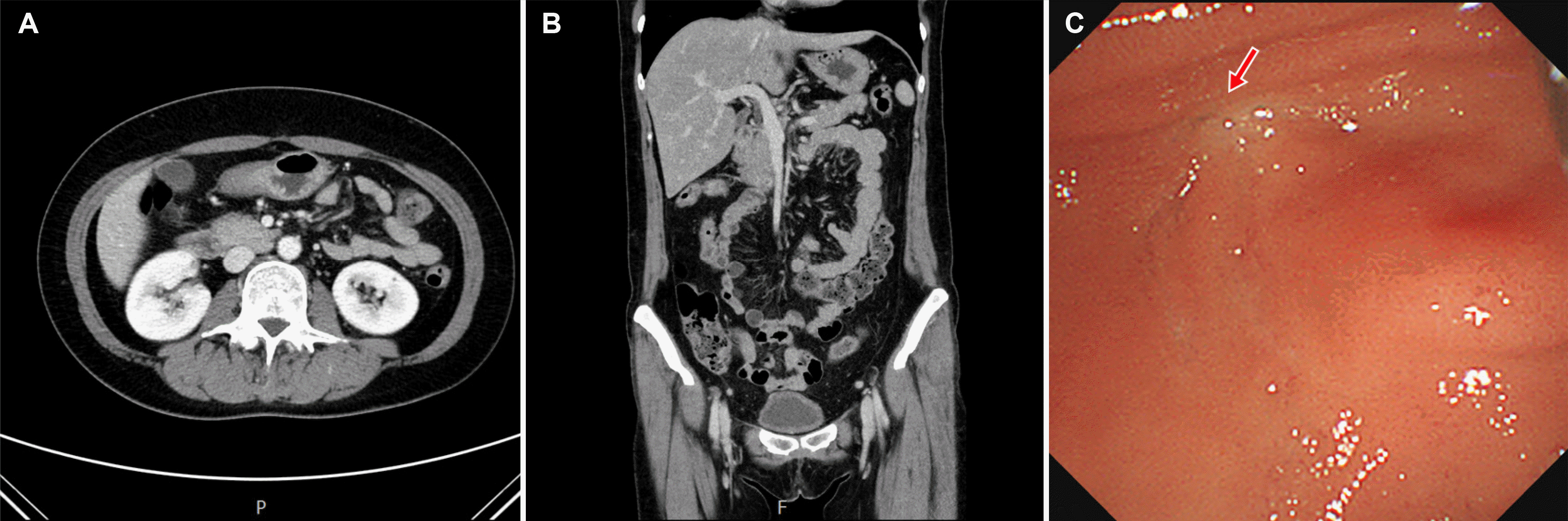 | Fig. 4(A, B) Complete resolution of portal vein thrombosis and duodenal diverticulitis was noted on the 1-month follow-up abdominopelvic computed tomography; (A) transverse image; (B) coronal image; (C) endoscopic resolution of duodenal diverticulitis and closed diverticular opening (arrow) was noted on the 1-month follow-up endoscopy.
|
Go to :
DISCUSSION
A duodenal diverticulum is an acquired outpouching of the mucosa and submucosa through an area of weakness in the duodenal wall. The condition is commonly reported in up to 20% of autopsy cases. The most common location is the second portion near the papilla of Vater, while less than 10% are located in the first and fourth parts of the duodenum.1 The incidence increases with age, with no sex difference.7
Duodenal diverticulitis is rarer than colonic diverticulitis. This is attributed to the following: 1) a duodenal diverticulum usually has a larger diameter and orifice, 2) duodenal passage is relatively fast, and 3) the duodenum is relatively bacteria-free.8 On the other hand, when duodenal diverticulitis occurs, perforation, abscess, secondary bile duct obstruction, cholecystitis, and pancreatitis may develop, with reported mortality rates of 33-44%.9
The main symptoms and signs of duodenal diverticulitis include abdominal pain, fever, and leukocytosis. On the other hand, these are nonspecific and similar to acute cholecystitis, acute pancreatitis, and peptic ulcer. Hence, it is important to consider duodenal diverticulitis in a differential diagnosis.8 In this present case, biliary tract disease was initially suspected due to right upper abdominal pain, fever, elevated WBC count, elevated CRP level, and jaundice. Abdominopelvic CT and MRCP showed no signs of biliary tract disease.
The diagnosis of duodenal diverticulitis is based on CT, MRCP, and endoscopic findings.10 Duodenal diverticulitis could be suspected if a thickened wall with internal air-fluid layers in the duodenal diverticulum is noted on a CT scan.10 MRCP is helpful for excluding biliary tract or pancreatic disease. An endoscopic examination with side-viewing duodenoscopy is helpful for a definite diagnosis and therapeutic intervention.11 Duodenal diverticulitis should be suspected when mucosal edema with redness and exudate is observed around the duodenal diverticulum.11 In the present case, a fluid and air-filled 2.3-cm-sized diverticulum with linear marginal enhancement and surrounding fat stranding was observed on CT, and mucosal edema with exudate from the diverticular orifice was observed on duodenoscopy, which was highly suggestive of acute duodenal diverticulitis.
The treatment of duodenal diverticulitis is based on the patient’s clinical manifestation. If symptoms are mild and there is no sepsis, conservative treatments, such as fasting and the administration of empirical antibiotics, can be effective. Surgical treatment may be considered when symptoms are severe. 10 The endoscopic procedure can be an effective treatment and safe alternative to surgical treatment.10 In the present case, the diverticular orifice was dilated with an extraction balloon, and subsequent irrigation helped the rapid improvement of the patient’s symptoms.
Septic thrombophlebitis or pylephlebitis is a rare complication of an intraperitoneal infection, which is characterized by suppurative thrombosis of the PV or its branches.5 The common cause of pylephlebitis in the MV and PV systems is diverticulitis, following appendicitis, pancreatitis, bowel perforation, and inflammatory bowel disease.5,12 SMV is the most commonly involved site, followed by PV and the inferior mesenteric vein (IMV).4 The pylephlebitis that occurs in the mesenteric vein is related to the colonic infection. In particular, sigmoid diverticulitis can cause a thrombus in the IMV and PV, which is called ascending septic thrombophlebitis.12
Until now, MVT and PVT related to the duodenum are rarely reported. There are case reports of SMVT associated with duodenal diverticulitis and PVT associated with duodenal ulcers.6,13 In this present case, the cause of the thrombus was presumed to be a local infection rather than ascending septic thrombophlebitis considering its adjacent anatomic location. The main symptoms of pylephlebitis were abdominal pain, fever, leukocytosis, and bacteremia, but these were nonspecific. Thus, a definite diagnosis is often delayed.14 According to a previous report, CT, MRI, abdominal ultrasound, angiography, and fluorodeoxyglucose (FDG)-PET helped diagnose duodenal diverticulitis.13,15
Antibiotics and anticoagulants are the treatment of choice for pylephlebitis.16 The treatment period of anticoagulation has not been clearly established, but is generally recommended up to 6 months. Treatment is required for a longer period if patients have coagulopathy or repeated thrombosis.6 In the present case, after the antibiotic and anticoagulation treatment for 1 month, the thrombus completely disappeared in the follow-up CT. The patient had no coagulopathy or thrombosis; thus, no additional maintenance treatment was performed.
In conclusion, this paper reported a rare case of duodenal diverticulitis with PVT. When examining patients with upper abdominal pain, the duodenal diverticulum and its related complications should be considered in a differential diagnosis. Minimally invasive endoscopic therapy, including dilatation, irrigation, and disimpaction, can be effective when conservative management fails and may be a viable alternative to surgery for treating duodenal diverticulitis.
Go to :
 XML Download