

 PDF
PDF Citation
 Print
Print


INTRODUCTION
Melanoma is very aggressive, highly metastatic malignancy that generally originates from cutaneous lesions. It is more likely to regress than all other cancers. However, distinguishing between primary and metastatic lesions can be difficult [1]. Regardless of whether it is primary or metastatic, its prognosis is extremely poor. Melanoma of the gallbladder is so rare that standard treatment guidelines have not been established. Here, we report a case of metastatic melanoma of the gallbladder and describe the approach we used to treat this patient.
Go to : 
CASE
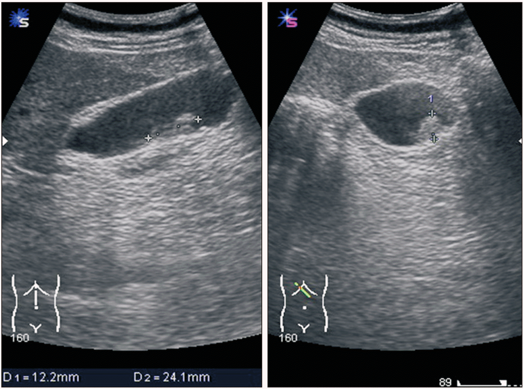
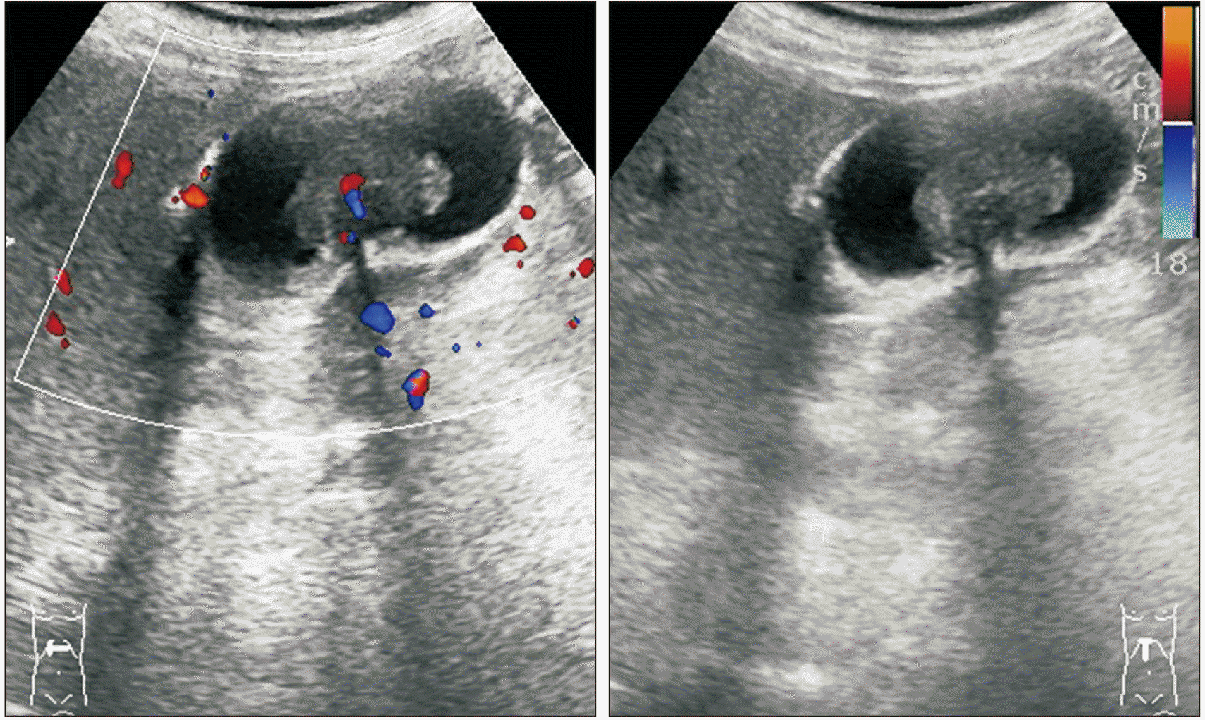
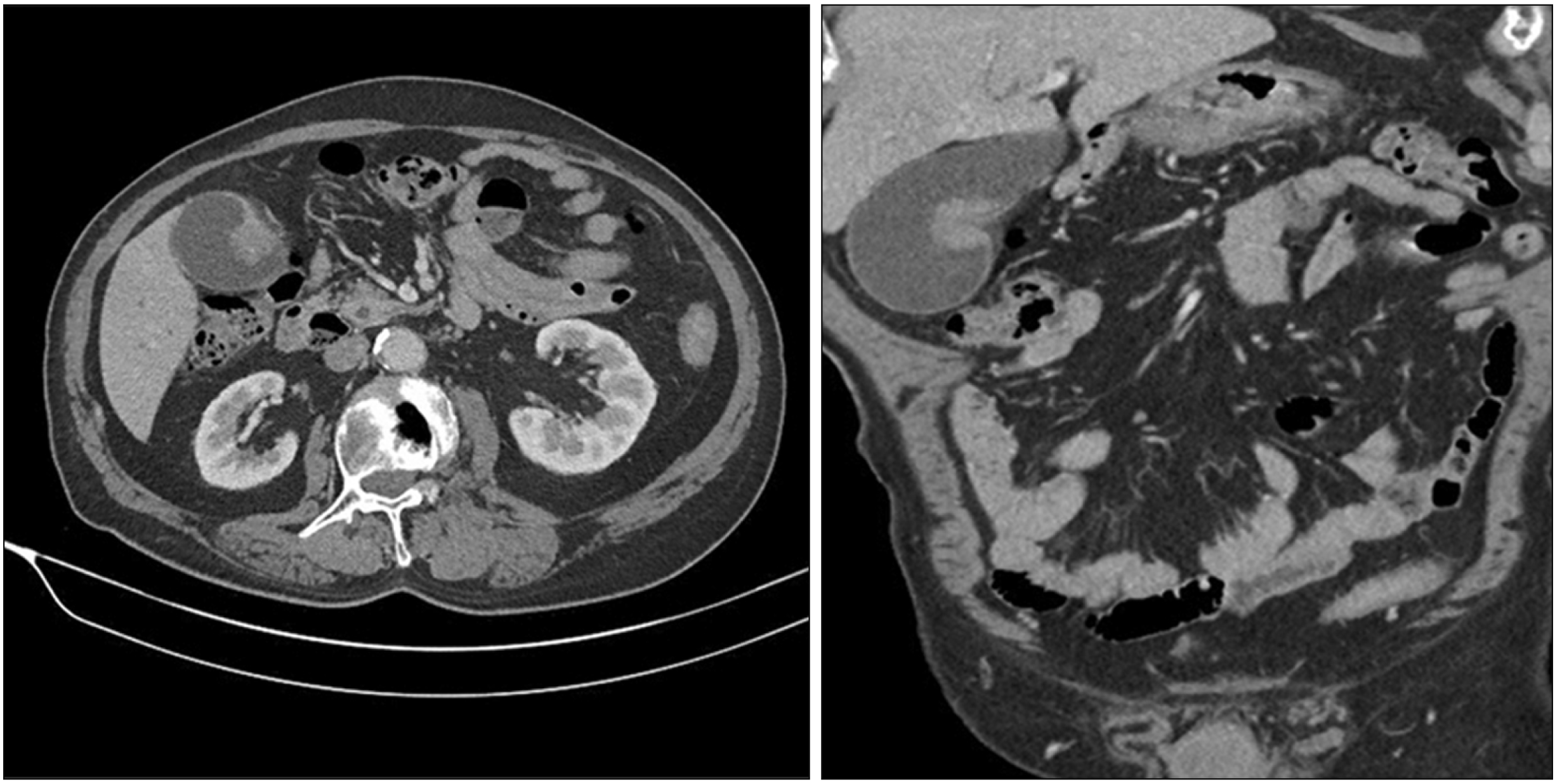
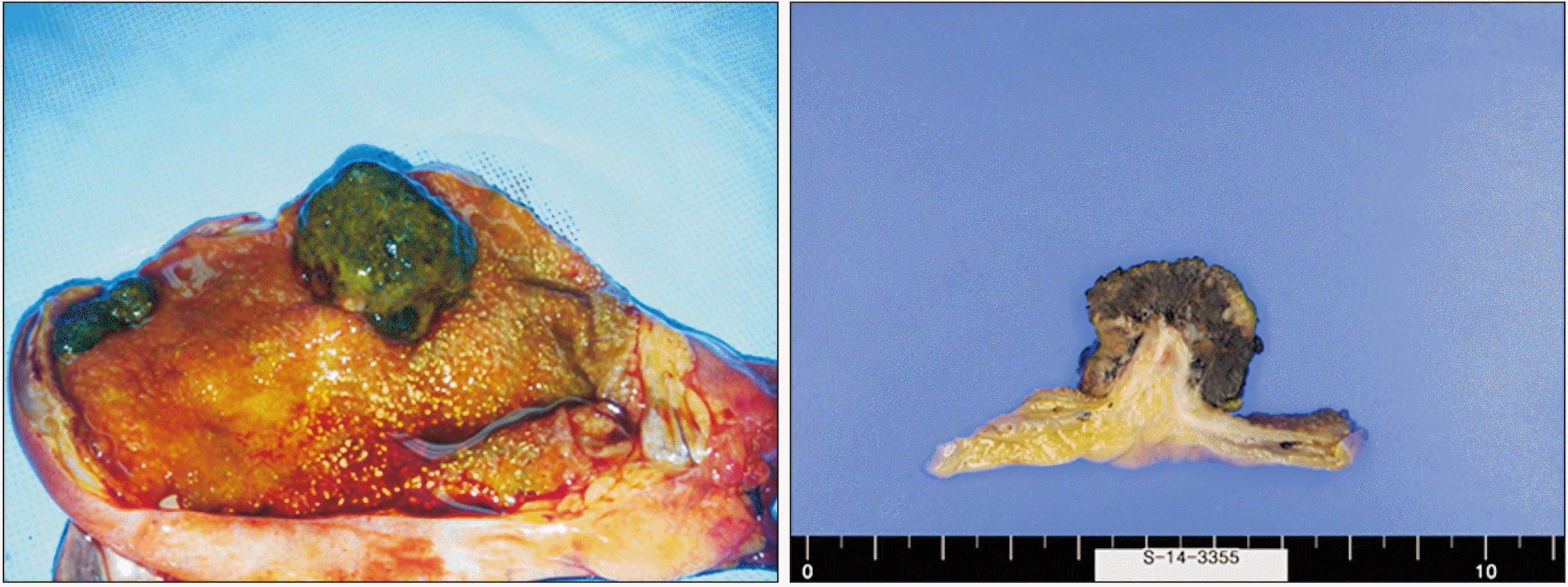
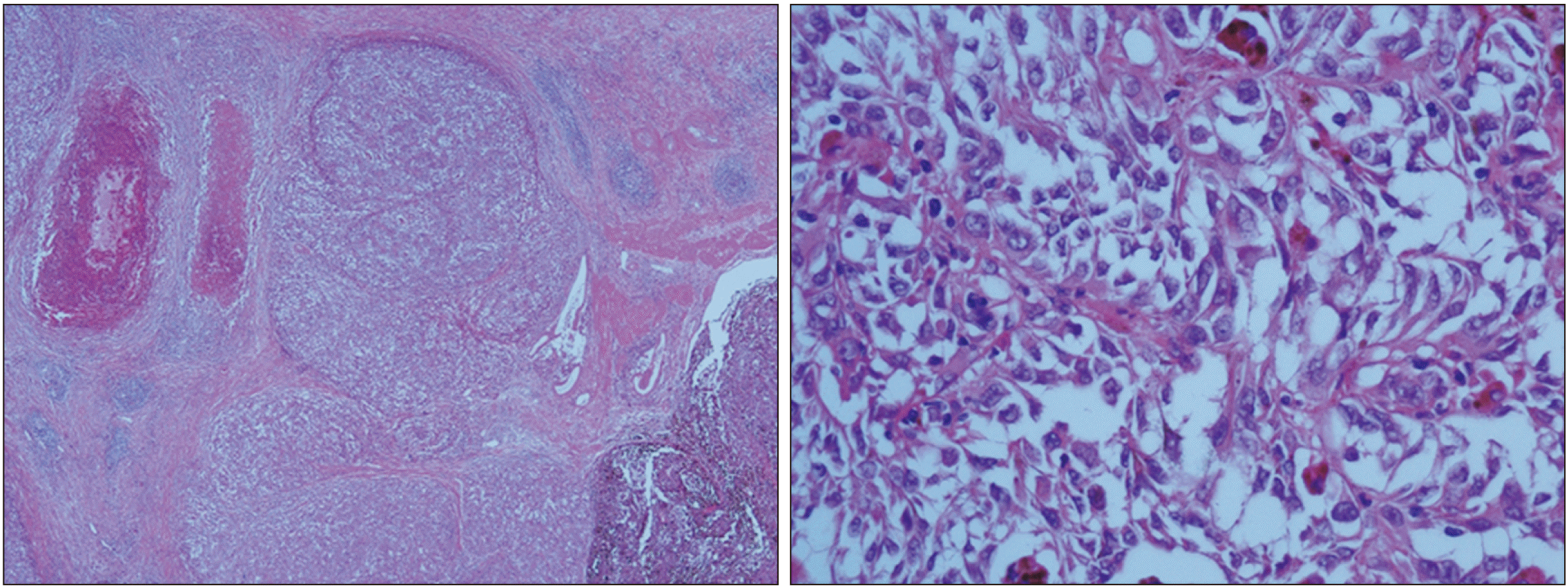
A 77-year-old male patient visited our hospital with abnormal findings on his health screening examination. In September 2012, endoscopic examination showed a black-pigmented lesion at the cardia of the stomach and biopsy revealed melanoma, which presumed to be metastatic (Fig. 1). The advice from the pathologist was to find the primary lesion. Further examinations were performed to determine the primary origin. However, no other focus was found after physical evaluation. It was concluded that the origin of primary lesion was an unknown melanoma or the stomach was the primary lesion. In addition, abdominal ultrasonography detected the presence of a 2.4-cm sized gallbladder nodule (Fig. 2). Whole-body positron emission tomography-computed tomography (PET-CT) scan revealed only a hypermetabolic lesion in the gallbladder (Fig. 3). Surgical exploration was recommended for the gallbladder nodule and based on the possibility of primary gastric melanoma. However, the patient refused surgery and subsequent outpatient care. In February 2014, the patient returned and health screening examination showed that the gallbladder lesion had increased to 4.0 cm in size (Fig. 4). On endoscopic examination, the previously diagnosed melanoma of the stomach was not identified. CT scan confirmed findings observed on the ultrasonography. The radiologist indicated a possible diagnosis of gallbladder cancer and estimated a clinical staging of T3N1M0 (Fig. 5). Because of this possible diagnosis of gallbladder malignancy, open radical cholecystectomy including liver wedge resection was performed. Macroscopic evaluation found a dark-brown polypoid lesion measuring approximately 3.0 cm in diameter and two large lymph nodes (approximately 1.2 cm in size) adherent to the gallbladder (Fig. 6). Histopathologic examination of the gallbladder tumor revealed sheets of atypical cells with abundant melanin pigments in the cytoplasm that extended through the lamina propria to the subserosa (Fig. 7). A total of 28 lymph nodes were acquired, among which three had metastatic melanoma cell involvement. Immunohistochemically, tumor cells were strongly positive for melanocytic markers S-100 and HMB45.
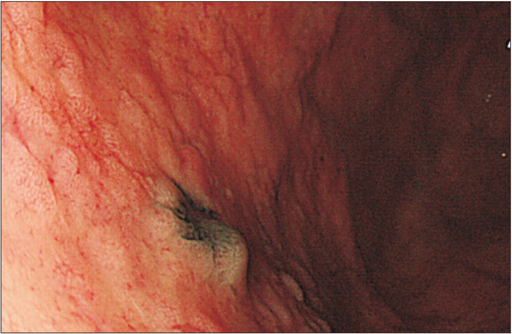 | Fig. 1Image of a black-pigmented lesion in the stomach cardia which was confirmed to be malignant melanoma by biopsy.
|
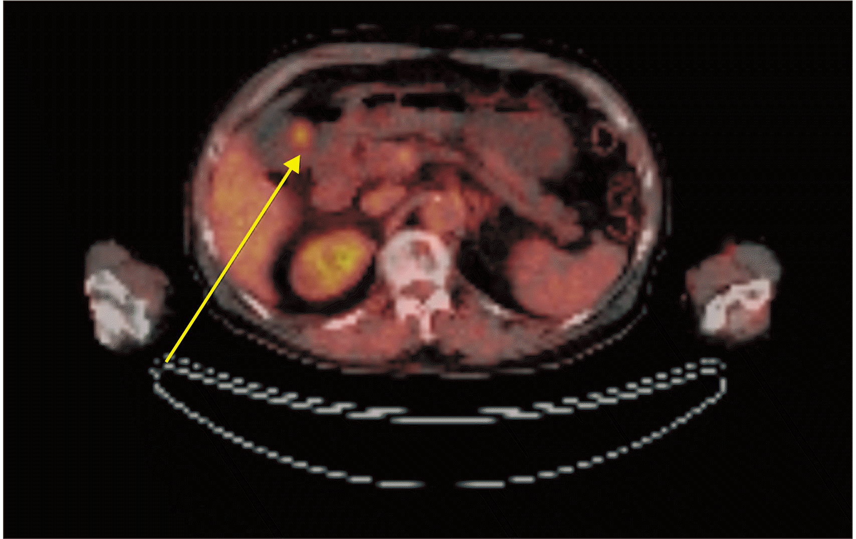 | Fig. 3Positron emission tomography-computed tomography confirming significant fluorodeoxyglucose uptake in the gallbladder.
|
The postoperative course of the patient was uneventful. He was discharged home after 10 days. The hemato-oncologist recommended chemoimmunotherapy. However, the patient decided to be observed for progression through outpatient care because of his old age. The patient underwent CT scanning every 6 months. In April 2017, a CT scan detected a very small hypodense lesion in the spleen and diffuse wall thickening of the gastric antrum. Because of concerns regarding melanoma metastasis, extensive examinations including PEC-CT and endoscopic examination were scheduled for the stomach and colon. On PET-CT scan, the spleen nodule noted on the CT scan did not show fluorodeoxyglucose uptake. The endoscopic examination identified a suspected malignancy lesion in the stomach lesser curvature of cardia and sigmoid colon, respectively. All lesions in the colon and stomach were removed by endoscopic submucosal dissection. The final biopsy revealed adenocarcinoma, not melanoma. After that, follow-up tests were performed regularly. No recurrence of melanoma was observed. The CT performed in March 2018 showed duodenal wall thickening. The patient was scheduled to undergo esophagogastroduodenoscopy. However, the patient did not appear in the outpatient clinic. It was found that the patient died naturally at the age of 82 years due to the aging process in July 2018, 52 months after the operation was performed.
Go to :
DISCUSSION
The tumor biology of malignant melanoma is highly unpredictable. Although melanoma may go into spontaneous remission, it can also cause systemic metastases after it is thought to be cured [2]. Melanoma is considerably more likely to regress than all other cancers. Approximately 4% to 6% of metastatic melanomas present with an unknown primary lesion [1]. In our case, the pathologist indicated that the lesion in the stomach was a metastatic disease and recommended us to look for the primary lesion. Extensive examinations were conducted, including ophthalmology, otorhinolaryngology, and dermatology exams. However, the causative lesion was not found. Dermatological examination revealed hypopigmentation in the anterior chest and left leg. Punch biopsy was performed to determine the possibility of complete regression of melanoma. However, chronic dermatitis was confirmed. The specimen suggested that the primary lesion was either an unknown melanoma or originated in the stomach.
The criteria for distinguishing whether a gallbladder tumor is a primary or metastatic lesion are difficult. Heath and Womack have proposed criteria for distinguishing primary melanoma from metastatic melanoma [3]. They proposed that primary tumors must be 1) solitary arising from the mucosal surface; 2) papillary or polypoid; 3) displaying junctional activity (the presence of pigmented dendritic cells at the junction of the epithelium and lamina propria); and 4) having other sites excluded as the primary site of origin. Among these criteria, junctional activity is considered the most indicative of a primary lesion. Our case presented with a solitary polypoid lesion. However, there was insufficient evidence to diagnose primary gallbladder melanoma because junctional activity could not be assessed. Cutaneous melanoma is known to metastasize to any organ. Metastatic disease from this malignancy to the gastrointestinal tract occurs at a frequency of 2% to 4%. Of these cases, the gallbladder is found to be a metastatic site at a frequency of 15% [4]. When referring to the previous literature, it is reasonable to consider that a gallbladder lesion is the metastatic lesion. The prognosis of melanoma of the gallbladder is very poor. Mean survival rates for patients with primary and metastatic lesions are 20.1 months and 8.4 months, respectively [5]. Primary melanoma of the gallbladder has a better prognosis than metastatic melanoma of the gallbladder because it is usually accompanied by symptoms such as acute cholecystitis confined to the gallbladder [6]. Treatment options for a metastatic disease depend on the extent of the disease and the clinical manifestation in the patient. Six case reviews of metastatic melanoma of the gallbladder have been reported (Table 1). Katz et al. [6] reported 13 cases of metastatic melanoma of the gallbladder in 2007. Patients who presented with disease confined to the gallbladder had a median survival of 39 months compared to a median survival of 10 months for patients presenting with multiple metastatic sites. Among patients who underwent cholecystectomy, there was a 10-month increase in survival compared to those who did not. The authors emphasized that factors associated with an improved survival rate were symptom manifestation, isolated metastases, and surgical approach. They suggested that, considering the aggressive nature and the biology of advanced melanoma, more extensive surgical resections such as lymphadenectomy did not seem to be warranted. In this study, three laparoscopic cholecystectomies were performed and two cases of port metastases occurred, although a retrieval bag was used to avoid dissemination of cancer cells. Tuveri and Tuveri [7] have reported a case of isolated metastatic melanoma to the gallbladder treated by laparoscopic cholecystectomy and lymphadenectomy of the hepatoduodenal ligament. They suggested that laparoscopic cholecystectomy could be used as an adequate treatment once the presence of a widespread disease was ruled out. The surgeon should use a retrieval bag for the removal of the gallbladder to avoid disruption of the gallbladder, which could cause port site or peritoneal metastases. Most gallbladder metastases of melanoma are intraluminal. Lymphadenectomy is not appropriate for stage IV melanoma [8]. However, this study notes that if the diagnosis of gallbladder lesions is unclear (primary or metastasis), the patient is young, or the extent of the tumor is large, lymphadenectomy may be considered. They recommended that lymphadenectomy should be performed only by experienced laparoscopic surgeons to reduce the rate of complications [7]. Christou et al. [9] have reported a case of metastatic malignant melanoma of the gallbladder accompanied by lymphadenopathy of the celiac axis and liver hilum. They performed open cholecystectomy and wedge liver resection and removed only suspected lymph nodes that were palpable intraoperatively. Marone et al. [10] and Giannini et al. [11] have also removed only suspected lymph nodes based on preoperative imaging or surgical findings. However, since the final biopsy results were different from their expectations and the exact lymph node metastasis could not be determined, adequate lymph node dissection is recommended. The patient in the present case report was presumed to have gallbladder cancer. Thus, surgery was performed. However, the lesion was diagnosed as melanoma with lymph node metastasis. It was considered a metastatic gallbladder melanoma because of his previous melanoma diagnosis history. In the case of stage IV melanoma, it is not appropriate to consider lymph node dissection for a palliative surgery. However, in our case, a long-term survival rate was confirmed. A reliable indicator for the differential diagnosis between primary and metastatic melanoma is junctional activity in the pathology report. However, this is difficult to accurately diagnose with frozen biopsy specimens during surgery. Considering the uncertainty of the diagnosis and the long-term survival of our patient, we think that performing lymphadenectomy after considering the general condition of the patient and/or tumor extent is a suitable option. Although lymphadenopathy was not identified in the preoperative imaging study, metastasis was confirmed for the lymph nodes obtained during the surgery. Therefore, we expect it would be better to have enough lymph node dissection. Recurrence of laparoscopic port sites has not been reported outside the review paper of Katz et al. [6]. Thus, if there are no other organ metastases and if the lesion is confined to the gallbladder, laparoscopic cholecystectomy may be considered. Endo-bags should be considered to prevent cancer cells from contaminating the abdominal cavity. However, if aggressive surgical treatment is planned after considering the patient’s age and/or general condition rather than palliative surgery for symptom control or prophylactic cholecystectomy, we think it would be better to have an open surgery for adequate lymph node dissection.
Table 1
Summary of clinical features of previous reports of metastatic melanoma of the gallbladder
| Author | Year | Age (yr) | Sex | Site of primary | Other metastatic organ | Treatment | Open or laparoscopic surgery | Lymphadenectomy | Lymph node metastasis | Overall survival after surgery |
|---|---|---|---|---|---|---|---|---|---|---|
| Guida et al. [5] | 2002 | 32 | F | Rt. shoulder | (-) | Cholecystectomy | Open | Locoregional lymphadenectomy | Hepatoduodenal ligament LN (+) | 4 mona) |
| Katz et al. [6] | 2007 | Not described | F: 6 M: 7 | Trunk 7, Head & neck 3, Extremity 1, Vulva 1, Unknown origin 1 | Multiple metastasis: 8 (liver, lung, bone, brain adrenal, thyroid) | Cholecystectomy: 9 Non-operative treatment: 4 | Open: 6 Lap.: 3 | Not described | Not described | Cholecystectomy– median 16 mon Non-operative- median 6 mon |
| Marone et al. [10] | 2007 | 54 | M | Trunk | Diaphragmatic peritoneum | Cholecystectomy, CTX, ITX | Lap. | Suspected LN | LN (+) | 11 mon |
| Tuveri and Tuveri [7] | 2007 | 37 | F | Rt. leg | (-) | Cholecystectomy | Lap. | Locoregional lymphadenectomy | LN (-) | More than 5 years |
| Christou et al. [9] | 2014 | 58 | M | Back | Liver hilum | Cholecystectomy Wide liver wedge resection | Open | Suspected LN | LN (-) | More than 6 mona) |
| Giannini et al. [11] | 2016 | 50 | M | Back | Spleen | Cholecystectomy Splenectomy | Lap. | Suspected LN | LN (-) | More than 6 mona) |
| 40 | F | Rt. hemithorax | (-) | Cholecystectomy | Lap. | Suspected LN | LN (-) | More than 6 mona) |
![]()
In conclusion, in case of melanoma found in the gallbladder, the original melanoma lesion has a possibility of regression. Since biopsy could determine whether it is a primary or metastatic lesion, making treatment planning very difficult. Treatment guidelines for melanoma found in the gallbladder are unclear due to its low occurrence. We report a rare case of metastatic malignant melanoma of the gallbladder treated with an open radical cholecystectomy, including lymphadenectomy of regional lymph nodes and liver wedge resection. Even for cases of metastatic melanoma discovered by chance, considering the long-term survival of our case following a surgical resection, extended cholecystectomy including regional lymph node dissection may be a good therapeutic approach for gallbladder melanoma.
Go to :
 XML Download