

 PDF
PDF Citation
 Print
Print


INTRODUCTION
Since 1960, portal vein arterialization (PVA) has been attracting attention due to its role as a salvage inflow technique in various clinical applications. It was initially performed in shunt surgery for portal hypertension to prevent a decreased hepatic inflow. Later, this indication was abandoned because of improvement of radiological procedures. PVA has been largely used in the transplantation domain or in the enlarged radical operations in case of hilar cancer invading the hepatic artery (HA). Moreover, it has been used in the treatment of fulminant hepatic failure.
Strongly promoted by enthusiasts, it is a surgical technique that must be used as a salvage surgery for HA disruption when surgical reconstruction is technically impossible [1] and when all other possible strategies have been ruled out [2].
We report a case of HA disruption complicating a left extended hepatectomy successfully treated with PVA.
Go to : 
CASE
A 62-year-old man without previous medical history was referred to our institution with a diagnosis of hilar cholangiocarcinoma involving the bile duct confluence and the left hepatic duct (Klatskin type IIIB). Percutaneous biopsy showed a poorly differentiated solid pattern that was CK 7 positive and CD10 negative.
Laboratory data showed severe anaemia, slight elevation of alkaline phosphatase and gamma-glutamyl transpeptidase (g-GT), increased blood level of total bilirubin (9.5 mg/dL) and elevated level of tumour markers: CA 19/9 of 3,061 U/mL and CEA of 8.5 ng/mL.
Prior to admission, he was treated for positioning of a metallic endoscopic biliary stent (10 mm × 60 mm) and two percutaneous transhepatic bilateral biliary drainages (8-Fr). The right drainage subsequently caused an intrahepatic haemorrhage because of the presence of a hepatic angioma. An angiographic procedure with super selective embolization was required.
At the admission, his general conditions were poor. He presented jaundice and itching. His nutritional assessment indicated poor conditioning.
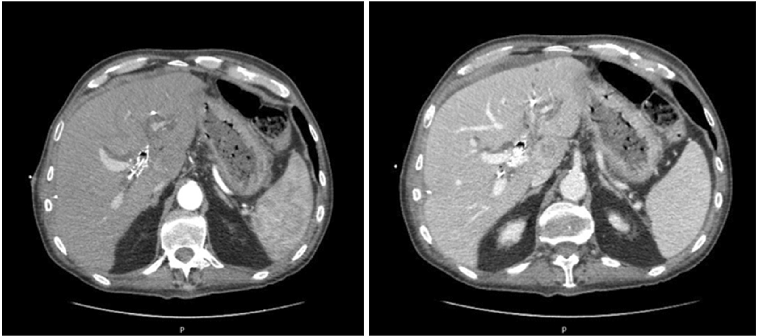
Preoperative total body computed tomography scan (CT) (Fig. 1) confirmed the presence of an infiltrating lesion originating from the biliary bifurcation extended to the second order left bile ducts as well as the caudate segment. The lesion had a diameter of 30 mm × 46 mm, showing contrast enhancement and proximal dilatation of the biliary tree. The left HA appeared thread. It was partially involved.
The patient underwent a radical left extended hepatectomy (including segment I) with extra-hepatic bile duct resection and right Roux-en-Y double duct hepaticojejunostomy through the suture of both the right anterior and posterior sectorial bile ducts. Since it was impossible to remove the metallic biliary stent, its distal part was left in the main bile duct stump, close to the head of the pancreas. During surgery, resected margins were assessed by frozen-section histology. Results were negative.
Hepatic hilum dissection was found to be difficult due to tumour location and size. In addition, there was an inflammatory reaction to the prosthetic device. To achieve a better control, the liver was completely mobilized.
The patient was transferred to our intensive care unit (ICU). On postoperative day (POD) 3, he was moved to the surgical ward. He was haemodynamically stable with the following blood results: GOT of 49 UI/L, GPT of 145 UI/L, total bilirubin of 2.01 mg/dL (direct bilirubin 1.49 mg/dL), g-GT of 161 UI/L, and INR of 1.3. He started a full liquid diet which was well tolerated.
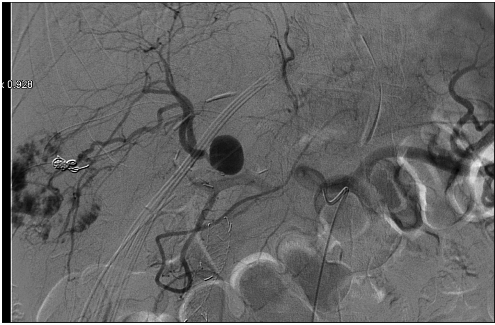
Postoperative course was uneventful until POD 5 when the patient complained vomiting. At the examination, his abdomen was distended and a pancreatic fistula was revealed by the abdominal surgical drains content, with increased level of amylases and lipases (amylases >7,500 U/L; lipases >3,500 U/L). An abdominal contrast CT-scan showed a small collection in gallbladder bed (3 cm × 1.5 cm), abdominal effusion, and right HA branch pseudo-aneurysm (18 mm) whose presence was confirmed through an angiography (Fig. 2). No active bleeding was revealed. Stent placement in the right HA was infeasible. Coiling of the pseudoaneurysm was associated with a risk of complete occlusion inducing critical liver failure. After the procedure, his general conditions suddenly deteriorated and an emergency laparotomy was performed.
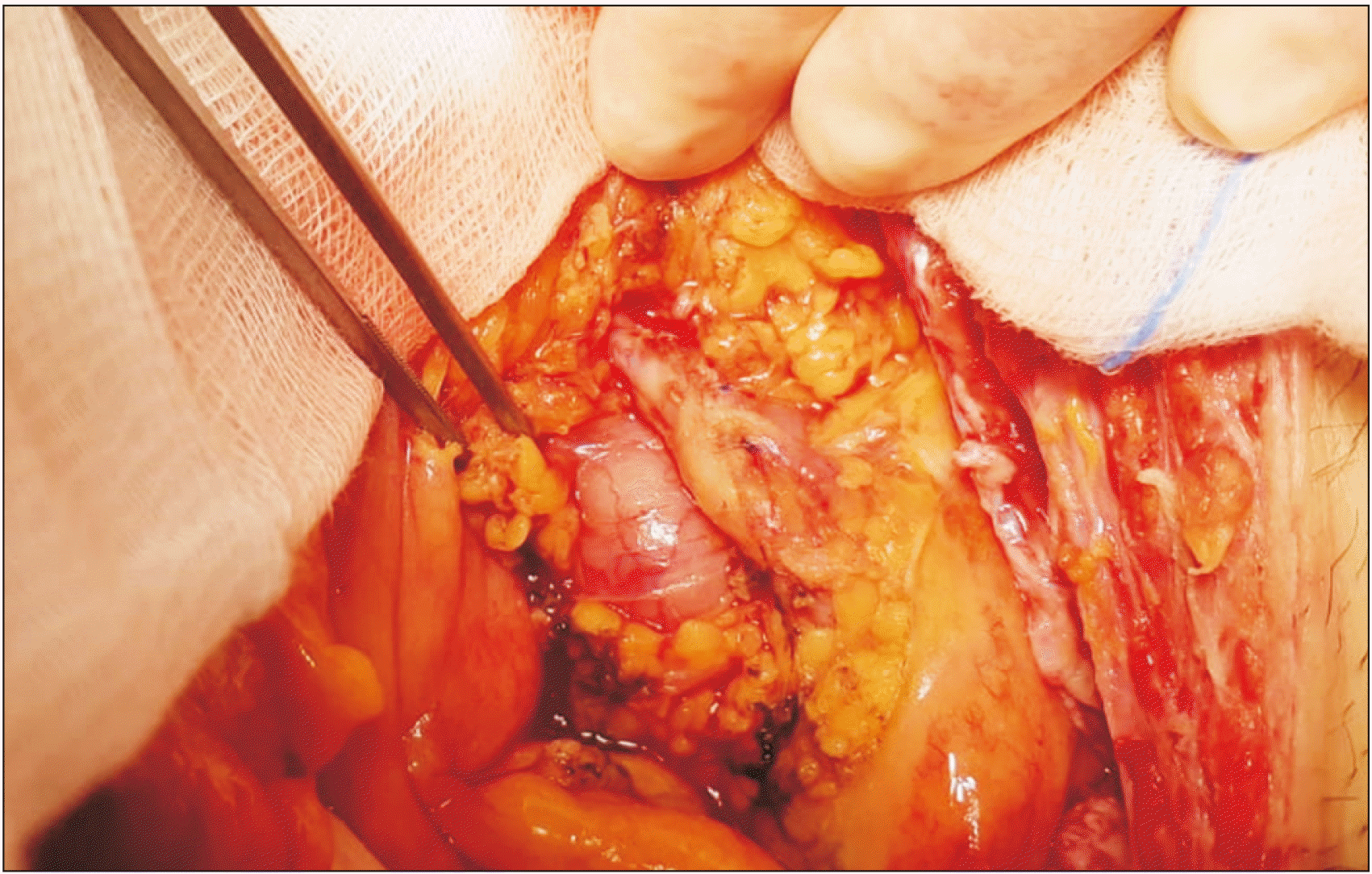
The liver was pale and ischemic. Intraoperative ultrasound showed no arterial flow. The right branch of the HA was disrupted in the area of the pseudo-aneurysm. Considering the local inflammatory situation, a HA reconstruction was impossible. Thus, PVA was used to provide arterial inflow to the remnant liver. A side-to-side arteriovenous shunt was created between the ileocolic artery and the ileocolic vein using two Prolene Suture 7–0 (Fig. 3).
At the end of the procedure, the liver was reddish-brown in colour. It felt rubbery to the touch. The intraoperative ultrasound showed satisfactory patency of the PVA with good portal flow in the absence of arterial flow. The patient was transferred to the ICU. On POD 3, blood tests revealed total bilirubin level of 4.88 mg/dL (direct bilirubin, 2.89 mg/dL), platelets of 123,000/mL, and INR of 2.23. An asymptomatic biliary fistula occurred. It was conservatively managed. He was extubated on POD 10. Doppler ultrasound on POD 15 showed that the cross-sectional area and blood flow of the portal vein were increased (main portal vein, 18 cm/sec). The patient was discharged on POD 54 in good general condition. The patient is alive and doing well at six-month follow-up without evidence of recurrence or systemic disease. At three months follow up, the patient was admitted for ascites (2 L/day) and portal hypertension was suspected as the major cause. The ascites was rapidly resolved with medical treatment and a single paracentesis. The patient was then discharged. No evidence of portal hypertension was found in Doppler ultrasound.
Written informed consent was obtained from the patient for publication of this case report and accompanying images. A copy of the written consent is available for review by the Editor-in-Chief of this journal upon request.
Go to :
DISCUSSION
We report a rescue case of PVA following major complications after a left extended hepatectomy.
As a matter of fact, HA disruption represents potentially lethal complications of hepatic, biliary, and pancreatic surgeries [3]. It often leads to biliary ischemia (bile leakage, bile infarcts, biloma formation) and partial liver necrosis.
In the case described here, previous arterial embolization and the challenging hepatic pedicle dissection due to inflammation and biliary stump fistula might have fragilized the HA wall, causing the HA pseudo-aneurysm. On the other side, complete liver mobilization hindered the development of arterial collaterals.
PVA was historically used in portal hypertension surgery in association with portocaval shunting. It seemed to attenuate the ischemic effect on cholangiocytes caused by a HA disruption, reducing cholestasis and proinflammatory cytokine production, ductular reaction, and fibrosis [4] in animal models.
Because of anastomotic arterioportal thrombosis and complications related to an excessive liver arterialization with the development of other therapeutic options, this surgical approach was abandoned in favor of radiological shunting therapies.
PVA is largely used in the field of liver transplantation [5]. It was first described in 1989 by Sheil et al. [6]. After a suprahepatic inferior vena cava anastomosis, they attached a cannula inserted in the recipient iliac artery to a donor portal vein cannula, resulting in early liver revascularization.
According to Bonnet et al. [7], it could be particularly useful in case of pre-existing diffuse portal vein thrombosis, notably a contraindication for orthotopic liver transplantation (OLT) or more rarely in case of portal vein thrombosis complicating a recent OLT.
In a patient with HA thrombosis after a liver transplantation, PVA presents the disadvantage of leaving the portal hypertension unchanged. Thus, an over arterialization of the liver can eventually lead to fibrosis. Some authors have reported an aneurysmal dilatation of the PV and its intrahepatic branches revealed by CT scan and various complications (most notably right-sided heart failure and portal hypertension) following PVA [8].
Current indications for PVA in the field of liver transplantation domain include post-OLT hepatic artery thrombosis (HAT), extended splanchnic vein thrombosis pre-OLT or post-OLT, and low portal flow pre-OLT according to anatomic variations such as the absence of portal and mesenteric veins.
Moreover, according to the literature, PVA can also be used in case of acute liver failure as a life-saving measure in order to provide temporary life support [9,10].
Beside OLT, PVA is also used to prevent HAT after cancer surgery, representing an alternative strategy when HA reconstruction after its accidental resection is impossible. In 2004, Kondo et al. [11] described 10 cases of biliary cancer treated with radical surgery performed through an en-bloc resection including HA and an end-to-side arterioportal reconstruction between the common hepatic or gastroduodenal artery and the PV.
Since then, only Young et al. [12] have reported the use of PVA in a cholangiocarcinoma surgery, describing 2 cases of hilar cholangiocarcinoma treated with PVA as a salvage strategy.
Finally, PVA can be a promising emergency intervention allowing the entire liver cells (hepatocytes and, specifically, cholangiocytes) to mitigate the hypoxic-ischemic damage by developing arterial collaterals. Results emerging from a Paris group [1], both in case of liver transplant or hepatic resection, have shown an overall survival rate of 63% at a median follow-up of 13 months.
In our case, PVA was performed in an emergency setting to avoid ischemic injury after HA pseudoaneurysm and disruption, thus decreasing serum transaminase levels and limiting the parenchymal ischemia.
Despite this enthusiasm, PVA is a procedure associated with high morbidity rates even if the natural course of a completely de-arterialized liver is death. Complications related to arteriovenous shunt thrombosis can lead to emergency liver transplantation as only a rescue option [13].
The aim of PVA is to create a time frame to allow collateral arterial vessels onset or a definitive surgery performance. Early thrombosis can be a catastrophic event due to acute liver ischemia, although a delayed occlusion is often well tolerated. Surveillance is recommended because an embolization of the arterioportal fistula could be necessary to treat or prevent the onset of portal hypertension.
In conclusion, complete arterial hepatic deprivation is known to be associated with severe ischemic damage, including bile infarcts, bilomas, and eventually liver abscesses after infection. PVA has a beneficial effect on ischemia-hypoxia-induced bile duct injury. However, increased portal blood represents a cause of portal hypertension and liver fibrosis. Thus, measures are needed to control blood flow and patients who have undergone PVA should be followed up.
Go to :
 XML Download