

 PDF
PDF Citation
 Print
Print


INTRODUCTION
Congenital absence of the portal vein (CAPV) is a rare venous malformation in which the mesenteric venous blood drains directly into the systemic circulation. The majority of patients with CAPV show no signs or symptoms of portosystemic encephalopathy. They only show slightly abnormal results in liver function tests. Liver transplantation (LT) is indicated for patients with symptomatic CAPV refractory to medical treatments [1-4].
The congenital portocaval shunt (PCS) drains the entire mesenteric venous blood either directly into the inferior vena cava (IVC) or the left renal vein. Thus, theoretically, there is no portal hypertension or collateral circulation [3-5]. As the liver with CAPV does not have sufficient portal inflow, the hepatic arterial flow is the main inflow of blood. If a patient cannot tolerate a medical treatment, LT should be taken into account for such a case. For patients with CAPV, their liver function profiles are not severely impaired with resultant low Pediatric End-stage Liver Disease scores. Because of the low chance for deceased donor liver transplantation in the current Korean setting, patients with CAPV need to consider living donor liver transplantation (LDLT). We herein present a case of pediatric LDLT using a left liver graft for CAPV and liver tumors.
Go to : 
CASE
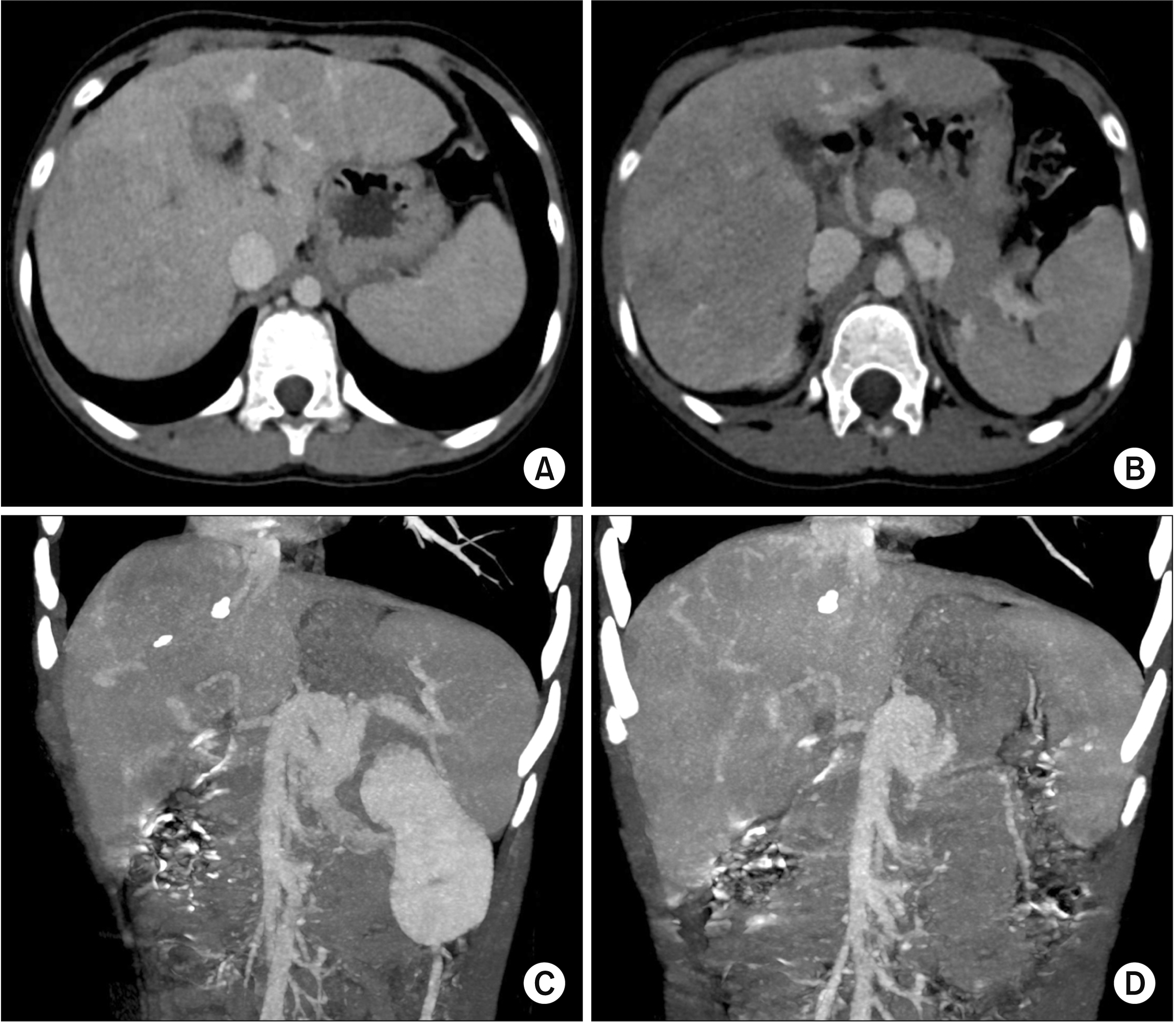
A 9-year-old girl was referred to our hospital due to the diagnosis of multiple liver mass. These liver lesions were diagnosed with focal nodular hyperplasia (FNH) on imaging studies and percutaneous liver biopsy. There was no abnormal finding on the diagnostic exome sequencing. The patient was prematurely born as the second baby of a twin. Imaging study findings were compatible with CAPV type I (Fig. 1). Her blood ammonia level was raised to 137 µg/dL. However, she did not complain of any symptoms. The patient was not indicated for Rex shunt operation due to poor development of the intrahepatic portal vein system. Thus, we decided to perform LDLT. The donor was a 39-year-old mother of the patient. The left liver volume was measured to be 508 mL on computed tomography volumetry (Fig. 2).
Recipient’s operation was performed according to standard procedures of pediatric LDLT. As the recipient’s native portal vein was completely absent, only the hepatic artery branches were meticulously dissected after transection of the common bile duct (Fig. 3A). After hepatic arteries were transected, Kocher’s maneuver was extensively performed to expose the left renal vein. Subsequently, the insertion site of the splenorenal shunt was isolated (Fig. 3B). The confluence portion of the superior mesenteric vein (SMV) and the splenic vein (SV) was meticulously dissected, and the SMV, SV, coronary vein branches, and gastric vein branches were isolated (Fig. 3C).
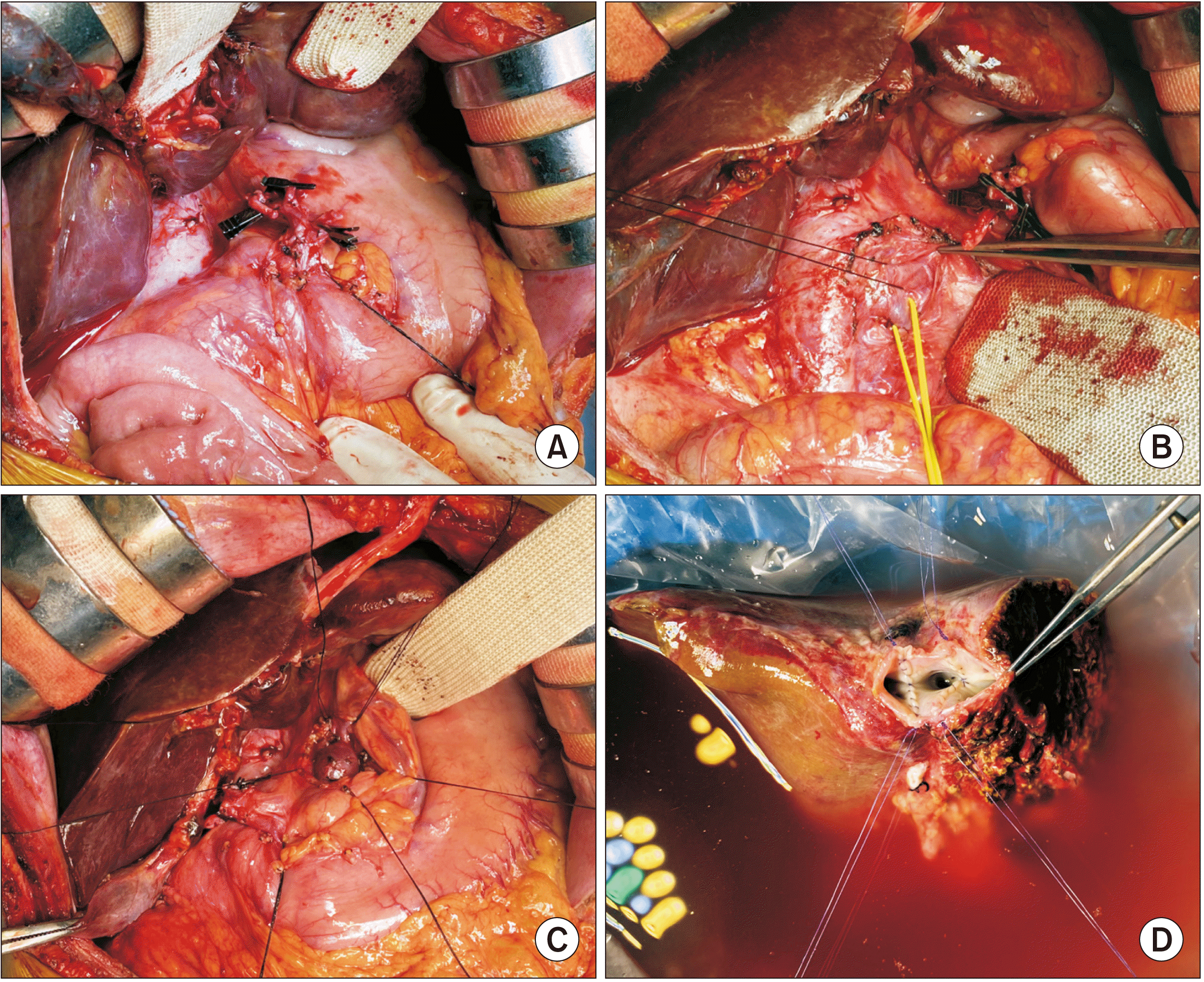 | Fig. 3Intraoperative photographs for recipient hepatectomy and graft bench work. (A) The hepatic artery branches are meticulously dissected after transection of the common bile duct. (B) The retropancreatic space is dissected and the left renal vein (yellow vessel loop) and the insertion site of the splenorenal shunt (black silk sling) are isolated. (C) The confluence portion of the mesentero-splenic vein is meticulously dissected. (D) The graft hepatic veins are separated into three openings, which are unified to make a wide single orifice.
|
In accordance with the recipient’s operation, a left liver graft measuring 400 g at the back table was recovered, yielding a graft-to-recipient ratio of 1.7%. The graft left portal vein was unusually small, measuring only 7 mm in diameter. The graft hepatic vein was separated into three openings, which were unified into a 3 cm-wide single orifice through unification venoplasty (Fig. 3D).
After the native liver was removed, the hepatic vein orifices at the recipient IVC were unified with venoplasty using a cryopreserved saphenous vein homograft patch because the right hepatic vein trunk was hypoplastic (Fig. 4A). The vein branches at the SMV-SV confluence portion were securely clamped (Fig. 4B) and a 1.5 cm-long longitudinal incision was made at the confluence portion (Fig. 4C). A 4 cm-long cold-stored iliac vein homograft conduit was anastomosed to the SMV-SV confluence in an end-to-side fashion using 6-0 Prolene (Fig. 4D–4F).
 | Fig. 4Intraoperative photographs of the hepatic vein and portal vein venoplasty. (A) The hepatic vein orifices at the recipient inferior vena cava are unified with venoplasty using a cryopreserved saphenous vein patch. (B) The vein branches at the mesentero-splenic confluence portion are securely clamped. (C) A 1.5 cm-long longitudinal incision is made at the confluence portion. (D–F) A 4 cm-long fresh-stored iliac vein conduit is anastomosed to the confluence portion in an end-to-side fashion.
|
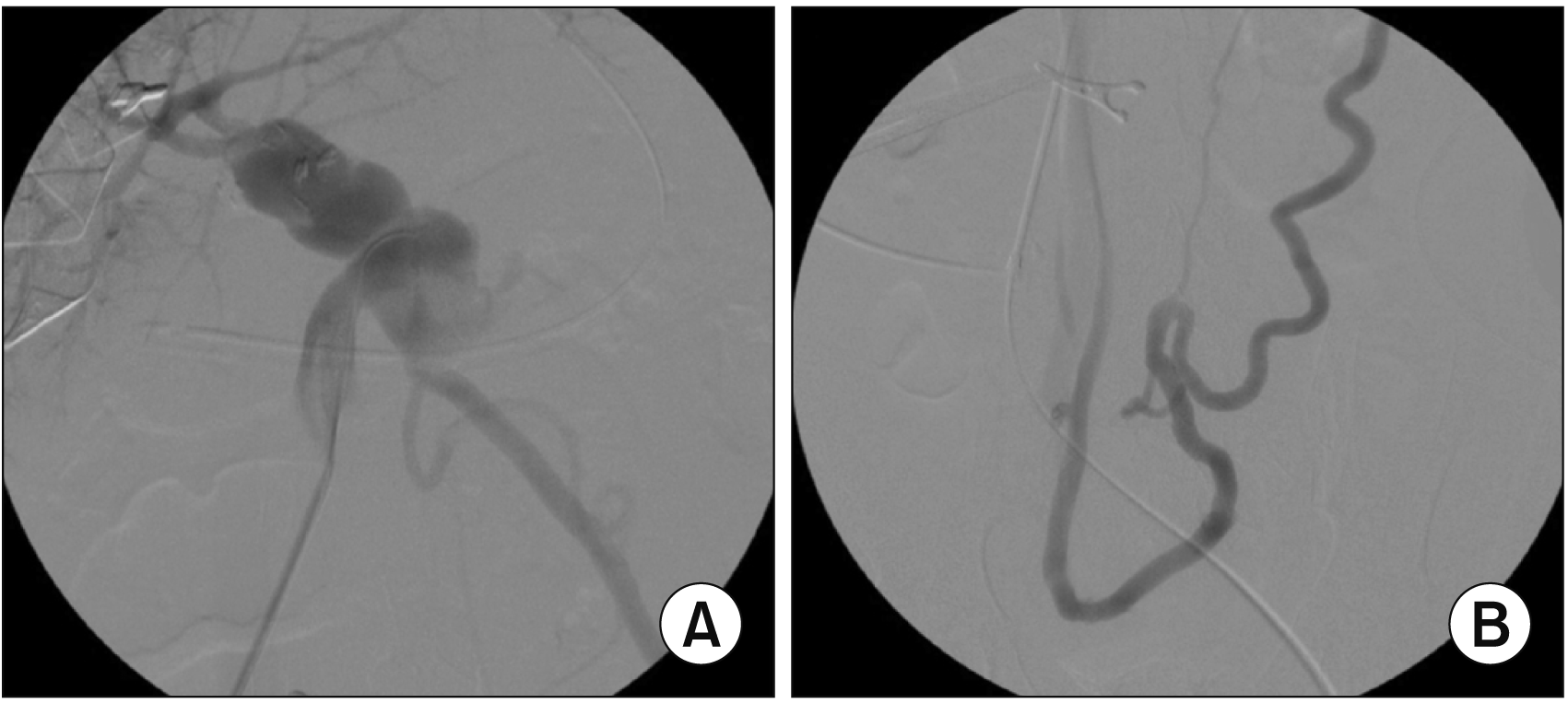
The graft hepatic vein orifice was anastomosed with the size-matched recipient IVC orifice (Fig. 5A, 5B). The interposed iliac vein conduit was anastomosed with the graft portal vein (Fig. 5C, 5D). Because the graft portal vein was much smaller than the conduit, a small niche was made each at the anterior and posterior walls of the graft portal vein to facilitate wide anastomosis. Thereafter, graft reperfusion was initiated. The splenorenal shunt was securely ligated (Fig. 5E), and then marked increase in the portal blood flow was observed (Fig. 5F). Surgical microscopy was used for graft hepatic artery reconstruction. Intraoperative direct portography was performed through a jejunal vein branch, in which the portal blood flow was well maintained (Fig. 6A). An enlarged collateral vein through the inferior mesenteric vein and the lumbar vein was identified (Fig. 6B) and ligated. Roux-en-Y hepaticojejunostomy was used for biliary reconstruction.
 | Fig. 5Intraoperative photographs of graft implantation. (A, B) The graft hepatic vein orifice is anastomosed with the size-matched recipient hepatic vein orifice. (C, D) The portal iliac vein conduit is anastomosed with the graft portal vein. (E) The congenital splenorenal shunt is securely ligated. (F) Portal blood flow is increased after ligation of the splenorenal shunt.
|
The pathology report of the explant liver showed two types of tumors. The first was a 2 cm-sized hepatocellular adenoma of beta-catenin mutated subtype at segment IV. Another one comprised of multiple (> 10) FNH lesions measuring up to 7.1 cm in size that scattered over both lobes, indicating multiple FNH syndrome. There were also multiple regenerative nodules measuring up to 2.7 cm in size in both lobes (Fig. 7).
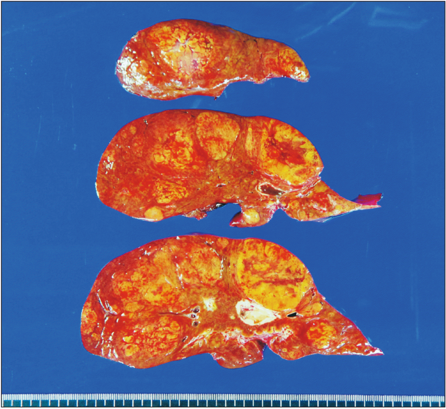 | Fig. 7Gross photographs of the explant liver. There are three types of liver nodules. A 2 cm-sized hepatocellular adenoma of beta-catenin mutated subtype is located at segment IV. Multiple focal nodular hyperplasia nodules measuring up to 7.1 cm in size are scattered over both lobes. Multiple regenerative nodules measuring up to 2.7 cm in size are also present in both lobes.
|
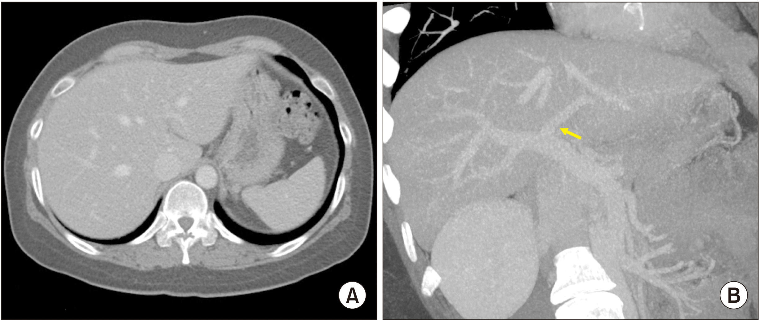
The patient recovered uneventfully from the LDLT operation. The reconstructed portal vein was maintained well without any hemodynamic abnormalities (Fig. 8, 9). This patient has been doing well for the past 6 months after the LDLT.
 | Fig. 8Posttransplant computed tomography scan taken at four days after the transplantation. (A, B) The graft portal vein reconstruction appears smooth and streamlined. (C, D) The iliac vein conduit from the mesentero-splenic vein junction is well visualized as a newly made main portal vein.
|
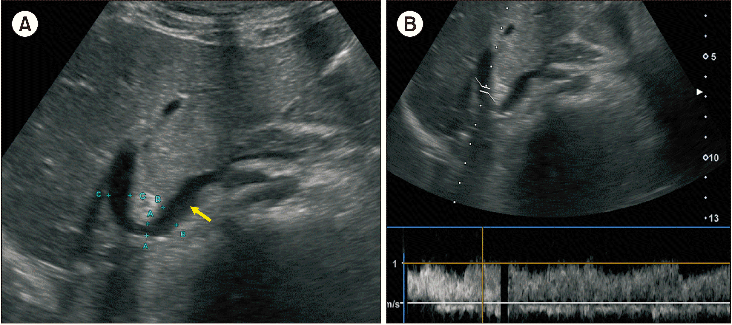 | Fig. 9Posttransplant Doppler ultrasonography at 21 days after transplantation. (A) The contour of portal vein anastomosis appears smooth and streamlined with slight stenosis at the anastomosis site. Arrow indicates the interposed iliac vein conduit. (B) The portal vein blood flow is well maintained.
|
Go to :
DISCUSSION
CAPV is a rare venous malformation in which mesenteric venous blood drains directly into the systemic circulation. There are two types of congenital PCS: intrahepatic PCS and extrahepatic PCS. Intrahepatic PCS is localized between the portal and hepatic veins [6]. Extrahepatic PCS is divided into type I and type II according to intrahepatic portal venous supply [7]. Type I PCS is an extrahepatic shunt without patent intrahepatic portal vein. Thus, the entire mesenteric venous blood drains directly into the systemic vein such as the IVC or the left renal vein. This type is called CAPV. Type II PCS is an extrahepatic shunt with patent intrahepatic portal vein. Thus, the patent portal vein perfuses the liver and the shunt vessel drains some mesenteric venous blood into the systemic circulation. Our patient had type I PCS because there was no portal venous structure and all splanchnic blood flow was drained through the splenorenal shunt.
The standard treatment for CAPV has not been established yet. Although PCS can be accompanied by hyperammonemia, the majority of patients with PCS have no signs of encephalopathy, as shown in the present case. Such patients only show slightly abnormal results in liver function tests. The majority of patients with CAPV receive conservative medical treatment for hyperammonemia, whereas only a small portion of patients with CAPV require surgical treatments including LT. Surgical treatment is indicated when hyperammonemia or portosystemic encephalopathy is refractory to medical treatment. CAPV is a venous malformation in which mesenteric venous blood drains directly into the systemic circulation. It might be accompanied by hepatopulmonary syndrome. Surgical treatment for CAPV can also be indicated for hepatopulmonary syndrome [8,9].
Pretransplant imaging studies in CAPV patients have usually demonstrated a large communication vein to the IVC through the splenorenal shunt, thus no evidence of portal hypertension has been observed in the imaging studies or intraoperative findings. However, we have previously reported an atypical case of CAPV showing portal hypertension with gastric and esophageal varix, which was the primary cause that led to LT [10].
Most patients with type I PCS are indicated for LT as surgical reconstruction of portal vein structures of the native liver is impossible, as in our patient. Although LT for symptomatic CAPV has been reported in the literature [1-3,5,9,11-13], techniques for portal vein reconstruction have not been well established yet. There are two methods of portal vein reconstruction in LT for CAPV. The first method is to anastomose the PCS directly to the graft portal vein in an end-to-end fashion [2,11]. The second method is to use a venous interposition graft through an end-to-side anastomosis to the PCS [3,5]. As the present case lacked any portal vein stump, direct anastomosis was technically impossible. Thus, we used vein conduit interposition as an end-to-side anastomosis to the PCS. The prerequisite for reconstruction with a vein conduit is the availability of an adequate vein allograft. To obtain suitable vein homograft, we had to wait for one month before the LDLT operation.
There is a strong association between the development of liver masses such as FNH and hepatocellular adenomas in patients with CAPV. This association has been hypothesized to be related to the lack of portal vein flow, which can lead to compensatory arterial hyperperfusion and the formation of regenerative nodules. Although rare, these neoplasms may degenerate to hepatocellular carcinoma [14-18]. Imaging characteristics of these hepatic neoplasms can be challenging due to the absence of intrahepatic portal systems and their associated portal venous or delayed imaging patterns. Apparently, a biopsy of these lesions is critical in cases showing an interval growth or a change in enhancement pattern [14-18].
In conclusion, as CAPV patients can have various vascular anomalies, such combined vascular anomalies should be thoroughly assessed before and during the LT operation. The most effective reconstruction techniques should be used to achieve satisfactory results following LT.
Go to :
 XML Download