Abstract
Graft size matching is essential for successful liver transplantation in infant recipients. We present our technique of graft dextroplantation used in an infant who underwent living donor liver transplantation (LDLT) using a reduced left lateral section (LLS) graft. The patient was an 11-month-old female infant weighing 7.8 kg with hepatoblastoma. She was partially responsive to systemic chemotherapy. Thus, LDLT was performed to treat the tumor. The living donor was a 34-year-old mother of the patient. After non-anatomical size reduction, the weight of the reduced LLS graft was 235 g, with a graft-to-recipient weight ratio of 3.0%. Recipient hepatectomy was performed according to the standard procedures of pediatric LDLT. At the beginning of graft implantation, the graft was temporarily placed at the abdomen to determine the implantation location. The graft portal vein was anastomosed with an interposed external iliac vein homograft. As the liver graft was not too large and it was partially accommodated in the right subphrenic fossa, thus the abdominal wall wound was primarily closed. The patient recovered uneventfully. An imaging study revealed deep accommodation of the graft within the right subphrenic fossa. The patient has been doing well for six months without any vascular complications. This case suggests that dextroplantation of a reduced LLS graft can be a useful technical option for LDLT in infant patients.
In liver transplantation (LT) for infant patients, matching of the graft size to the recipient’s abdomen is critically important because implantation of a large-for-size graft can induce various vascular complications and hinder primary closure of the abdomen. To overcome size mismatching in infants using adult partial liver grafts, several variants of left lateral section (LLS) grafts have been introduced [1-7]. Infant portal vein is usually much smaller than the vascular stumps of an adult LLS graft. In addition, during the posttransplant follow-up period, regeneration and rotation of the LLS graft towards the right side can cause an additional issue for pediatric recipients as graft regeneration directs to the empty space on the right side [8,9]. Rotation to the right side of a left-sided graft can induce twist or compression of the graft’s vascular structures [9]. To prevent venous vascular complications in infant recipients, dextroplantation of a left liver graft as a unique technique has been recently introduced [9]. We have also developed the concept of dextroplantation technique of LLS grafts in pediatric recipients. However, our technique for graft dextroplantation is practically different from the abovementioned technique. We herein present our technique of graft dextroplantation used in an infant patient who underwent living donor liver transplantation (LDLT) with a reduced LLS graft.
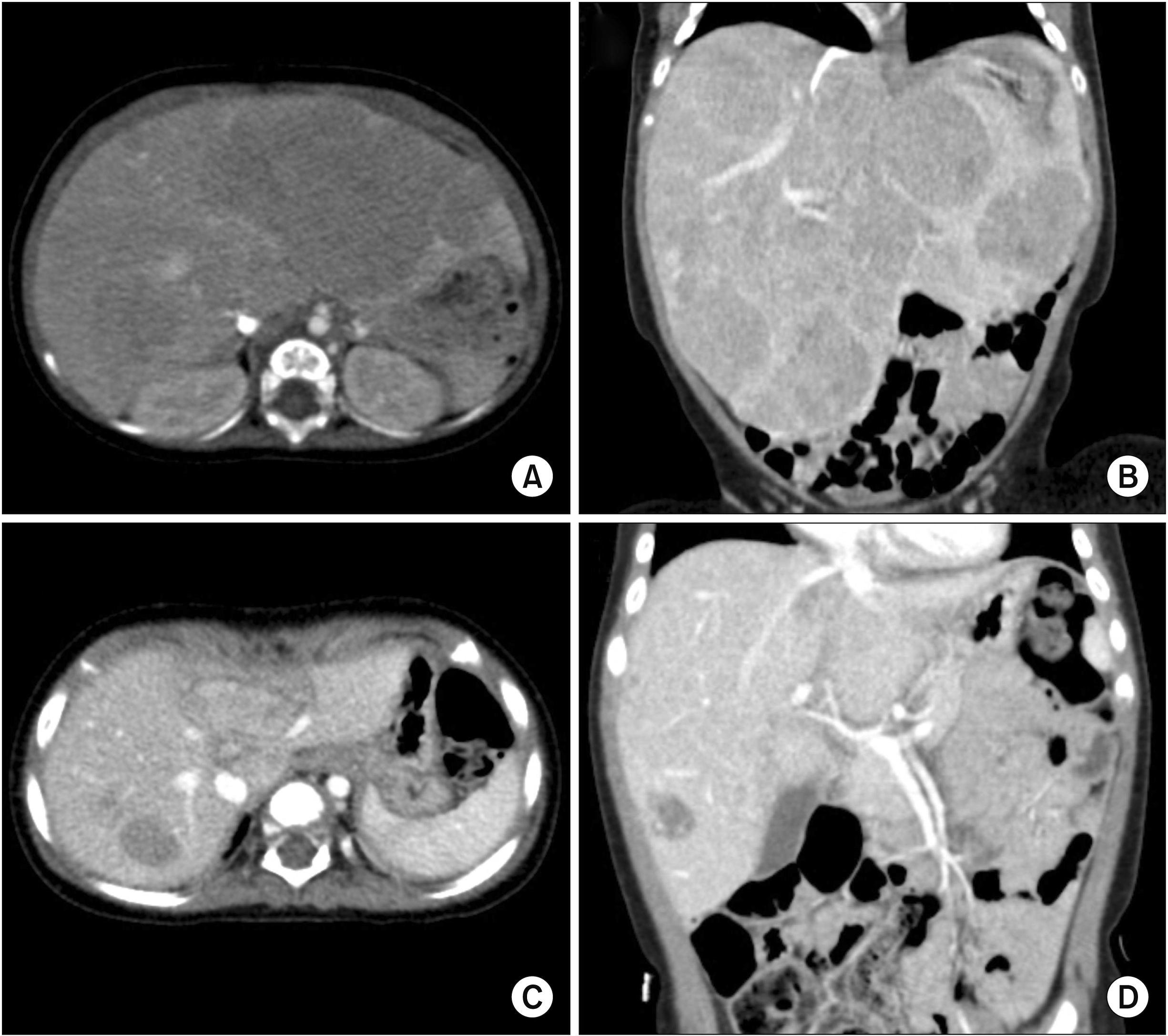
The patient was a 11-month-old 7.8 kg-weighing female infant with hepatoblastoma. She was born at 39 weeks through normal full-term spontaneous delivery. At 7 months after birth, she started experiencing multiple vomiting. Thus, workup studies were performed. Imaging studies and percutaneous liver biopsy confirmed the diagnosis of hepatoblastoma (Fig. 1A, 1B), which was classified as pediatric liver tumor staging (PRE-Treatment EXTent of tumor [PRETEXT] grouping system) stage IV. She underwent systemic chemotherapy (Children’s Oncology Group study AHEP0731). The tumor size was gradually reduced. However, some parts of the tumor remained due to partial responsiveness to systemic chemotherapy (Fig. 1C, 1D). Thus, we decided to perform LDLT to remove the tumor completely.
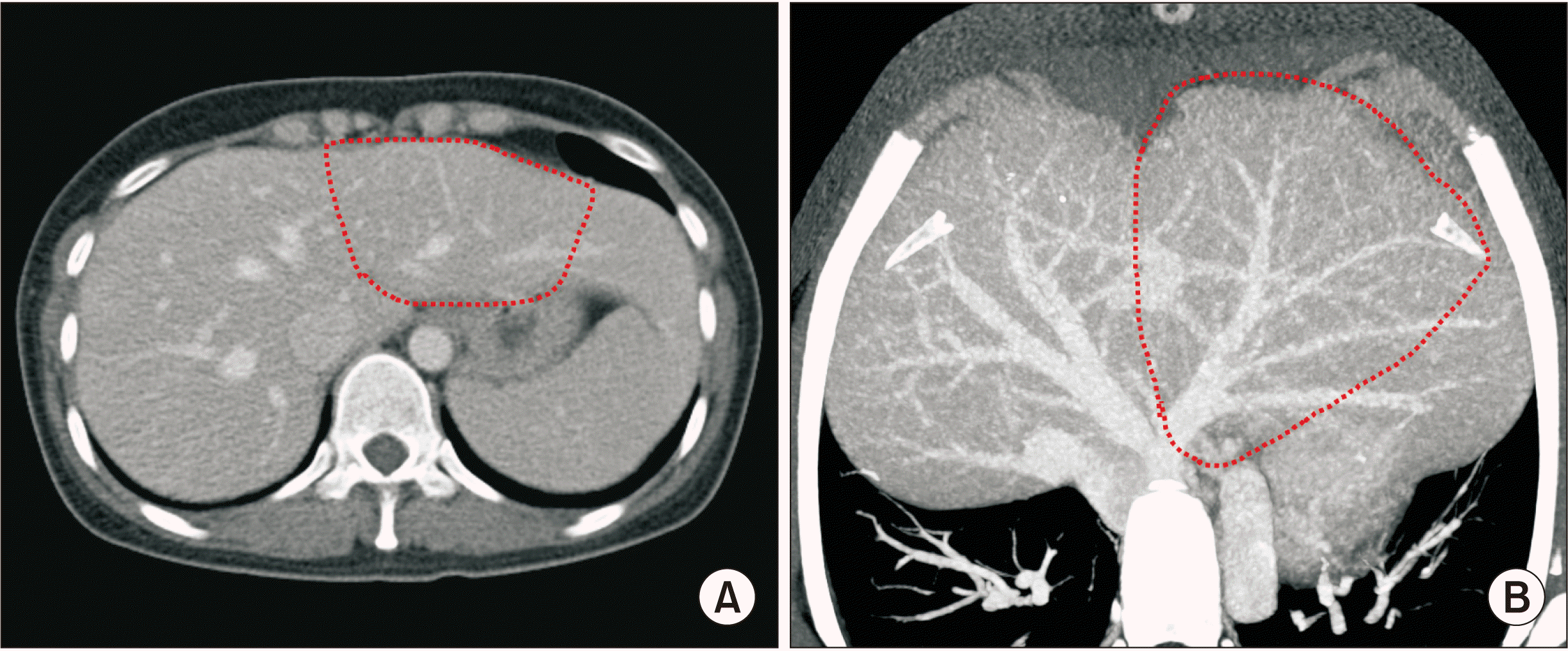
The living donor was a 34-year-old mother of the patient. The whole LLS volume was measured to be 350 mL on computed tomography volumetry (Fig. 2), which was equivalent to a graft-to-recipient weight ratio (GRWR) of 4.5%. The ratio of graft thickness to the recipient abdominal cavity was 1.3. Although the abdominal cavity of the recipient was enlarged due to hepatomegaly with multiple hepatoblastomas, the whole LLS graft appeared to be too large for secure implantation. Thus, we planned to perform size reduction to obtain a reduced or hyper-reduced LLS graft.
Recipient liver dissection for total hepatectomy was performed according to standard procedures of pediatric LDLT. The residual tumor invaded the retrohepatic inferior vena cava (IVC) close to the common trunk of the middle and left hepatic veins.
In accordance with recipient operation, donor operation was performed to recover the LLS graft. After parenchymal transection, the LLS volume was assessed by manual palpation, which was comparable to the preoperatively-measured volume of 350 mL. Thus, in situ non-anatomical size reduction was performed to remove the flat lateral portion (Fig. 2). The resected liver parenchyma weighed 95 g. The reduced LLS graft was harvested according to the standard procedure. The weight of the graft was 235 g, which had a GRWR of 3.0%. The graft hepatic vein was single and measured 1.2 cm in diameter. Thus, incision-and-patch plasty using a small patch of iliac vein homograft was performed at the medial wall of the graft left hepatic vein, by which the diameter of the graft hepatic vein was enlarged to 2 cm.
Recipient hepatectomy was performed according to standard procedures of pediatric LDLT. After clamping of the supra- and infra-hepatic IVC portions, the IVC wall close to the tumor was additionally excised and a 2 cm × 1 cm-sized vein homograft patch was attached to the IVC defect. An external iliac vein conduit homograft was anastomosed to the hypoplastic recipient portal vein.
After finishing venoplasty in the recipient and the graft, the reduced LLS graft was temporarily placed at the abdomen to determine the implantation location. The graft was partially accommodated in the right subphrenic fossa, thus we decided to perform dextroplantation of the graft. The axial orientation of the graft was marked with 6-0 Prolene stay sutures. Afterwards, graft implantation was performed in the order of hepatic vein, portal vein, hepatic artery, and Roux-en-Y hepaticojejunostomy (Fig. 3). The graft portal vein was anastomosed with the interposed external iliac vein conduit homograft. The liver graft was not too large for the size of the recipient abdomen and it was partially accommodated in the right subphrenic fossa. Therefore, the abdominal wall wound was primarily closed under minimal tension.
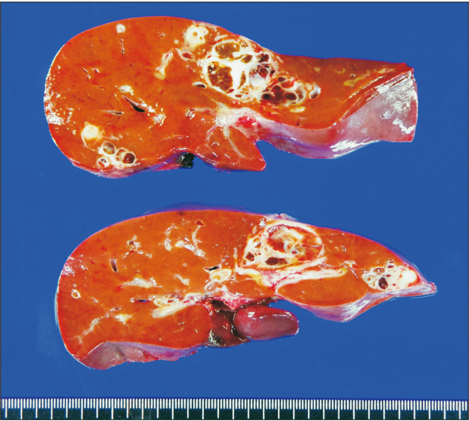
The resected liver specimen showed multiple viable hepatoblastomas measuring up to 5 cm in size (Fig. 4). Necrosis and foam cell infiltration were identified in 60% of tumors. Lymphovascular invasion and perineural invasion were absent.
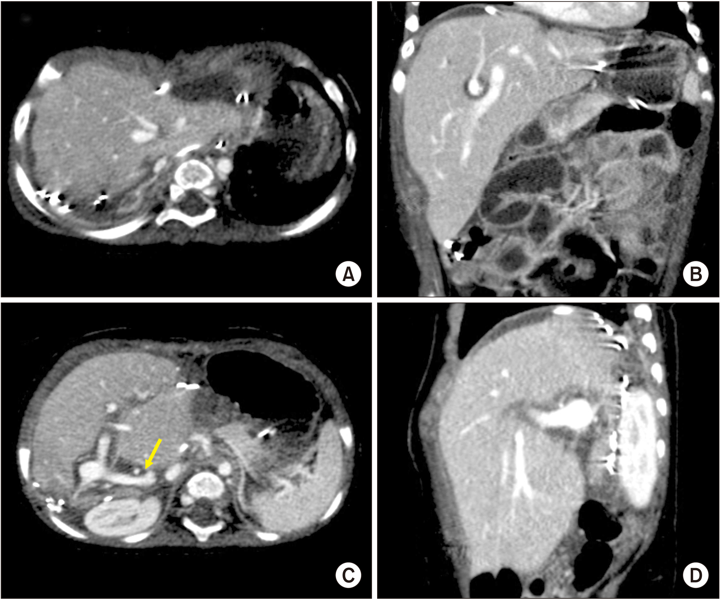
The patient recovered uneventfully (Fig. 5). Computed tomography images taken at four days after the transplantation revealed that the reduced LLS graft was accommodated within the right subphrenic fossa like the usual right liver graft. The patient has been doing well for the past 6 months. She is undergoing scheduled adjuvant chemotherapy.
Although experience with LT for infant patients has been accumulating worldwide, it is still regarded as challenging because large-for-size graft-related problems are big huddles for successful LT. The main problems of large-for-size grafts include the risk of abdominal compartment syndrome caused by the recipient’s small abdominal cavity, size discrepancies in vessel size, insufficient portal circulation, and insufficient tissue oxygenation [6,10]. It is essential to solve these problems simultaneously through an integrated planning for graft implantation.
The first step of our integrated planning for graft implantation was the reduction of the size of the LLS graft to the maximum extent possible [1-7]. Non-anatomical in situ size reduction technique was used in the present case as the donor’s LLS appeared like a flat fish [6], by which the graft size was effective reduced from 4.5% to 3.0% in terms of GRWR. The target size in graft size reduction is to obtain an estimated GRWR of less than 4% in infant patients. The thickness of an LLS graft is also of importance because non-anatomical resection is ineffective for reducing the graft thickness [2]. We have intentionally selected donors with a graft thickness-to-anteroposterior diameter in the recipient’s abdominal cavity ratio less than 1.0 [11].
The second step of our integrated planning involved venoplasty for the LLS graft hepatic veins and recipient IVC orifice. A few LT surgeons have used the middle-left hepatic or the right-middle vein trunk at the recipient IVC for hepatic vein reconstruction of an LLS graft [9]. However, we have made a wide hepatic vein orifice through the unification of the three major hepatic vein trunks [12]. This unified IVC orifice is usually twice larger than the diameter of the recipient IVC. The hepatic vein orifice of an LLS graft also gets enlarged through incision and patch venoplasty to match with the enlarged IVC orifice [12]. Reconstruction of such enlarged vascular orifices can effectively prevent anastomotic stricture and provide tolerance to twisting and compression of the recipient IVC or graft hepatic vein.
The third step of our integrated planning involved venoplasty of the recipient portal vein with the interposition of an iliac vein homograft unless the portal vein was large enough. The interposed vein homograft provides wide tolerance to anastomotic stenosis and twisting of the portal vein [13].
The fourth step of our integrated planning involved dextroplantation of an LLS graft if the graft could be comfortably accommodated in the right subphrenic fossa. The reduced LLS graft was well accommodated in the right subphrenic fossa in the present case, which enabled us to perform primary closure of the abdomen. Meanwhile, in practice, a relatively thick LLS graft is usually not accommodated deeply in the right subphrenic fossa of an infant recipient unless the GRWR is less than 3.0%. Thus, the decision should be made on a case-by-case basis concerning the application of dextroplantation. Our current techniques for reconstruction of graft hepatic vein and portal vein have already been optimized for both conventional implantation and dextroplantation. Thus, only minor adjustment of the axis of the vascular reconstruction is necessary after initial temporary placement of an LLS graft in the destined position.
The basic concept of our technique for LLS graft dextroplantation is practically different from that of a recently introduced technique [9]. The latter technique is intended to solve the risk of vascular complications through a unique design of vascular reconstruction. However, we intended to prevent vascular complications through venoplasty using vein homografts. Thus, the prerequisite to perform our integrated planning for LLS graft implantation is the availability of vein homografts. If an adequate iliac vein homograft is not available at the tissue bank of our institution, we might have to delay the infant LDLT operation until it is available. For patch venoplasty, any small-sized vein segment that can be recovered from the living donor’s abdomen such as ovarian vein, gonadal vein, and inferior mesenteric vein can be used as a beneficial small vein patch [14]. When performing implantation of a split LLS graft, we have often adjusted the start time of the pediatric recipient operation to obtain a vein homograft from the deceased donor without significant delay.
Vein homografts used in the LT program were obtained from the institutional tissue bank of our institution and the Korea Public Tissue Bank. All human tissues stored at the tissue bank were donated after obtaining informed consent of donors’ family members. All procedures for vascular tissue procurement and processing were complied with Korean legislation and conformed to the ethical and safety concerns for therapeutic use [15].
In conclusion, dextroplantation of a reduced LLS graft is a useful technical option for implantation of a relatively large-sized graft in a small infant patient.
REFERENCES
1. Kasahara M, Sakamoto S, Sasaki K, Uchida H, Kitajima T, Shigeta T, et al. 2017; Living donor liver transplantation during the first 3 months of life. Liver Transpl. 23:1051–1057. DOI: 10.1002/lt.24743. PMID: 28220684.

2. Kanazawa H, Sakamoto S, Fukuda A, Uchida H, Hamano I, Shigeta T, et al. 2013; Living-donor liver transplantation with hyperreduced left lateral segment grafts: a single-center experience. Transplantation. 95:750–754. DOI: 10.1097/TP.0b013e31827a93b4. PMID: 23503505.
3. Kasahara M, Sakamoto S, Shigeta T, Uchida H, Hamano I, Kanazawa H, et al. 2013; Reducing the thickness of left lateral segment grafts in neonatal living donor liver transplantation. Liver Transpl. 19:226–228. DOI: 10.1002/lt.23572. PMID: 23172804.

4. Kasahara M, Sakamoto S, Umeshita K, Uemoto S. 2014; Effect of graft size matching on pediatric living-donor liver transplantation in Japan. Exp Clin Transplant. 12 Suppl 1:1–4. DOI: 10.6002/ect.25Liver.L5. PMID: 24635782.

5. Mizuta K, Yasuda Y, Egami S, Sanada Y, Wakiya T, Urahashi T, et al. 2010; Living donor liver transplantation for neonates using segment 2 monosubsegment graft. Am J Transplant. 10:2547–2552. DOI: 10.1111/j.1600-6143.2010.03274.x. PMID: 20977646.

6. Namgoong JM, Hwang S, Song GW, Kim DY, Ha TY, Jung DH, et al. 2020; Pediatric liver transplantation with hyperreduced left lateral segment graft. Ann Hepatobiliary Pancreat Surg. 24:503–512. DOI: 10.14701/ahbps.2020.24.4.503. PMID: 33234754. PMCID: PMC7691208.

7. Namgoong JM, Hwang S, Kim DY, Song GW, Ahn CS, Kim KM, et al. 2020; Pediatric split liver transplantation using a hyperreduced left lateral segment graft in an infant weighing 4 kg. Korean J Transplant. 34:204–209. DOI: 10.4285/kjt.2020.34.3.204.

8. Moon SB, Moon JI, Kwon CH, Kim SJ, Seo JM, Joh JW, et al. 2011; Graft rotation and late portal vein complications in pediatric living donor liver transplantation using left-sided grafts: long-term computed tomography observations. Liver Transpl. 17:717–722. DOI: 10.1002/lt.22262. PMID: 21618693.

9. Ahn SW, Yi NJ, Kim HC, Ahn HY, Hong SK, Lee JM, et al. 2021; Dextroplantation of left liver graft in infants. Liver Transpl. 27:222–230. DOI: 10.1002/lt.25883. PMID: 32897624.

10. Raices M, Czerwonko ME, Ardiles V, Boldrini G, D'Agostino D, Marcó Del Pont J, et al. 2019; Short- and long-term outcomes after live-donor transplantation with hyper-reduced liver grafts in low-weight pediatric recipients. J Gastrointest Surg. 23:2411–2420. DOI: 10.1007/s11605-019-04188-y. PMID: 30887299.

11. Sakamoto S, Kanazawa H, Shigeta T, Uchida H, Sasaki K, Hamano I, et al. 2014; Technical considerations of living donor hepatectomy of segment 2 grafts for infants. Surgery. 156:1232–1237. DOI: 10.1016/j.surg.2014.05.003. PMID: 24909347.

12. Namgoong JM, Hwang S, Park GC, Ahn CS, Kim KH, Kim KM, et al. 2021; Outflow vein venoplasty of left lateral section graft for living donor liver transplantation in infant recipients. Pediatr Transplant. 25:e13970. DOI: 10.1111/petr.13970. PMID: 33433048.

13. Hwang S, Kim DY, Ahn CS, Moon DB, Kim KM, Park GC, et al. 2013; Computational simulation-based vessel interposition reconstruction technique for portal vein hypoplasia in pediatric liver transplantation. Transplant Proc. 45:255–258. DOI: 10.1016/j.transproceed.2012.05.090. PMID: 23375311.

14. Hasegawa T, Kimura T, Sasaki T, Okada A. 2002; Living-related liver transplantation for biliary atresia associated with polysplenia syndrome. Pediatr Transplant. 6:78–81. DOI: 10.1034/j.1399-3046.2002.1c045.x. PMID: 11906647.

15. Kwon H, Kwon H, Hong JP, Han Y, Park H, Song GW, et al. 2015; Use of cryopreserved cadaveric arterial allograft as a vascular conduit for peripheral arterial graft infection. Ann Surg Treat Res. 89:51–54. DOI: 10.4174/astr.2015.89.1.51. PMID: 26131446. PMCID: PMC4481033.

Fig. 1
Pretransplant computed tomography findings of the recipient. At five months before the liver transplantation operation, huge multiple tumors occupied the whole liver. (A, B) The abdomen was distended by the enlarged liver. (C, D) At one month before liver transplantation, the tumor size was markedly regressed.

Fig. 2
Pretransplant computed tomography findings of the donor. (A) The left lateral section looked like a flat fish. (B) The anatomy of the left hepatic vein is visualized to design the extent of non-anatomical size reduction. Dotted lines indicate the territory of the reduced left lateral section graft.

Fig. 3
Intraoperative photographs showing dextroplantation of the reduced left lateral section graft. (A) The portal vein is reconstructed with the interposed vein homograft. (B) The flat lateral part of the graft was resected and a small medial portion of segment III was removed. (C) The graft is partially accommodated in the right subphrenic fossa.

Fig. 4
Gross photograph of the explanted liver. There are multiple viable tumors with extensive necrosis.

Fig. 5
Posttransplant computed tomography findings taken at four days after the transplantation. (A, C) The reduced left lateral graft is deeply located at the right subphrenic fossa. (B, D) It looks like a usual right liver graft. The elongated interposed portal vein is visualized (arrow). No vascular complication is identified.


 PDF
PDF Citation
 Print
Print


 XML Download