

 PDF
PDF Citation
 Print
Print


INTRODUCTION
Hepaticojejunostomy (HJ) is a common surgical procedure. However, leak/dehiscence of anastomosis has an incidence ranging from 0.4% to 12% [1-3]. This clinical situation requires a rapid treatment as it is a major cause of postoperative morbidity [1]. Endoscopic and “rendez-vous” procedures are usually infeasible in these patients due to modified bowel anatomies after surgeries. Percutaneous biliary interventions have been described for the treatment of this post-surgical biliary disease [1-5]. We present a case of post-surgical complete HJ dehiscence that was treated by percutaneous techniques (drainage and bio-resorbable stenting) with trans-hepatic rescue and HJ neo-creation.
CASE REPORT: TECHNIQUE
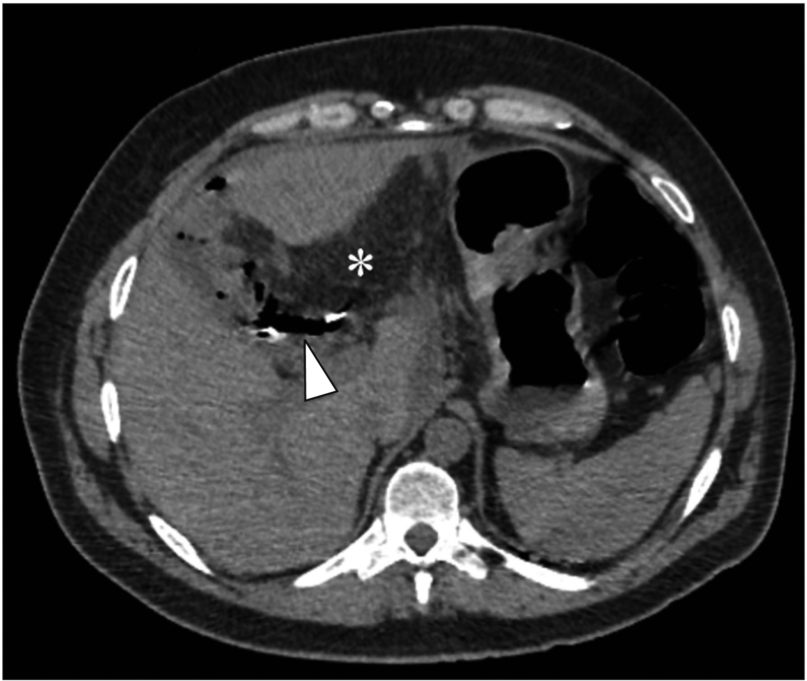
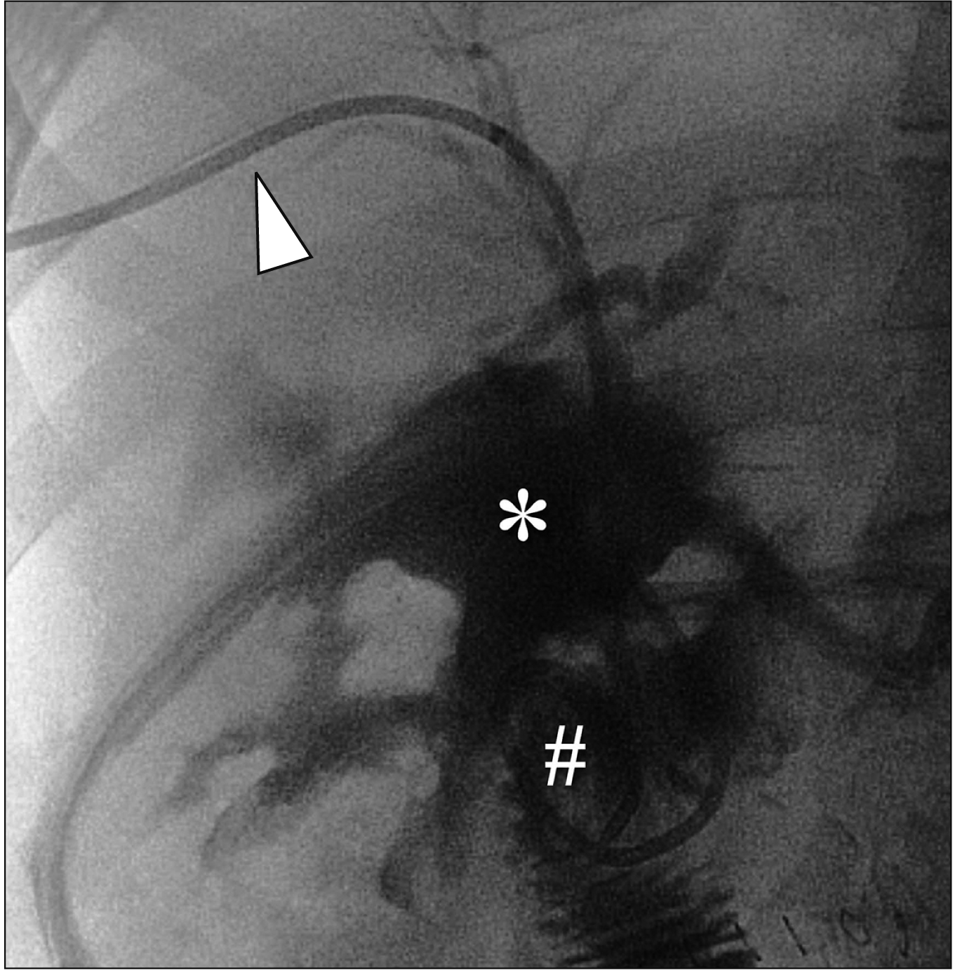
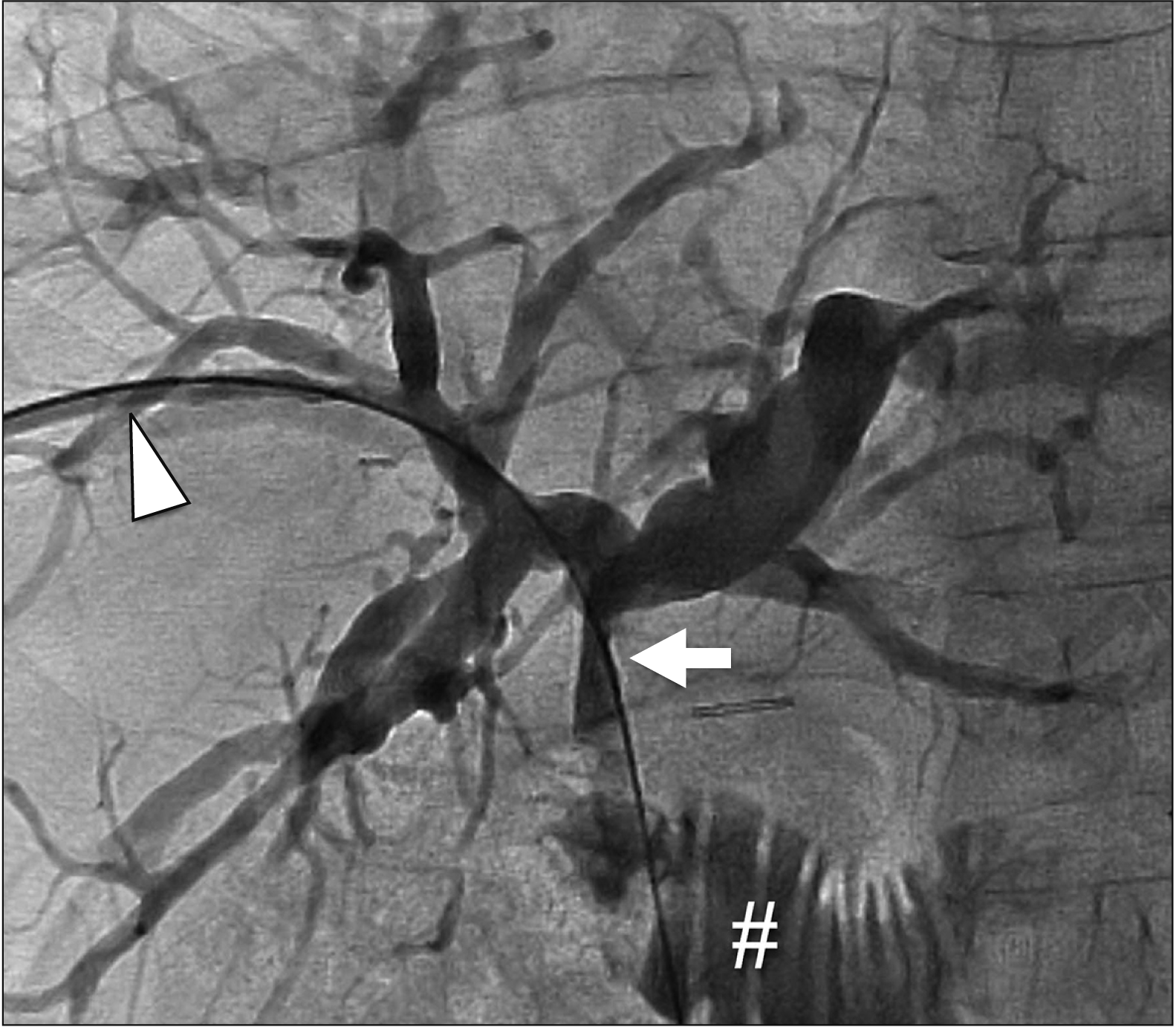
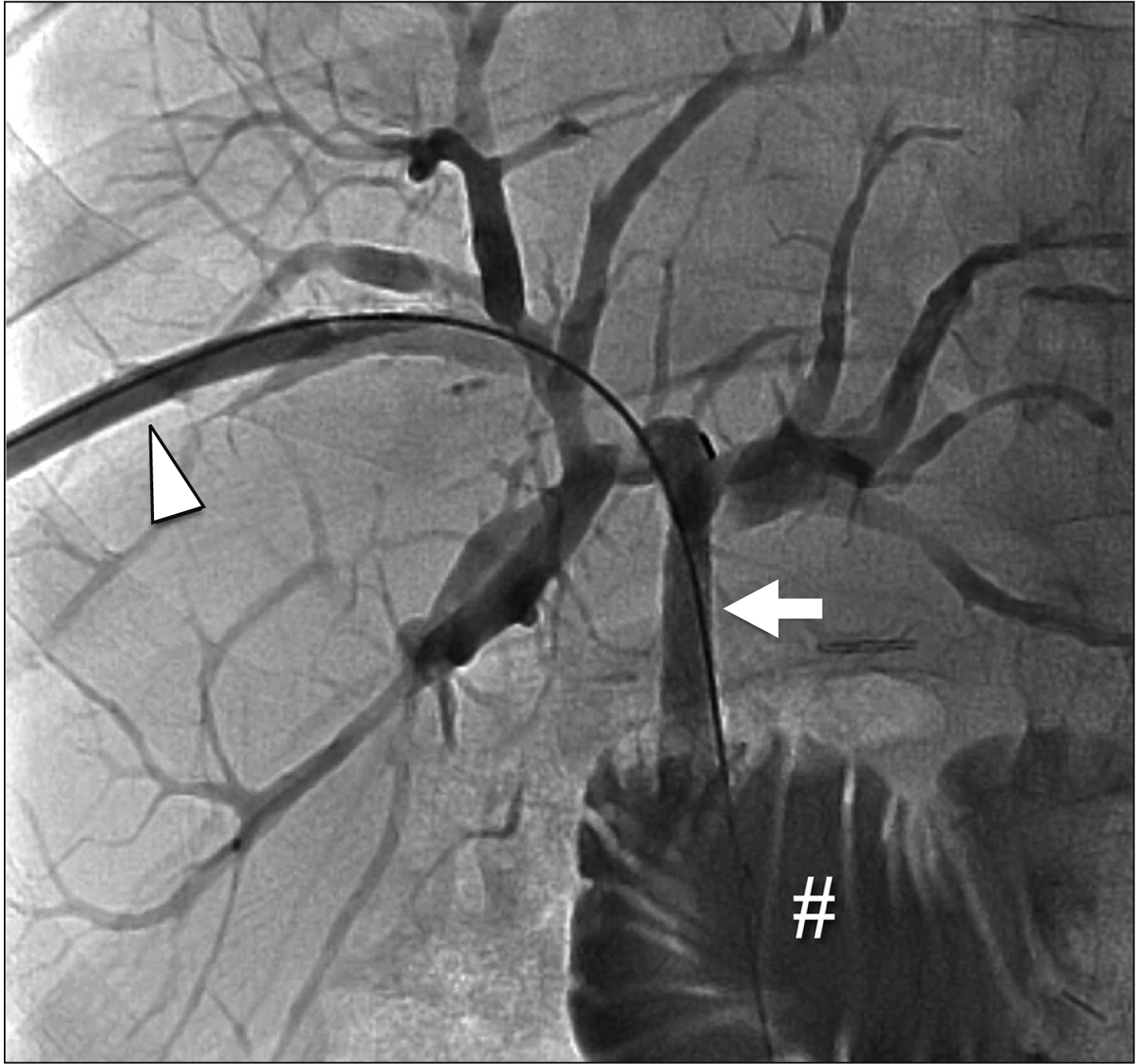
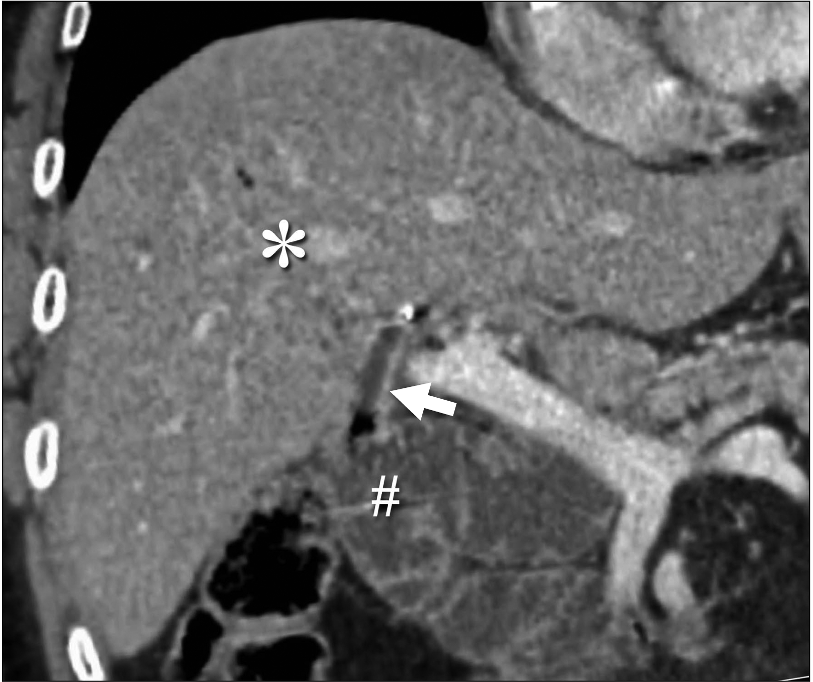
A 48-year-old male underwent pancreatic surgery with HJ for a locally advanced pancreatic adenocarcinoma. On the 10th postoperative day, the patient started to have upper abdominal pain, fever, and evidence of bilious output from surgical drain. He underwent urgent multi-detector computed tomography that revealed a collection, with bubble of air at the level of HJ without intrahepatic biliary ducts dilatation (Fig. 1). Complete HJ dehiscence was suspected. It was confirmed by a percutaneous catheter contrast study (Fig. 2). His liver function values were within normal limits except for gamma-glutamyl transpeptidase of 93 U/L (range, 8–61 U/L). After a multidisciplinary discussion, the patient was a candidate for a first-line percutaneous approach. From right surgical drainage, a retrograde opacification of non-dilated intrahepatic biliary tree was achieved (Fig. 2). The opacification of the intrahepatic biliary tree allowed us to indicate the possible percutaneous manoeuvre, while knowing that the puncture of non-dilated biliary ducts could be burdened by a high risk of technical failure with possible liver complications [1]. With a Seldinger’s technique, right percutaneous trans-hepatic internal-external biliary drainage (8 Fr) was inserted under fluoroscopic guidance, with its tip inserting into the anastomotic bowel loop (Fig. 3). The drainage ensured regular outflow of bile from the liver to the bowel, leading to the creation a retroperitoneal fibrosis with consequent a neo-HJ in one month. At three months follow-up, a HJ stenosis was noted on cholangiography (Fig. 4). Endobiliary interventional radiology approach was chosen. Given the failure of three high pressure (16 Atm) balloon bilioplasty attempts for recoiling of the stenosis, a 10 mm × 45 mm bio-resorbable biliary stent (SX-ELLA DV biliary; ELLA-CS, Hradec Kralove, Czech Republic) was positioned which ensured regular patency of the neo-HJ without complication (Fig. 5). Follow-up studies at 32 months after stenting, including clinical observation and laboratory tests, showed no complications and regular patency of the neo-HJ (Fig. 6).
DISCUSSION
Biliary leaks remain a significant source of morbidity in patients undergoing pancreatic surgery [1-5]. The incidence of HJ dehiscence after pancreatic open surgery ranges from 3% to 8% [1]. HJ dehiscence is generally due to bile duct ischemia with consequent necrosis leading to a fistula [2]. The most complicate biliary leak to treat is complete HJ dehiscence [1,2,6]. Clinical signs of HJ dehiscence may include leucocytosis, fever, abdominal pain, and presence of bile-looking from surgical drain [1-3]. This complication requires prompt diagnosis and subsequent treatment. This diagnosis can be confirmed by imaging ultrasound, computed tomography, and magnetic resonance that can identify bilomas and/or the biliary leak [1]. A multidisciplinary approach is essential in these cases to evaluate the previous pathology, post-surgical anatomy, types of biliary leakage, and possible treatments (from the less invasive one to the most invasive one). Traditionally, biliary leakages have been treated by surgery. However, endoscopic and/or interventional radiology procedures are adopted recently. Endoscopic procedures are frequently technically unfeasible in these patients due to a modified bowel anatomy [1,2]. According to the International Study Group of Liver Surgery of 2011, for patients with a moderately compromised clinical condition, percutaneous procedures are indicated as the first line treatment [7]. Percutaneous trans-hepatic cholangiography followed by percutaneous trans-hepatic biliary drainage (PTBD) can confirm the diagnosis and treat biliary collection and fistula by leakage healing [1-3]. However, when HJ dehiscence is completed, PTBD may be technically unfeasible due to the absence of intrahepatic biliary duct dilatation, unlike an occlusive pathology [8]. This decompression of intrahepatic biliary ductal does not allow an ultrasound-guided puncture. Therefore, percutaneous transhepatic access can be done by retrograde visualization of the intrahepatic biliary tree from surgical drainage or by a direct puncture of a biliary duct near the hepatic hilum with consequent contrast media filling and opacification of the intrahepatic biliary tree. Despite this technical limitation of biliary tract decompression for interventional radiological approach and possible technical unsuccessful, a percutaneous attempt can be indicated in most cases before a more invasive surgical approach [2].
Only a few authors have described successful percutaneous approach in patients with altered anatomies due to previous surgeries [1-7,9-12]. Percutaneous manoeuvres performed range from internal-external biliary drainage to stent deployment: plastic, metallic, and bioresorbable.
Percutaneous internal-external biliary drainage is the first-line manoeuvre that allows the drainage of the bile (both internally and externally) with the aim to detain and seal the biliary leak. Timing of this approach is directly proportional to the extent of the anastomotic dehiscence. Thus, the resolution period with percutaneous treatment of HJ dehiscence is highly variable, with a minimum time of about 30 days (as in our case) and an average of 180 days [1-3]. Cholangiography is the diagnostic imaging exam that can reveal successful healing of HJ. However, if no signs of anatomical anastomotic and clinical improvement are shown during follow-up controls, surgical intervention is mandatory. Antibiotic therapy and daily trans-catheter saline solution syringe injection (10–20 mL) are necessary to reduce the risk of infection and occlusion of the catheter itself [13].
Plastic stent has the advantage of possible removal. However, their small diameters (maximum up to 14 Fr) give them the possible risk of occlusion, especially in case of biliary sludge [1,2,5]. Metallic bare and covered stents are effective but irreversible solutions. Over time, metallic stents can produce a “foreign body” effect with fibrosis that may need or complicate a possible percutaneous or surgical re-intervention [5,9,11]. Bio-resorbable stents have advantages of both plastic and metal devices. With a large diameter (up to 10 mm), they allow a long-term dilatation without needing additional manoeuvres. Stent degradation occurs by hydrolysis within six months, without needing its removal or the risk of a “foreign body” effect [9,11].
In conclusion, complete HJ dehiscence is a severe and critical complication of open surgery. In selected cases of HJ dehiscence, percutaneous interventional radiological manoeuvres are even more important than ever. A percutaneous approach can solve this post-surgical complication with minimally invasive techniques using drainage and stents.

 XML Download