Abstract
Backgrounds/Aims
A superior mesenteric artery first approach (SFA) technique can improve the complete resection rate. It can be used to determine whether an operation can be performed by invading the superior mesenteric artery before performing a pancreatic transection in patients with pancreatic ductal adenocarcinoma (PDAC). The aim of this study was to compare perioperative outcomes between laparoscopic and open SFA for PDAC.
Methods
Between January 2017 and August 2019, consecutive patients who underwent laparoscopic and open pancreaticoduodenectomy (PD) for PDAC using SFA procedures were included and compared between laparoscopic and open procedures.
Results
Fourteen and 83 patients underwent laparoscopic and open surgeries, respectively. In perioperative outcomes, there were no significant differences in the amount of intraoperative blood loss or transfusion rate between the two groups. In the laparoscopic group, the operation time was longer with less patients showing wound infection. R0 resection rate and the number of retrieved lymph nodes showed no significant difference. The average time to adjuvant chemotherapy was longer in the open group. There was no significant difference in the mean survival time or the recurrence free period.
Pancreatic ductal adenocarcinoma (PDAC) is known as one of high mortality cancers [1,2], accounting for about 85% of all pancreatic cancers [2]. To date, PDAC has a 5-year survival rate of less than 5% [2,3]. Its prevalence is increasing due to an aging society [3,4]. Genetic, diabetes, obesity, and smoking are known as causes of pancreatic cancer [1,5,6]. There are no useful screening tests known for pancreatic cancer or recommended [7]. Therefore, the stage is often advanced at the time of diagnosis, which affects the prognosis of PDAC [2].
Surgery is the only known treatment to cure PDAC. Additional adjuvant chemotherapy can improve survival [1]. In PDAC, minimally invasive surgery (MIS) can reduce the number of hospital days with a benefit for pain reduction. In distal pancreatectomy, MIS shows similar results to open surgery. In pancreaticoduodenectomy (PD), many studies have revealed the safety of MIS [8,9].
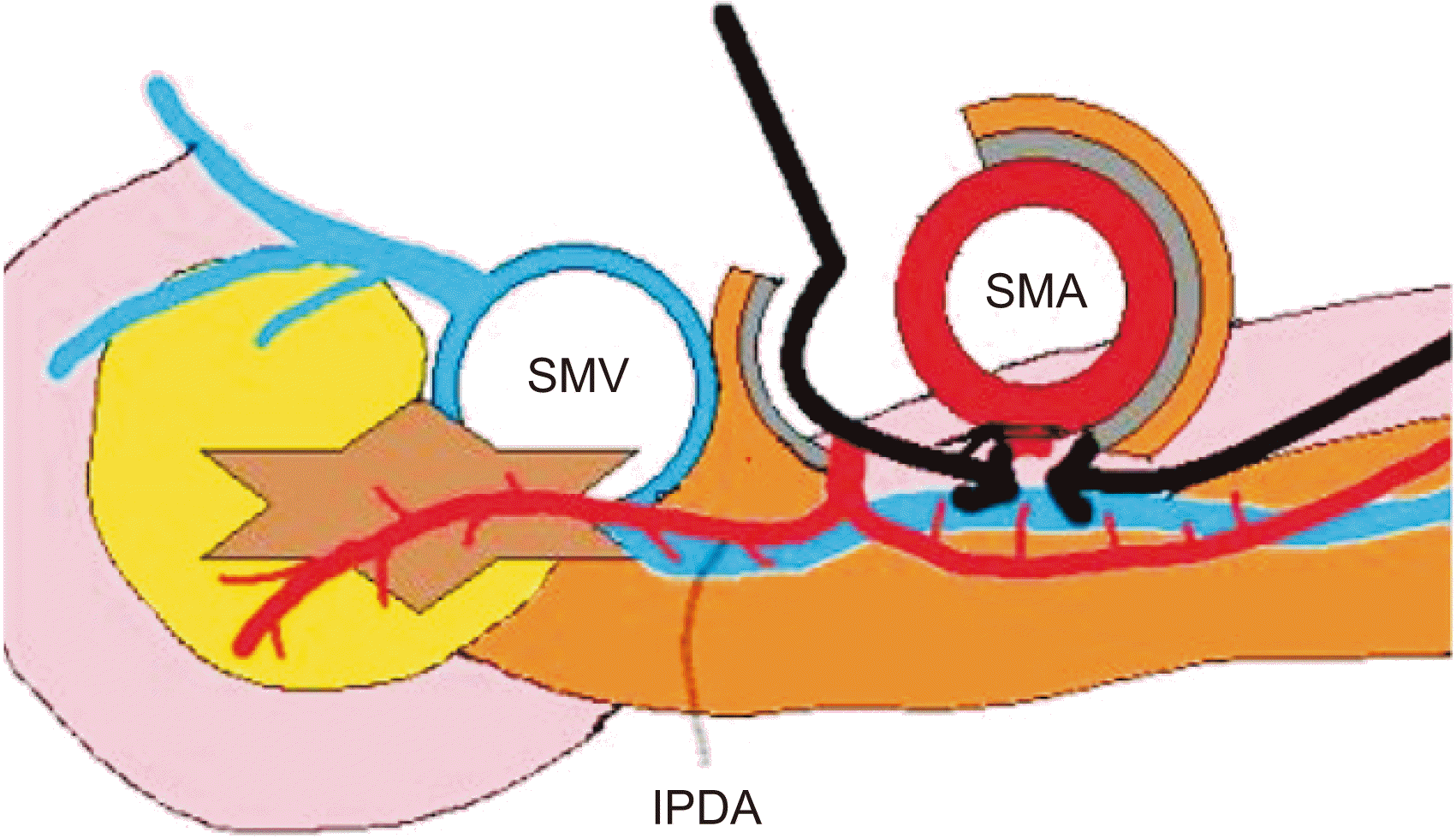
It is known that tumor invasion at resection margin affects the prognosis after surgical resection. As a result, many efforts have been made to improve surgical outcomes and improve the complete resection rate with the development of surgical techniques and equipment [10-12]. In this context, the superior mesenteric artery first approach (SFA) technique has emerged. Conventional PD first ligates the inferior pancreaticoduodenal artery (IPDA) before pancreatic resection. Blocking the blood flow can reduce the amount of bleeding and prevent damage to nearby blood vessels such as the right hepatic artery (RHA) from superior mesenteric artery (SMA). Complete removal of the surrounding soft tissue can obtain a sufficient number of lymph nodes and increase the rate of complete resection of the tumor [13-15].
With the development of surgical methods, there has been an intermediate classification between resectable pancreatic cancer and locally advanced pancreatic cancer (LAPC) that cannot be surgically resected. It is called borderline resectable pancreatic cancer (BRPC) [16,17]. The definition of BRPC means when the tumor covers part of the hepatic artery or it is attached to the SMA, the superior mesenteric vein (SMV) or part of the portal vein is narrowed by the adjacent tumor [16,18]. In this case, surgery may be possible by reconstructing some of the vessels after a combined resection. However, it can increase the incidence of incomplete resection and postoperative complications [19,20]. The most common invasion of the tumor, which was confirmed by postoperative specimens, was the cutsurface to the SMA [13]. To overcome these limitations, the SFA was introduced. Patients with advanced pancreatic cancer are unsuitable to undergo surgical resection if there is a tumor invasion in the adventitia of the SMA during surgery. In fact, the presence of vascular invasion can only be determined intraoperatively. The SFA can be used to determine the invasion of the SMA before pancreatic resection and to decide if surgery is possible [15].
The SFA technique is widely used in open surgery. Various studies have shown that surgical complications of SFA do not differ from those of a conventional procedure [13,21]. It can also bring about complete resection rate, increase survival rate [22], and reduce bleeding amount [23]. However, research about the safety of SFA technique in laparoscopic procedures is limited. Thus, the purpose of this study was to determine whether a surgery using the SFA technique in laparoscopic PD would differ in safety and prognosis compared to an open surgery.
Between January 2017 and August 2019, patients at a tertiary referral center who underwent laparoscopic and open PD using SFA procedures for PDAC were analyzed in this study. Surgical techniques were reviewed through recorded video for patients undergoing laparoscopic surgery. Patients included in this study were those considered to have resectable PDAC. Patients with clinically stage T4, concurrent distant metastasis, concurrent malignancy, and prior history of malignancy were excluded from this study.
Preoperative imaging was performed to determine the feasibility of surgery based on the criteria for BRPC and LAPC following the National Comprehensive Cancer Network (NCCN) guideline [17]. The SFA technique was used for the resection after confirming that there was no tumor invasion at the SMA. Complete resection of the tumor was possible by hanging the SMA first. It can be divided into three levels according to the range of soft tissue dissection around the pancreas. The first level simply refers to the separation of soft tissues around the pancreas from blood vessels without lymph node dissection. The second level is a method of ligation of the IPDA at the baseline to remove the surrounding lymph nodes in en-bloc while preserving nerve bundles around the SMA. The third level is a method of obtaining complete resection of the tumor from perineural spread by removing both soft tissues and nerve bundles located 180° to the right side of the SMA [15,24]. In this study, the level 3 method was used to remove all soft tissues and nerves on the right side of the SMA (Fig. 1; Supplementary Video 1). Data about the characteristics of patients (age, sex, American Society of Anesthesiologists (ASA) score [25], neoadjuvant chemotherapy, body mass index), preoperative laboratory findings (blood carcinoembryonic antigen [CEA], carbohydrate antigen 19-9 [CA19-9], total bilirubin), preoperative patient performance, blood loss during surgery, operation time, postoperative complications (pancreatic fistula, bile leakage, diarrhea, delayed gastric emptying, bleeding, surgical wound infection), and oncologic outcome were collected. Based on the collected data, results were compared between the laparoscopic group and the open SFA group. This study was approved by the Institutional Review Board of Asan Medical Center (No. 2020-1330).
After abdominal access was established, the greater omentum was divided using an energy device and the right colon was separated and fully mobilized from the liver and duodenum. The retropancreatic SMV was then exposed and the right gastroepiploic vessels were transected. After removing soft tissues from around the SMV and the SMA, each was hung with a vessel loop. Mobilization of the duodenum to the Treitz ligament was performed with traction of the duodenum by a surgical assistant. The stomach and the duodenum were divided using a linear stapler.
After cholecystectomy, dissection of the hepatoduodenal ligament and isolation of the common bile duct were performed. The right and left hepatic arteries were identified and isolated. Lymph node dissection was then performed. The gastrohepatic ligament was opened to visualize the superior border of the pancreas and to identify the common hepatic artery. The right gastric artery and the gastroduodenal artery were identified and transected using a Hem-o-lock clip. The pancreas was divided above the SMV using an energy device. After retracting the resected pancreas to the right side of the patient’s abdomen, the portal vein was identified and hung with a vessel loop. The jejunum was divided approximately 10–15 cm distal to the Treitz ligament with a linear stapler. An energy device and an electrocautery were used to separate the remaining soft tissues and branches from the SMA between the uncinate process of the pancreas and the SMA to complete the resection. Pancreatojejunostomy was performed using a double-layered, end-to-side, duct-to-mucosa method. A polyethylene internal stent was inserted into the pancreatic duct. End-to-side hepaticojejunostomy was performed using continuous suturing at the posterior wall with interrupted or continuous suturing at the anterior wall. Duodenojejunostomy or gastrojejunostomy with jejunojejunostomy was performed. Two or three closed suction drains were placed at the superior and inferior borders of the pancreatojejunostomy site.
For statistical analysis, chi-square test was used for univariate analysis of discontinuity variables and Student’s t-test was used for univariate analysis of continuity variables. Both methods were interpreted as statistically significant results when p-value was less than 0.05. Multivariate analysis was performed for variables with p-value < 0.1 in univariate analysis, including MIS. The odds ratios (ORs) and 95% confidence intervals (CIs) were identified using a logistic regression model. The overall survival and disease-free survival were analyzed using the Kaplan-Meier technique. All statistical analyses were performed using IBM SPSS version 26.0 (IBM Corp., Armonk, NY, USA).
There were 14 patients in the laparoscopic group and 83 patients in the open group. The median age of the two groups was same as 65 years old (p = 0.493). The median body mass index (BMI) was 21.5 kg/m2 (17.5–24.0 kg/m2) in the laparoscopic group and 22.5 kg/m2 (17.5–29.3 kg/m2) in the open group (p = 0.025). Preoperatively identified ASA classification system categories belonged to category II in both groups, showing no significant difference (92.9% vs. 94.0%, p = 0.136). According to the blood test for preoperative tumor markers, CEA showed a median of 2.0 ng/mL in the laparoscopic group and a median of 3.4 ng/mL in the open group (p = 0.198). CA19-9 was not significantly different between the two groups either, with a median of 48.7 U/mL in the laparoscopic group and a median of 72.8 U/mL in the open group (p = 0.376). According to the NCCN classification criteria confirmed by preoperative imaging, 9 patients (64.3%) were resectable in the laparoscopic group and 5 patients (35.7%) were borderline resectable. In the open group, 64 patients (77.1%) were resectable and 19 patients (22.9%) were borderline resectable. One patient (7.1%) in the laparoscopic group and 16 patients (19.3%) in the open group underwent neoadjuvant chemotherapy, showing no significant difference between the two groups (p = 0.309; Table 1).
There was no significant differences in blood loss (340 mL vs. 386 mL; p = 0.688) or transfusion rate (7.1% vs. 14.5%; p = 0.526) between laparoscopic and open groups. The total operation time was significantly longer in the laparoscopic group (422 vs. 336 minutes; p < 0.001). However, the number of cases requiring combined resection of portal vein showed no statistically significant difference (7.1% vs. 9.6%; p = 0.769; Table 2).
Complication rate was not significantly different (28.6% vs. 38.6%; p = 0.480). There was no significant difference in severe complication of Clavien-Dindo classification 3 or more between the two groups (1 vs. 0, p = 0.684). There was no patient (0%) with wound infection in the laparoscopic group whereas there were 8 patients (9.6%) in the open surgery group (p = 0.004). Pancreatic fistulas were found in 2 patients (14.3%) in the laparoscopic group and 18 patients (21.6%) in the open group. Bile leakage was absent in both groups. Bleeding was noted in 3 patients (3.6%) in the open group. Of these three patients, two were found to have intraperitoneal hematoma on regular computed tomography (CT) follow-up after surgery without requiring an additional treatment such as intervention. Only one patient needed intensive care because of volume overload following respiratory failure. Delayed gastric emptying was found in 4 patients (4.8%) in the open group that required conservative treatment (4 vs. 0; p = 0.407).
Symptoms of diarrhea that required medication were found in 4 patients (28.6%) in the laparoscopic group and 9 patients (10.8%) in the open group. None of these patients died within 90 days after surgery. One patients (7.1%) in the laparoscopic group and five patients (6.0%) in the open group were required to be re-admitted within 60 days. The mean hospital stay was 10 days in the laparoscopic group and 12.7 days in the open group (p = 0.319).
Postoperative adjuvant chemotherapy in our center was performed for 12 patients (85.7%) in the laparoscopic group and 61 patients (73.5%) in the open group. The average time to postoperative adjuvant chemotherapy in the laparoscopic group was shorter than that in the open group (36.1 vs. 53.6 days; p = 0.016).
After surgery, all patients took intravenous patient-controlled analgesia and per os analgesics. The average number of additional IV analgesics was 11 times in the laparoscopic group and 12 times in the open group without statistical significance (p = 0.806; Table 2). Univariate analysis revealed factors affecting postoperative complications. Resectability according to NCCN guideline of CT image, MIS, neoadjuvant chemotherapy, sex, body mass index, CA19-9, and serum bilirubin levels did not significantly affect postoperative complications. Only the correlation between age and complication rate was significant (OR, 1.054; 95% CI, 1.003–1.106; p = 0.036). Multivariate analysis revealed that whether MIS was done (OR, 0.638; 95% CI, 0.181–2.241; p = 0.479), age (OR, 1.054; 95% CI, 1.003–1.106; p = 0.036), and BMI (OR, 1.164; 95% CI, 0.990–1.368; p = 0.065) were significant variables affecting postoperative complications. MIS did not affect complications (OR, 0.857; 95% CI, 0.227–3.242; p = 0.819). BMI was not a significant variable affecting postoperative complications either (OR, 1.160; 95% CI, 0.978–1.376; p = 0.088). Only age showed a statistically significant association with complication rates (OR, 1.054; 95% CI, 1.002–1.109; p = 0.041; Table 3).
The average size of tumors after surgery was 2.3 cm in the laparoscopic group and 2.8 cm in the open group (p = 0.274). There was no statistically significant difference in cell differentiation. More than 70% of patients showed moderate differentiation in both groups (p = 0.517). Lymphovascular invasion was observed with 9 patients (64.3%) in the laparoscopic group and 46 patients (55.4%) in the open group (p = 0.604). Perineural invasion was also observed with 10 patients (71.4%) in the laparoscopic group and 61 patients (73.5%) in the open group (p = 0.761). Total number of lymph nodes harvested during surgery as a factor affecting the survival of patients was 17 in the laparoscopic group and 20 in the open group, showing no significant difference between the two groups (p = 0.073). R1 resection rate was 21.4% in the laparoscopic group and 20.5% in the open group, showing no significant difference between the two groups (p = 0.936; Table 4).
Univariate analysis revealed that R0 resection was not affected by MIS or tumor resectability determined by preoperative imaging test, neoadjuvant chemotherapy, age, sex, BMI, CA19-9, and serum bilirubin levels. In the multivariate analysis, there was no difference in complete resection rate according to the surgical method (OR, 1.180; 95% CI, 0.283–4.915; p = 0.818; Table 5).
The median duration of follow-up was 15 months and the median survival time was 18 months. There was no significant difference in mean survival between the laparoscopic group (30.8 months; 95% CI, 25.1–36.5 months) and the open group (30.5 months; 95% CI, 37.8–33.2 month) (p = 0.997; Fig. 2).
The median duration of recurrence free survival was 13 months. There was no significant difference in the mean recurrence free survival between the laparoscopic group (18.8 months; 95% CI, 13.4–24.1 months) and the open group (23.2 months; 95% CI, 19.9–26.6 months) (p = 0.621; Fig. 2). For distant metastasis, about 80% of them showed hepatic metastasis and the remaining 20% showed lung metastasis. For patients with recurrent findings, peritoneal seeding was the most common (40.0%), followed by recurrence of lymph nodes around the mesenteric artery (22.0%). Common hepatic artery, inferior vena cava, periaortic lymph nodes, and remaining pancreatic recurrence were also observed.
In resectable pancreatic cancer, SFA technique has the advantage of improving complete resection rate, sufficient lymph node removal, and major peripheral blood vessel damage [13-15]. In BRPC or LAPC after neoadjuvant chemotherapy, prior confirmation of SMA invasion can confirm the possibility of surgical resection before dividing the pancreas [22]. Previous studies have shown that the SFA in open surgery can be safely achieved without increasing complications compared to standard procedures [26]. However, a laparoscopic technique with SFA is not well known. This study compared the laparoscopic group and the open group of patients who underwent surgery using the SFA.
This study was performed on patients who underwent PD using the SFA technique for PDAC in recent two years. The operation time was significantly longer in the laparoscopic group. However, there was no significant difference in blood loss between the two groups. There was no difference in the incidence of complications between the two groups either after surgery. There were no statistically significant factors affecting the incidence of complications including MIS. However, there was a difference in the time period after surgery and before adjuvant chemotherapy between the two groups. The period from the operation date to adjuvant chemotherapy and the period from discharge date to adjuvant chemotherapy was also longer in the open group (46.9 vs. 30.6 days, p = 0.022). This was thought to be because the time to recover the general condition was longer in the open group than in the laparoscopic group. There was no difference in the number of additional IV analgesic medications between the two groups.
The average number of harvested lymph nodes confirmed after surgery and the rate of R0 resection were similar between the two groups. The R0 resection rate was not affected by MIS. The median duration of follow up was short. However, there was no significant difference in overall survival or recurrence free survival between the two groups.
To date, many studies have shown that laparoscopic PD for the treatment of PDAC can reduce the number of hospital stay compared to open PD, with reduced pain and comparable postoperative complications [27]. Laparoscopic surgery has been recognized as a safe procedure that can be realized as open as a treatment for PDAC [8,9]. We analyzed the learning curves of skillful experts in laparoscopic pylorus-preserving pancreaticoduodenectomy. Results showed that after laparoscopic pylorus-preserving pancreaticoduodenectomy for 55 patients with benign disease, the period needed for stabilization and the time required for surgery gradually decreased. After about 100 surgeries, the patient group was expanded to malignant tumors. From about 180 surgical cases, it was confirmed that the operation time was shortened with lymph node dissection in malignant cases and the prognosis was better in complications. In terms of oncological prognosis, these results were similar to those of open surgery [28].
With continuous development of surgical technique, increasing the survival rate by improving R0 resection has been recognized as an important issue in the treatment of pancreatic head cancer [10-12]. In this context, SFA technique has emerged. The SFA can increase the number of harvested lymph nodes and secure a complete resection margin [13-15]. In addition, it is possible to confirm complete surgical resection for tumors with borderline resectable patients [15]. Currently, many studies have revealed that the SFA can be safely implemented without showing any significant difference in complications or prognosis compared to previous procedures [13,21]. With a laparoscopic procedure, there is a lack of studies. We compared results of laparoscopic SFA with those of open surgery. Postoperative results, complications, and prognosis were not significantly between the two groups. Therefore, SFA can be safely realized by laparoscopy if it is performed by an expert surgeon with sufficient experience in laparoscopic PD.
This study has some limitations. First, it was retrospective in nature. Second, the number of patients included in this study was not sufficient. Due to the small number of patients, propensity score matching could not be performed and the possibility of bias could not be excluded. Third, this study included only patients who underwent PD with SFA. Therefore, we could not compare results between standard PD and SFA. In addition, the average follow up period was 16 months, which was a short-term that could not make a definitive conclusion.
In conclusion, PD using SFA technique can also be considered in a laparoscopic surgery. Perioperative outcomes are comparable between laparoscopic and open SFA groups. Therefore, if practiced by an expert surgeon, SFA is judged to be a safe technique. However, this study has some limitations. A prospective research with more patients is needed in the future.
Supplementary data related to this article can be found at https://doi.org/10.14701/ahbps.2021.25.3.358
Notes
REFERENCES
1. McGuigan A, Kelly P, Turkington RC, Jones C, Coleman HG, McCain RS. 2018; Pancreatic cancer: a review of clinical diagnosis, epidemiology, treatment and outcomes. World J Gastroenterol. 24:4846–4861. DOI: 10.3748/wjg.v24.i43.4846. PMID: 30487695. PMCID: PMC6250924.
2. Ilic M, Ilic I. 2016; Epidemiology of pancreatic cancer. World J Gastroenterol. 22:9694–9705. DOI: 10.3748/wjg.v22.i44.9694. PMID: 27956793. PMCID: PMC5124974.
3. Ferlay J, Soerjomataram I, Dikshit R, Eser S, Mathers C, Rebelo M, et al. 2015; Cancer incidence and mortality worldwide: sources, methods and major patterns in GLOBOCAN 2012. Int J Cancer. 136:E359–E386. DOI: 10.1002/ijc.29210. PMID: 25220842.
4. World Health Organization. 2015. World health statistics 2015. World Health Organization;Geneva:
5. Bosetti C, Lucenteforte E, Silverman DT, Petersen G, Bracci PM, Ji BT, et al. 2012; Cigarette smoking and pancreatic cancer: an analysis from the International Pancreatic Cancer Case-Control Consortium (Panc4). Ann Oncol. 23:1880–1888. DOI: 10.1093/annonc/mdr541. PMID: 32937733. PMCID: PMC3629994.
6. Wang YT, Gou YW, Jin WW, Xiao M, Fang HY. 2016; Association between alcohol intake and the risk of pancreatic cancer: a dose-response meta-analysis of cohort studies. BMC Cancer. 16:212. DOI: 10.1186/s12885-016-2241-1. PMID: 26968702. PMCID: PMC4788838.
7. Shin EJ, Canto MI. 2012; Pancreatic cancer screening. Gastroenterol Clin North Am. 41:143–157. DOI: 10.1016/j.gtc.2011.12.001. PMID: 22341255. PMCID: PMC3670598.
8. Correa-Gallego C, Dinkelspiel HE, Sulimanoff I, Fisher S, Viñuela EF, Kingham TP, et al. 2014; Minimally-invasive vs open pancreaticoduodenectomy: systematic review and meta-analysis. J Am Coll Surg. 218:129–139. DOI: 10.1016/j.jamcollsurg.2013.09.005. PMID: 24275074.
9. de Rooij T, Lu MZ, Steen MW, Gerhards MF, Dijkgraaf MG, Busch OR, et al. 2016; Minimally invasive versus open pancreatoduodenectomy: systematic review and meta-analysis of comparative cohort and registry studies. Ann Surg. 264:257–267. DOI: 10.1097/SLA.0000000000001660. PMID: 26863398.
10. Demir IE, Jäger C, Schlitter AM, Konukiewitz B, Stecher L, Schorn S, et al. 2018; R0 versus R1 resection matters after pancreaticoduodenectomy, and less after distal or total pancreatectomy for pancreatic cancer. Ann Surg. 268:1058–1068. DOI: 10.1097/SLA.0000000000002345. PMID: 28692477.
11. Geer RJ, Brennan MF. 1993; Prognostic indicators for survival after resection of pancreatic adenocarcinoma. Am J Surg. 165:68–72. discussion 72–73. DOI: 10.1016/S0002-9610(05)80406-4. PMID: 8380315.
12. Nitecki SS, Sarr MG, Colby TV, van Heerden JA. 1995; Long-term survival after resection for ductal adenocarcinoma of the pancreas. Is it really improving? Ann Surg. 221:59–66. DOI: 10.1097/00000658-199501000-00007. PMID: 7826162. PMCID: PMC1234495.
13. Weitz J, Rahbari N, Koch M, Büchler MW. 2010; The "artery first" approach for resection of pancreatic head cancer. J Am Coll Surg. 210:e1–e4. DOI: 10.1016/j.jamcollsurg.2009.10.019. PMID: 20113929.
14. Sabater L, Cugat E, Serrablo A, Suarez-Artacho G, Diez-Valladares L, Santoyo-Santoyo J, et al. 2019; Does the artery-first approach improve the rate of R0 resection in pancreatoduodenectomy? a multicenter, randomized, controlled trial. Ann Surg. 270:738–746. DOI: 10.1097/SLA.0000000000003535. PMID: 31498183.
15. Inoue Y, Saiura A, Oba A, Kawakatsu S, Ono Y, Sato T, et al. 2019; Optimal extent of superior mesenteric artery dissection during pancreaticoduodenectomy for pancreatic cancer: balancing surgical and oncological safety. J Gastrointest Surg. 23:1373–1383. DOI: 10.1007/s11605-018-3995-3. PMID: 30306451.
16. Varadhachary GR, Tamm EP, Abbruzzese JL, Xiong HQ, Crane CH, Wang H, et al. 2006; Borderline resectable pancreatic cancer: definitions, management, and role of preoperative therapy. Ann Surg Oncol. 13:1035–1046. DOI: 10.1245/ASO.2006.08.011. PMID: 16865597.
17. Tempero MA, Malafa MP, Al-Hawary M, Asbun H, Bain A, Behrman SW, et al. 2017; Pancreatic adenocarcinoma, version 2.2017, NCCN Clinical Practice Guidelines in Oncology. J Natl Compr Canc Netw. 15:1028–1061. DOI: 10.6004/jnccn.2017.0131. PMID: 28784865.
18. Katz MH, Pisters PW, Evans DB, Sun CC, Lee JE, Fleming JB, et al. 2008; Borderline resectable pancreatic cancer: the importance of this emerging stage of disease. J Am Coll Surg. 206:833–846. discussion 846–848. DOI: 10.1016/j.jamcollsurg.2007.12.020. PMID: 18471707. PMCID: PMC5901743.
19. Ramacciato G, Nigri G, Petrucciani N, Pinna AD, Ravaioli M, Jovine E, et al. 2016; Pancreatectomy with mesenteric and portal vein resection for borderline resectable pancreatic cancer: multicenter study of 406 patients. Ann Surg Oncol. 23:2028–2037. DOI: 10.1245/s10434-016-5123-5. PMID: 26893222.
20. Ravikumar R, Sabin C, Abu Hilal M, Bramhall S, White S, Wigmore S, et al. 2014; Portal vein resection in borderline resectable pancreatic cancer: a United Kingdom multicenter study. J Am Coll Surg. 218:401–411. DOI: 10.1016/j.jamcollsurg.2013.11.017. PMID: 24484730.
21. Varty PP, Yamamoto H, Farges O, Belghiti J, Sauvanet A. 2005; Early retropancreatic dissection during pancreaticoduodenectomy. Am J Surg. 189:488–491. DOI: 10.1016/j.amjsurg.2005.01.007. PMID: 15820467.
22. Ironside N, Barreto SG, Loveday B, Shrikhande SV, Windsor JA, Pandanaboyana S. 2018; Meta-analysis of an artery-first approach versus standard pancreatoduodenectomy on perioperative outcomes and survival. Br J Surg. 105:628–636. DOI: 10.1002/bjs.10832. PMID: 29652079.
23. Horiguchi A, Ishihara S, Ito M, Nagata H, Shimizu T, Furusawa K, et al. 2007; Pancreatoduodenectomy in which dissection of the efferent arteries of the head of the pancreas is performed first. J Hepatobiliary Pancreat Surg. 14:575–578. DOI: 10.1007/s00534-006-1198-x. PMID: 18040623.
24. Inoue Y, Saiura A, Yoshioka R, Ono Y, Takahashi M, Arita J, et al. 2015; Pancreatoduodenectomy with systematic mesopancreas dissection using a supracolic anterior artery-first approach. Ann Surg. 262:1092–1101. DOI: 10.1097/SLA.0000000000001065. PMID: 25587814.
25. Daabiss M. 2011; American Society of Anaesthesiologists physical status classification. Indian J Anaesth. 55:111–115. DOI: 10.4103/0019-5049.79879. PMID: 21712864. PMCID: PMC3106380.
26. Negoi I, Hostiuc S, Runcanu A, Negoi RI, Beuran M. 2017; Superior mesenteric artery first approach versus standard pancreaticoduodenectomy: a systematic review and meta-analysis. Hepatobiliary Pancreat Dis Int. 16:127–138. DOI: 10.1016/S1499-3872(16)60134-0. PMID: 28381375.
27. Cho A, Yamamoto H, Nagata M, Takiguchi N, Shimada H, Kainuma O, et al. 2009; Comparison of laparoscopy-assisted and open pylorus-preserving pancreaticoduodenectomy for periampullary disease. Am J Surg. 198:445–449. DOI: 10.1016/j.amjsurg.2008.12.025. PMID: 19342003.
28. Song KB, Kim SC, Lee W, Hwang DW, Lee JH, Kwon J, et al. 2020; Laparoscopic pancreaticoduodenectomy for periampullary tumors: lessons learned from 500 consecutive patients in a single center. Surg Endosc. 34:1343–1352. DOI: 10.1007/s00464-019-06913-9. PMID: 31214805.