

 PDF
PDF Citation
 Print
Print


INTRODUCTION
Biliary tract cancer (BTC), comprising intrahepatic cholangiocarcinoma (IHC), extrahepatic cholangiocarcinoma (EHC), and gallbladder cancer (GBC), is a rare and highly fatal malignancy [1]. Surgical resection is the only curative treatment for BTC [2]. Most patients with BTC do not develop symptoms and signs in the early stages; therefore, the disease is usually diagnosed at an advanced stage. BTC is considered unresectable based on several factors, including vascular invasion and lymph node metastasis, which are strongly associated with poor prognosis [1]. Similarly, perihilar cholangiocarcinoma (PHC) is most often considered inoperable due to the complex liver anatomy involved [3]. As a result, the resectability rates of BTCs generally remain low, suggesting the feasibility of curative treatment in a limited number of cases [4]. Since treatment guidelines for neoadjuvant chemotherapy have yet to be formulated, palliative chemotherapy is generally considered as the treatment of first choice for unresectable advanced BTC. Unfortunately, initially unresectable BTC treated with isolated palliative chemotherapy is associated with poor prognosis [5].
Recent studies have yielded promising results demonstrating the effectiveness of chemotherapeutic agents such as gemcitabine (GEM) or its combination with other agents in BTC [6]. Administration of these agents has facilitated surgical resection in a few downsized patients [7-9]. This strategy of downsizing chemotherapy and subsequent surgical resection, termed conversion surgery, has generated encouraging results for pancreatic cancer, hepatic metastases of colorectal cancer, and even gastric cancer [10-12]. However, evidence supporting the role of conversion surgery in BTC has yet to be reported.
Many of the existing studies are case reports. Most of the retrospective analytical studies show disease heterogeneity and treatment. The majority of the studies are from East Asian countries, such as South Korea, Japan, and China, probably due to the geographic variation in the incidence of BTC incidence globally with a significantly higher burden in Asia compared with the western world [13]. Thus, further studies are needed in areas with a relatively higher incidence of BTC; however, before conducting large-scale studies, we first intended to present our experience from a single center. This study demonstrates the feasibility of conversion surgery after palliative chemotherapy as a therapeutic alternative in initially unresectable extrahepatic BTC patients.
MATERIALS AND METHODS
We conducted a retrospective review of electronic medical records of patients with initially unresectable BTC, who underwent palliative chemotherapy and subsequent surgery at Seoul National University Hospital from 2013 to 2019. Imaging studies including computed tomography (CT), magnetic resonance imaging (MRI), and positron emission tomography (PET) scans were conducted serially during the course of patients’ treatment. Prospective data including patient and tumor characteristics, changes in tumor markers, chemotherapy regimens and number of cycles, type of operation, and clinical outcomes including postoperative complications, recurrence, disease-free survival (DFS) and overall survival (OS) were collected. Death status was provided by the National Health Insurance Registry involving cases which were not associated with in-hospital mortality. The Institutional Review Board at Seoul National University Hospital granted approval for all the data collection, storage, and analysis (SNUH-2003-197-1112).
RESULTS
Patient characteristics
A total of 12 patients underwent palliative chemotherapy and subsequent surgery at Seoul National University Hospital between 2013 and 2019. The mean age at the time of diagnosis was 59.7 years, and the male-to-female ratio was 10 : 2. The final pathological diagnosis comprised six PHCs, four distal common bile duct (CBD) cancers, and two GBCs. All the patients underwent palliative chemotherapy initially for unresectable cancers, and the median time duration between first chemotherapy and surgery was 5.6 months (range of 2.2–33.1 months). Key image findings associated with reasons for initial unresectability for each of the patients are shown in Supplementary Fig. 1. A multidisciplinary conference including a surgeon, gastroenterologist, oncologist, radiologist, radiotherapist, and pathologist was held to decide if the medical downsizing through chemotherapy was adequate to render the disease operable. After being considered resectable, five patients underwent extended right hemihepatectomy, four underwent pancreaticoduodenectomy, and the remaining three underwent extended left hemihepatectomy, left trisectionectomy, and extended cholecystectomy, respectively. Four of the 12 patients experienced postoperative complications; three had intra-abdominal fluid collections, and one had a wound problem. The median postoperative hospital stay was 13.5 days (range, 8–61 days). Seven patients were treated with adjuvant chemotherapy. Cancer recurred in four patients during follow-up. Patient characteristics are listed in Table 1.
Palliative chemotherapy and radiotherapy prior to surgery
The two most common reasons for unresectability were suspected distant lymph nodal metastasis and major vascular invasion. One patient was initially judged inoperable due to suspicious peritoneal seeding. Five of the six PHC patients were also initially considered unresectable because of the Bismuth type IV PHC lesions based on imaging studies. Most patients’ cancers were unresectable due to more than one reason.
All patients were treated with GEM-based chemotherapy at some point before surgery. Nine of the 12 patients were treated with a combination of GEM and cisplatin (GEM + CDDP). Other GEM-based combination therapies included a combination of GEM and erlotinib (GEM-E) and a substantial dose of GEM combined with durvalumab (GEM-Durva). Two patients were treated with multiple regimens: one patient received FOLFIRINOX (combination of 5-fluorouracil, leucovorin, irinotecan, and oxaliplatin) followed by GEM monotherapy, and the other received GEM + CDDP followed by iFAM (combination of 5-fluorouracil, doxorubicin, and mitomycin-C), FL (combination of 5-fluorouracil and leucovorin), and UFT (tegafur/uracil). The patients who were treated with GEM-E, GEM-Durva, or FOLFIRINOX were initially thought to have been diagnosed with pancreatic cancer, which explains the chemotherapy regimens used. The total number of chemotherapy cycles administered to each patient ranged from 2–38 cycles, and a majority of the patients received fewer than 10 cycles. The shortest duration between first chemotherapy and surgery was 67 days, and the longest was 33.1 months. Preoperative radiotherapy, with a radiation dose of 56.0/44.8 Gy over 28 fractions, was performed only in 2 of the 12 patients (Table 2, 3).
Treatment response
A comprehensive radiological evaluation of the radiological and PET scans, and the difference in tumor marker levels was conducted to determine the degree of response after palliative chemotherapy. For radiological analysis, the response evaluation criteria in solid tumors (RECIST) criteria were used to determine the patients’ response to palliative chemotherapy prior to surgery. Of the 12 cases, 7 had stable disease (SD), 3 had partial response (PR), and 2 had complete response (CR) after preoperative chemotherapy, according to the RECIST criteria. One of the patients with CR, who went through the longest period and cycles of chemotherapy, ultimately showed no residual tumor based on final biopsy results after surgical resection.
Another method for evaluating the therapeutic response to chemotherapy entailed analysis of the metabolic response via PET scans. In eight out of 12 patients, PET scans were performed before treatment and either during or after chemotherapy, whereas in the other four patients either only one or none was done. In six of the eight patients who underwent PET scans before and after chemotherapy, reduced hypermetabolism was noted in the main tumor and lymph nodes. The largest measured difference in the available maximum standardized uptake value (SUVmax) was observed in patient 12, based on an SUVmax of 12.5 in the primary tumor before chemotherapy to an SUVmax of 4.9 after chemotherapy.
Differences in tumor marker levels were also observed before and after palliative chemotherapy in some patients. Although the carcinoembryonic antigen (CEA) levels showed minimal differences, the carbohydrate antigen 19-9 (CA 19-9) levels declined significantly after chemotherapy, especially in patients with high initial CA 19-9 levels. Patient 3 showed the largest difference with an initial CA 19-9 level of 5,919 µg/mL and a CA 19-9 level 22 µg/mL immediately before surgery (Table 2, 3).
Conversion surgery
All the patients eventually underwent conversion surgery after re-evaluation of resectability post-palliative chemotherapy. Since the indications for unresectabilty included distant lymph nodal metastasis and major vascular invasion, the reasons for secondary resectability were reduced lymph nodal size and lower involvement of the major vessels. In PHC, tumor respectability was also based on decreased tumor size and degree of involvement, and change in the PHC classification from Bismuth type IV to Bismuth type IIIa or IIIb. The reasons for conversion surgery are listed in Supplementary Table 1.
Four of the six PHC patients underwent extended right hemihepatectomy, one of them was treated via extended left hemihepatectomy, and the last one underwent trisectionectomy. All patients with distal CBD cancer underwent pancreaticoduodenectomy. Of the two GBC patients, one was exposed to extended right hemihepatectomy, and the other treated via extended cholecystectomy with bile duct resection due to severe adhesions around the CBD. In half of the cases, surgical difficulty was reported due to severe adhesions, and combined vessel resection was performed only in two cases. Both cases of combined vessel resection involved patients with PHC who underwent extended right hemihepatectomy, and in both cases, portal vein (PV) segmental resection and end-to-end anastomosis of the left PV to the main PV was performed.
Although some of the cases were initially considered unresectable due to suspicious arterial invasion, preoperative chemotherapy reduced arterial involvement significantly in these patients. Thus, no arterial resection was required during surgery. R0 resection was achieved in all cases (Table 2, 3).
Outcome
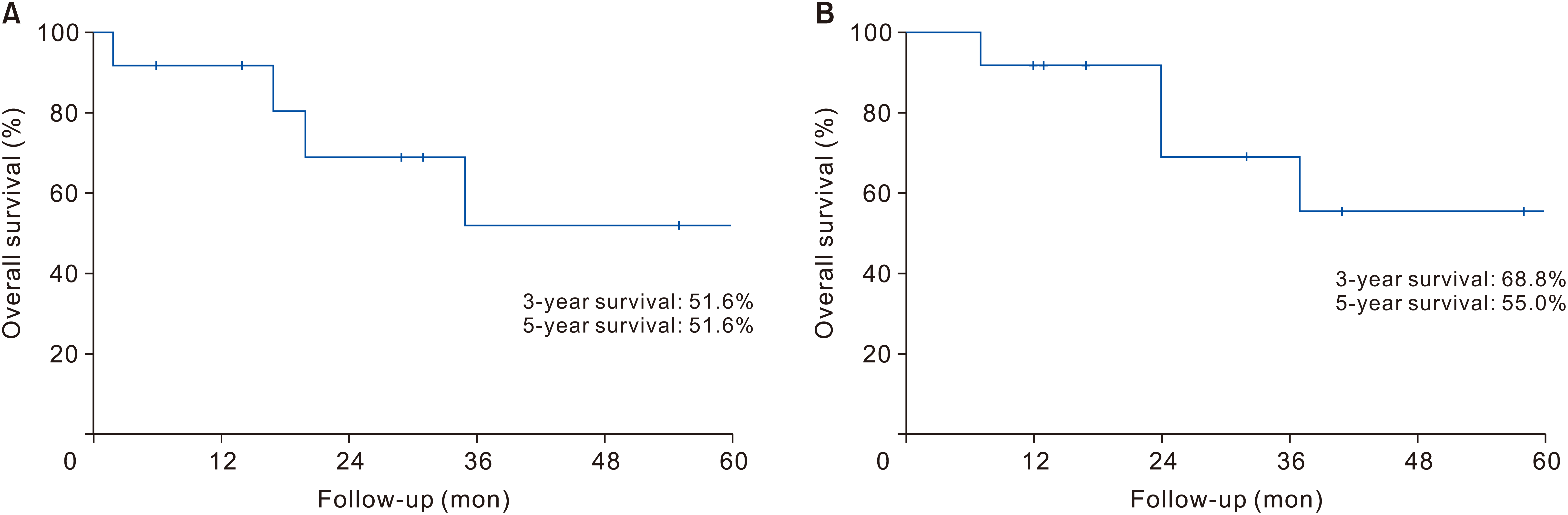
Of the four patients who experienced postoperative complications, three underwent percutaneous drainage for intra-abdominal fluid collection, and one was treated via wound revision surgery under general anesthesia. The median postoperative hospital stay was 13.5 days. Adjuvant chemotherapy was administered in seven patients. Recurrence was observed in four patients during follow-up. The 3-year and 5-year survival rates were 68.8% and 55.0%, respectively. OS rates are shown in Fig. 1. The treatment course and clinical outcomes of the patients are summarized in Table 2, 3.
Representative cases
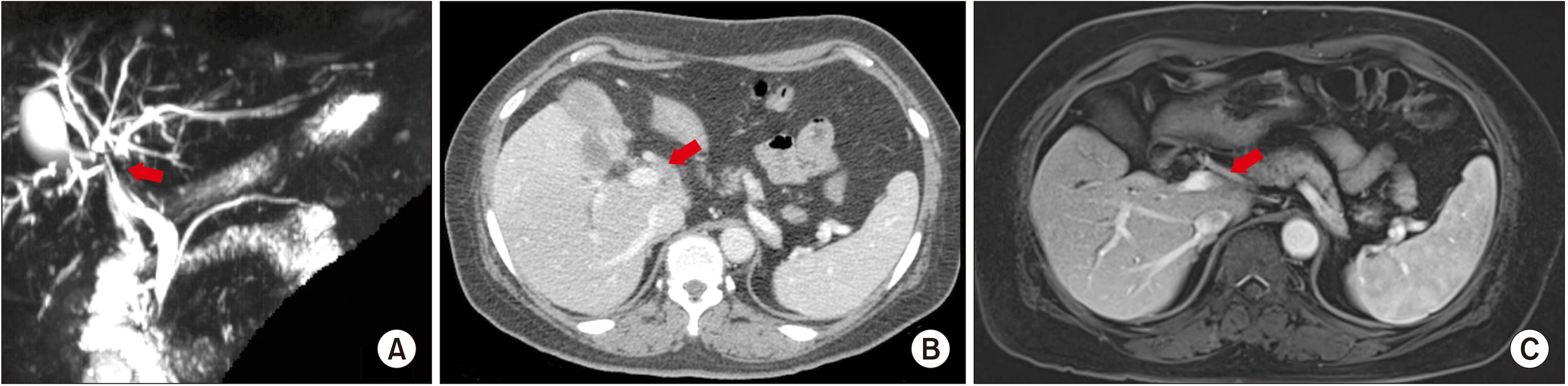
Two representative cases have been described in this study. One of the cases represents PHC with a long period of chemotherapy and long-term survival. Patient 6 presented with a history of abdominal pain and weight loss, and a 7 cm liver mass was detected on the CT scan. Liver biopsy revealed adenocarcinoma and was clinically diagnosed as PHC. The tumor was considered unresectable due to the involvement of both right and left intrahepatic ducts up to the second confluence, categorized as a PHC Bismuth type IV (Fig. 2A) with probable metastatic lymph nodes that appeared to invade the common hepatic artery and PV (Fig. 2B). Thus, 40 cycles of palliative chemotherapy with GEM + CDDP were administered over a period of approximately 30 months. After chemotherapy, imaging studies revealed reduced size of lymph nodes and lower left intrahepatic ductal and vascular involvement (Fig. 2C). The PET scan also revealed decreased uptake from an SUVmax of 5.8 to 4.0 at the initial site of hypermetabolism. Following the evaluation of secondary resectability, the patient underwent extended right hemihepatectomy with combined PV segmental resection. During immediate postoperative period, percutaneous drainage intervention was performed for fluid collection. The final postoperative pathological diagnosis revealed no residual tumor. The patient is currently being followed up in the outpatient department, with no evidence of cancer recurrence after more than 73 months after surgery.
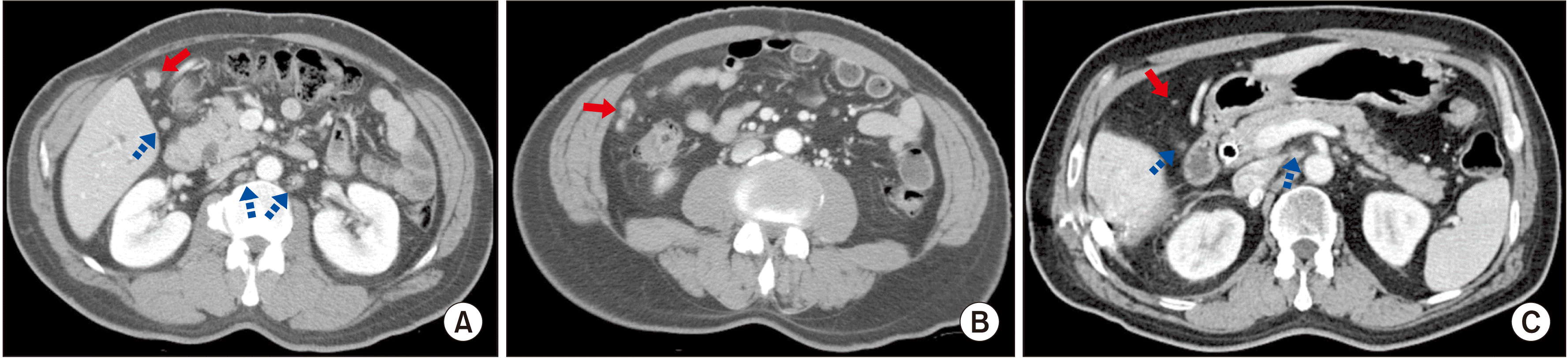
Another representative case in this study involves GBC treated with multiple chemotherapy regimens. Patient 11 was diagnosed with GBC associated with CBD invasion, along with peritoneal seeding and lymph node metastasis (Fig. 3A, 3B). The patient underwent palliative chemotherapy and multiple biliary stent interventions for recurrent cholangitis due to biliary obstruction. A total of 13 cycles of GEM + CDDP, three cycles of iFAM, one cycle of FL, and six cycles of UFT were administered to this patient. Multiple chemotherapy regimens were administered to address the recurring side effects following chemotherapy. Follow-up CT scans showed no difference in the size of main mass of the GBC and CBD, but a slight decrease in the retroperitoneal lymph nodal size was detected (Fig. 3C). In addition, pre-chemotherapy PET scans showed hypermetabolism in the gallbladder, portocaval lymph nodes, and peritoneal nodules in the right upper quadrant of the abdomen; however, the PET scans after chemotherapy showed hypermetabolism only in the CBD around the internal biliary stent and no hypermetabolism elsewhere, including the gallbladder. The GBC was thus considered resectable and treated via extended right hemihepatectomy with CBD resection. The pathological diagnosis after surgical resection revealed a well-differentiated adenocarcinoma of the gallbladder with a TNM staging of pyT2N0M0, and no residual tumor was detected in the CBD. A percutaneous drain was inserted postoperatively to drain fluid collection. Adjuvant chemotherapy was not performed. The patient is currently being followed up in the outpatient department without disease recurrence till date during the more than 64 months after the surgery.
DISCUSSION
BTCs are the second most common type of hepatobiliary cancer worldwide, and the global incidence of BTCs varies according geographically [13]. While BTCs are rare in Europe and North America, there is a high incidence in some regions of Latin America and Asia, which explains the active investigation into BTCs in these regions. The Global Burden of Disease study estimates 174,000 deaths due to BTCs worldwide in 2017, a 25% increase from the estimated deaths in 2007 [14]. The study using the Korean National Health Information Database, based on data from national health insurance that covers over 98% of South Korea’s population, reports that the overall 5-year survival rates for IHC, EHC, and GBC were 15.9%, 27.8%, and 30.0%, respectively [15]. Surgery is currently the only potentially curative treatment available for BTCs; however, only about 20% are resectable at diagnosis [16].
Recent studies investigating chemotherapy for BTC have reported positive data, especially for GEM-based chemotherapy. Phase II studies involving GEM + CDDP as first-line chemotherapy in advanced BTC reported that the combination chemotherapy was effective, safe and well-tolerated by the patients [17]. The results of the ABC-02 trial showed that BTC patients treated with GEM + CDDP had longer median OS, progression-free survival (PFS), and better rate of tumor control than those involving BTC treated with GEM alone [18]. Okusaka et al. [19] also reported better disease control rate, OS, and PFS in the GEM + CDDP group as compared to GEM-only group. Due to advances in intensive chemotherapy, the possibility of conversion from an unresectable BTC to a resectable disease is increasing. Since surgery remains the only curative treatment regardless of the effectiveness of chemotherapy, attempts are being made to treated patients with BTC via surgery whenever resection is feasible.
The concept of conversion surgery has already been applied to other cancers. Surgical resection after downsizing chemotherapy for advanced gastric tumors is known as “salvage,” “adjuvant,” or “secondary” gastrectomy. However, the concept of conversion surgery was described by Yoshida et al. [11] to define a treatment via R0 resection after chemotherapy in initially unresectable patients. Likewise, there have conversion surgery after downsizing chemotherapy has been reported in patients with colorectal cancer with liver metastasis [10]. Lastly, although surgical resection is the only curative treatment available for pancreatic cancer, only 20% to 30% of them are resectable at the diagnostic stage. After the introduction of novel chemotherapy regimens such as FOLFIRINOX for unresectable pancreatic cancer, OS improved dramatically and so has the possibility of conversion surgery after chemotherapy [12]. In a systemic review, Morganti et al. [20] reported that 8%–64% of patients with unresectable locally advanced pancreatic cancer at diagnosis undergo successful conversion surgery after chemotherapy, 57%–100% of the operated patients achieved R0 resection, and the resected patients manifested higher median survival than unresected patients after chemotherapy. Although no consensus has been reached regarding the ideal management of initially unresectable gastric cancer, colon cancer with liver metastasis, and pancreatic cancer, the positive outcomes of downsizing chemotherapy and subsequent surgical resection in some of these patients are encouraging signs for the application of conversion surgery.
Similarly, conversion surgery for BTCs is becoming increasingly feasible with the recent advances in chemotherapeutic regimes, but the actual clinical benefits have yet to be sufficiently investigated [9]. Therefore, prior to performing large-scale studies or clinical trials, we first evaluated a few cases of patients with BTC who underwent conversion surgery at our center, which manages a relatively large number of BTC cases every year. Our study demonstrated promising results of conversion surgery in BTC. The DFS of patients in our study ranged from 2 to 73 months, and the OS from the first chemotherapy ranged from 7 to 100 months. The 3-year and 5-year survival rates were 68.8% and 55.0%, respectively. These results are higher than the median OS in unresectable BTC patients treated with only palliative chemotherapy without surgery, which has been reported to range between 11.2 and 14.4 months in previous studies [18,19,21-23].
A literature review was performed to support our idea that conversion surgery is a potential treatment standard for some patients with BTC (Table 4). We conducted an extensive literature search of PubMed, Embase, and Google Scholar databases for articles published up to June 2020. We identified 10 published studies, all of which except for one study were published after 2009 [7-9,24-30]. Study subjects included all types of BTCs, including IHC, PHC, distal CBD cancer, and GBC. Most of the studies focused on a single type of BTC, but some studies unified all the different types under BTC. Different chemotherapy regimens were used in these studies. Fluorouracil-based chemotherapy was used in the earlier years, but eventually, GEM-based chemotherapy regimens were predominantly used. The studies reported R0 resection rates of 30.8%–100%. The median DFS was 14.4–26 months, and the median OS was 10.8–50.1 months.
One limitation of this study is that there was no control group to compare the results to, and thus the patients’ treatment protocols may not apply to the general BTC population. Nonetheless, the purpose of this study was to report the outcomes of an uncommon but emerging treatment strategy. Another study limitation is the heterogeneous group of BTCs. Since different types of BTCs may have different biological behaviors, separate studies managing individual cancer type are required. Lastly, the follow-up period of study patients was not consistent and was relatively short compared with other studies.
Conversion surgery is a feasible and effective therapeutic strategy in certain cases of initially unresectable BTCs. However, conversion surgery is not a standard treatment currently, suggesting the need for additional research and clinical trials investigating the optimal treatment strategy for initially unresectable BTCs. Additionally, further studies and efforts for developing new and effective chemotherapy regimens are also required. In any case, the active role of surgeons is essential throughout the treatment and decision-making process.
SUPPLEMENTARY DATA
Supplementary data related to this article can be found at https://doi.org/10.14701/ahbps.2021.25.3.349.
 XML Download