Journal List > Ann Lab Med > v.42(1) > 1147385

Kim, Park, Cho, Kim, Chae, Kim, Seong, and Park: Clinical Utility of Methylation-Specific Multiplex Ligation-Dependent Probe Amplification for the Diagnosis of Prader–Willi Syndrome and Angelman Syndrome

Abstract

Background

Prader–Willi syndrome (PWS) and Angelman syndrome (AS) are genomic imprinting disorders that are mainly caused by a deletion on 15q11-q13, the uniparental disomy of chromosome 15, or an imprinting defect. We evaluated the utility of methylation-specific multiplex ligation-dependent probe amplification (MS-MLPA) as a diagnostic tool and for demonstrating the relationship between molecular mechanisms and clinical presentation.

Methods

We performed MS-MLPA using DNA samples from 93 subjects (45 PWS, 24 AS, and 24 non-PWS/AS controls) who had previously undergone MS-PCR for the diagnosis of PWS/AS. We compared the results of both assays, and patients’ clinical phenotypes were reviewed retrospectively.

Results

MS-MLPA showed a 100% concordance rate with MS-PCR. Among the 45 PWS patients, 26 (57.8%) had a deletion of 15q11-q13, and the others (42.2%) had uniparental disomy 15 or an imprinting defect. Among the 24 AS patients, 16 (66.7%) had a deletion of 15q11-q13, 7 AS patients (29.2%) had uniparental disomy 15 or an imprinting defect, and one AS patient (4.2%) showed an imprinting center deletion.

Conclusions

MS-MLPA has clinical utility for the diagnosis of PWS/AS, and it is superior to MS-PCR in that it can identify the molecular mechanism underlying the disease.

INTRODUCTION

Prader–Willi syndrome (PWS, OMIM 176270) and Angelman syndrome (AS, OMIM 105830) are caused by the loss of expression of imprinted genes at 15q11-q13 [1]. PWS results from the absence of the paternal allele of 15q11-q13, whereas AS results from the absence of the maternal allele in the same region [2]. This phenomenon is called genomic imprinting. PWS and AS occur in one in 10,000–30,000 live births [3].
Both syndromes are neurodevelopmental disorders; however, their clinical phenotypes differ [4]. PWS is characterized by neonatal hypotonia, feeding problems, failure to thrive, hypogonadism, and childhood-onset obesity [5]. AS patients present with seizures, microcephaly, and severe developmental delay [6]. When a neonate shows neonatal hypotonia or developmental delay, PWS or AS should be considered as a part of the differential diagnosis.
Several molecular mechanisms lead to PWS and AS: deletion, uniparental disomy (UPD), imprinting defect (ID), and balanced translocation [7]. Deletion of 15q11-q13 accounts for approximately 70% of cases and is the leading cause of both syndromes. Deletions are subdivided into typical type I or II deletion, which respectively range from breakpoint (BP)1 to BP3 or from BP2 to BP3, and atypical deletion [8]. UPD is mostly due to maternal meiotic non-disjunction and accounts for 3%–30% of cases, whereas ID causes 1%–5% [9]. Loss of UBE3A function causes AS in 10%–20% of patients [10].
Because the molecular mechanisms of PWS and AS determine the recurrence risk, prognosis, and clinical phenotypes, understanding the genetic profiles of these diseases can help clinicians make an accurate diagnosis and counsel patients and their families appropriately [11]. Methylation-specific multiplex ligation-dependent probe amplification (MS-MLPA) is a diagnostic method for the simultaneous detection of copy number abnormalities and methylation status [12]. It can distinguish deletional types from non-deletional types of PWS and AS. We evaluated the clinical utility of MS-MLPA in comparison with that of methylation-specific (MS)-PCR in Korean PWS and AS patients. In addition, we investigated the relationship between clinical phenotypes and molecular mechanisms determined by MS-MLPA.

METHODS

Patients and samples

We retrospectively reviewed patients who underwent MS-PCR for PWS and AS in the Seoul National University Hospital (SNUH), Seoul, Korea between March 2007 and July 2018. We selected 45 PWS and 24 AS patients who provided informed consent for secondary utilization and also selected 24 patients who showed negative MS-PCR results and normal karyotypes. The medical records, including diagnosis, chief complaints, laboratory results, and other clinical information, were reviewed retrospectively. For PWS, Holm diagnostic criteria were calculated (Table 1) [5]. AS 1995 diagnostic criteria were applied for the diagnosis of AS (Table 2) [6]. This study was performed in accordance with the Declaration of Helsinki and was approved by the IRB of SNUH (IRB approval number 1811-075-985).

MS-MLPA

MS-MLPA was performed using archived genomic DNA and the standard protocol of the SALSA MLPA Probemix ME028-C1 PWS/AS kit according to the manufacturer’s guideline (MRC-Holland, Amsterdam, Netherlands). In brief, 200 ng of genomic DNA was denatured at 98°C for five minutes and hybridized with ME028 probe mix at 60°C for 16 hours. The product was aliquoted into two tubes: one for copy number analysis and one for methylation analysis using methylation-sensitive endonuclease. The PCR products were analyzed using an ABI 3130xl capillary sequencer (Applied Biosystems, Foster City, CA, USA) and the data were analyzed using GeneMarker v.1.51 (SoftGenetics, State College, PA, USA). To normalize peak intensities, we used internal control probe normalization, and the intensity ratios of identical probes from the sample were compared with controls.

Statistical analysis

The concordance rate between MS-PCR and MS-MLPA was calculated using Cohen’s kappa coefficient. Continuous variables were compared using Student’s t-test and Mann–Whitney U-test. All statistical tests were two-tailed and performed using SPSS version 25.0 (IBM, Armonk, NY, USA). Results were considered statistically significant at P<0.05.

RESULTS

Comparison of MS-PCR and MS-MLPA

There were no discordant results between MS-PCR and MS-MLPA, with 45 patients diagnosed as having PWS, 24 patients as having AS, and 24 non-PWS/AS controls. Therefore, the concordance rate was 100%, and Cohen’s kappa was 1.0, which indicates perfect agreement.

Genetic subtypes of PWS and AS

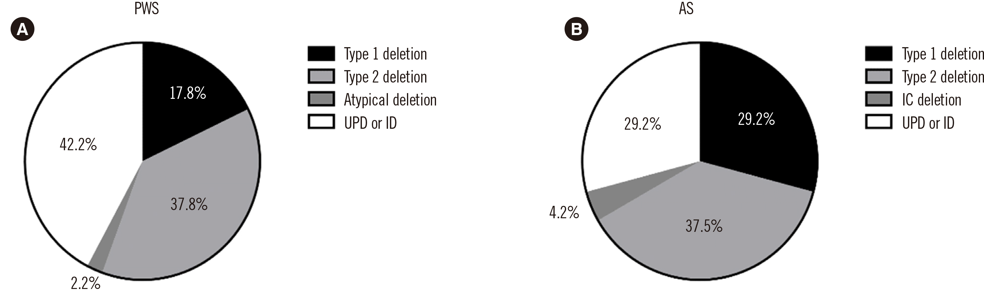
Unlike MS-PCR, MS-MLPA could discriminate between the deletion and non-deletion types (Figs. 1 and 2). Among the 45 PWS patients, 26 (57.8%) had deletions on the q arm of chromosome 15: eight had a type I deletion, 17 had a type II deletion, and one had an atypical deletion (Fig. 3A). The atypical deletion ranged from SNRPN to GABRB3, which is shorter than typical types. Nineteen PWS patients (42.2%) had UPD/ID. Among the 24 AS patients, 16 (66.7%) had deletions, seven (29.2%) had UPD/ID, and one (4.2%) showed microdeletion of the AS-shortest region of deletion overlap, which corresponds to an imprinting center (IC) deletion (Fig. 2C). Of the 16 patients with deletions, seven had type I deletions, and nine had type II deletions (Fig. 3B).
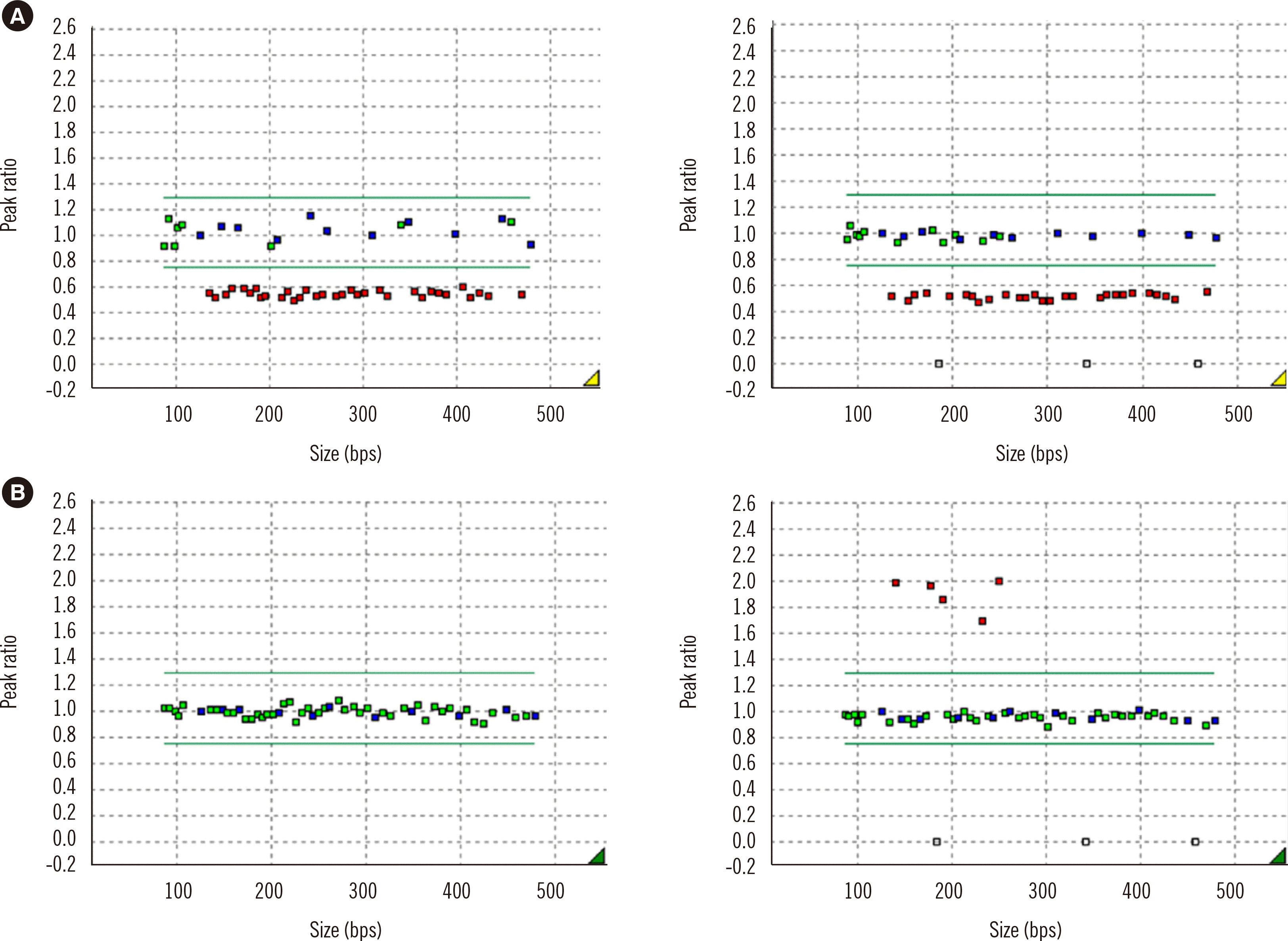
Clinical characteristics and molecular mechanisms of PWS and AS

The male:female ratio of PWS was 1.2:1, and that of AS was 0.6:1 (Table 3). The median age of PWS patients at diagnosis was four months (1–187 months), and that of AS patients was 24.5 months (9–95 months). In AS, the age at diagnosis differed according to the molecular mechanism: patients with the deletion type were diagnosed earlier than those with UPD/ID types (23.8 months vs. 55.7 months, P=0.002). Birth weight also significantly differed according to the molecular mechanism: patients with the deletion type weighed more at birth than those with the UPD type in PWS and vice versa in AS (2.7 kg vs. 2.4 kg, P=0.039 and 2.9 kg vs. 3.5 kg, P=0.035).
PWS patients mainly showed neonatal hypotonia, developmental delay, altered mentality, failure to thrive, waddling gait, or obesity. Most AS patients (91.7%) visited our hospital for developmental delay, except two patients who had seizures and torticollis as the main problem. In PWS cases, the mean Holm score was 4.46, which is below the diagnostic criteria (Holm score 6). We observed no notable difference in the Holm scores of PWS according to the molecular mechanism or deletion range.

DISCUSSION

Chromosomal microarray (CMA) is currently considered the first-tier diagnostic genetic test for neurodevelopmental disorders [14, 15]. However, there remains a necessity for methylation analysis, especially for imprinting disorders, because CMA is not sufficient for diagnosis of these disorders [16]. We showed that MS-MLPA can not only diagnose PWS and AS, but also reveal the underlying molecular mechanisms. We also demonstrated the relationship between molecular mechanisms and clinical characteristics of Korean PWS and AS patients. Our findings support that MS-MLPA is a useful diagnostic test for PWS and AS.
We detected the deletion type in 57.8% of PWS cases, which is in contrast to results in a previous Korean study in 2004, in which deletion accounted for 80% of PWS cases [17]. In line with our results, recent studies have demonstrated that the deletion type in PWS constitutes approximately 60% of total cases [18, 19]. Butler, et al. [18] reported that in PWS, the maternal age was higher in UPD cases than in deletion cases. Although we were not able to assess maternal age for all patients, the average maternal age in Korea is increasing yearly, from 30.0 years in 2004 to 32.4 years in 2016, according to the Korea Statistics [20]. Thus, the increase in the non-deletion type may be because UPD is more prone to occur as maternal age increases.
PWS patients with type I deletions show a more severe phenotype than those with type II deletions [21]; however, there were no significant differences in the diagnostic scores of our patients according to the deletion range. The mean Holm score of total PWS was even lower than the diagnostic score. This may be explained by poor clinical evaluation or diagnosis before clinical symptoms present due to advanced molecular diagnosis.
An atypical deletion, ranging from SNRPN to GABRB3, was detected in one PWS patient (case 39). Additional CMA results confirmed that the patient had a 2.8 Mb deletion from PWRN2 to GABRB3. He visited our hospital for lymph node enlargement at 12 years of age. At that time, he had mild learning disabilities and obesity [22, 23]. His Holm score was 5, which is higher than the mean score of PWS patients. This deletion type showed that MKRN3, MAGEL2, NDN, and C15orf2 are not indispensable for PWS symptoms [24].
One AS patient with an IC deletion (case 66) visited our hospital for developmental delay at 36 months of age. He showed a facial dysmorphism, developmental delay and generalized seizures, which indicated a diagnosis of AS. Although the probability of recurrence of IC is up to 50%, which is the highest recurrence rate for PWS/AS, his younger sister, who is the second-born child, is healthy [25].
To the best of our knowledge, this study is the largest cohort covering both PWS and AS, with clinical information. A few previous studies dealt with the molecular diagnosis of these syndromes, but most of them focused only on PWS or presented no clinical findings [2, 26].
Our study has some limitations. First, there is an intrinsic limitation to MS-MLPA in that it cannot distinguish UPD from ID. To distinguish these two mechanisms, microsatellite analysis or single nucleotide variant analysis is required [27]. However, we successfully differentiated the IC deletion type, which has a 50% recurrence risk, using MS-MLPA. In addition, we could not evaluate the behavioral or psychological status of the patients due to the lack of such information in the medical records. Lastly, this was a retrospective and single-center study. Thus, a selection bias may exist, and further large-scale prospective studies for PWS and AS are needed.
AS caused by a UBE3A variant, which accounts for 20% of AS cases, cannot be diagnosed using MS-MLPA, and further diagnostic testing is needed. To date, only two patients have been diagnosed as having AS due to UBE3A pathogenic variants in our laboratory. Therefore, we propose the use of MS-MLPA as a first-line diagnostic tool for PWS or AS, in accordance with the EMQN/ACGS guidelines [28]. The next step would be UBE3A variant analysis of samples from patients who are highly suspected of having AS, as well as maternal samples.
In conclusion, MS-PCR and MS-MLPA show perfect diagnostic concordance, and MS-MLPA can substitute for MS-PCR. In addition, MS-MLPA provides more information about the molecular mechanisms underlying the diseases and may be a helpful tool for genetic counseling of families with PWS and AS. Finally, patients who are strongly suspected of having AS and show negative MS-MLPA results should undergo additional testing.

ACKNOWLEDGEMENTS

None.

Notes

AUTHOR CONTRIBUTIONS

Kim B interpreted and statistically analyzed data and wrote the main manuscript; Park Y and Cho SI performed the experiments and filled out the clinical research form. Kim MJ, Chae JH, Kim JY, and Seong MW participated in study design and reviewed the final manuscript; Park SS conceived the study and study design and reviewed and approved the final manuscript. All authors have read and approved the manuscript.

CONFLICTS OF INTEREST

None declared.

RESEARCH FUNDING

None declared.

REFERENCES

1. Õiglane-Shlik E, Talvik T, Zordania R, Põder H, Kahre T, Raukas E, et al. 2006; Prevalence of Angelman syndrome and Prader-Willi syndrome in Estonian children: sister syndromes not equally represented. Am J Med Genet A. 140:1936–43. DOI: 10.1002/ajmg.a.31423. PMID: 16906556.
crossref
2. Botezatu A, Puiu M, Cucu N, Diaconu CC, Badiu C, Arsene C, et al. 2016; Comparative molecular approaches in Prader-Willi syndrome diagnosis. Gene. 575:353–8. DOI: 10.1016/j.gene.2015.08.058. PMID: 26335514.
crossref
3. Ferreira IR, Costa RA, Gomes LHF, Dos Santos Cunha WD, Tyszler LS, Freitas S, et al. 2020; A newborn screening pilot study using methylation-sensitive high resolution melting on dried blood spots to detect Prader-Willi and Angelman syndromes. Sci Rep. 10:13026. DOI: 10.1038/s41598-020-69750-0. PMID: 32747801. PMCID: PMC7400512.
crossref
4. Ehrhart F, Janssen KJM, Coort SL, Evelo CT, Curfs LMG. 2019; Prader-Willi syndrome and Angelman syndrome: visualisation of the molecular pathways for two chromosomal disorders. World J Biol Psychiatry. 20:670–82. DOI: 10.1080/15622975.2018.1439594. PMID: 29425059.
crossref
5. Holm VA, Cassidy SB, Butler MG, Hanchett JM, Greenswag LR, Whitman BY, et al. 1993; Prader-Willi syndrome: consensus diagnostic criteria. Pediatrics. 91:398–402. PMID: 8424017. PMCID: PMC6714046.
crossref
6. Williams CA, Angelman H, Clayton-Smith J, Driscoll DJ, Hendrickson JE, Knoll JH, et al. 1995; Angelman syndrome: consensus for diagnostic criteria. Am J Med Genet. 56:237–8. DOI: 10.1002/ajmg.1320560224. PMID: 7625452.
crossref
7. Nicholls RD, Saitoh S, Horsthemke B. 1998; Imprinting in Prader-Willi and Angelman syndromes. Trends Genet. 14:194–200. DOI: 10.1016/S0168-9525(98)01432-2. PMID: 9613204.
crossref
8. Butler MG, Miller JL, Forster JL. 2019; Prader-Willi syndrome - clinical genetics, diagnosis and treatment approaches: an update. Curr Pediatr Rev. 15:207–44. DOI: 10.2174/1573396315666190716120925. PMID: 31333129. PMCID: PMC7040524.
crossref
9. Buiting K. 2010; Prader-Willi syndrome and Angelman syndrome. Am J Med Genet C Semin Med Genet. 154C:365–76. DOI: 10.1002/ajmg.c.30273. PMID: 20803659.
crossref
10. Ramsden SC, Clayton-Smith J, Birch R, Buiting K. 2010; Practice guidelines for the molecular analysis of Prader-Willi and Angelman syndromes. BMC Med Genet. 11:70. DOI: 10.1186/1471-2350-11-70. PMID: 20459762. PMCID: PMC2877670.
crossref
11. Smith A, Hung D. 2017; The dilemma of diagnostic testing for Prader-Willi syndrome. Transl Pediatr. 6:46–56. DOI: 10.21037/tp.2016.07.04. PMID: 28164030. PMCID: PMC5253259.
crossref
12. Nygren AO, Ameziane N, Duarte HM, Vijzelaar RN, Waisfisz Q, Hess CJ, et al. 2005; Methylation-specific MLPA (MS-MLPA): simultaneous detection of CpG methylation and copy number changes of up to 40 sequences. Nucleic Acids Res. 33:e128. DOI: 10.1093/nar/gni127. PMID: 16106041. PMCID: PMC1187824.
crossref
13. Kubota T, Das S, Christian SL, Baylin SB, Herman JG, Ledbetter DH. 1997; Methylation-specific PCR simplifies imprinting analysis. Nat Genet. 16:16–7. DOI: 10.1038/ng0597-16. PMID: 9140389.
14. Shin S, Yu N, Choi JR, Jeong S, Lee KA. 2015; Routine chromosomal microarray analysis is necessary in Korean patients with unexplained developmental delay/mental retardation/autism spectrum disorder. Ann Lab Med. 35:510–8. DOI: 10.3343/alm.2015.35.5.510. PMID: 26206688. PMCID: PMC4510504.
crossref
15. Jang W, Kim Y, Han E, Park J, Chae H, Kwon A, et al. 2019; Chromosomal microarray analysis as a first-tier clinical diagnostic test in patients with developmental delay/intellectual disability, autism spectrum disorders, and multiple congenital anomalies: a prospective multicenter study in Korea. Ann Lab Med. 39:299–310. DOI: 10.3343/alm.2019.39.3.299. PMID: 30623622. PMCID: PMC6340852.
crossref
16. Liu S, Zhang K, Song F, Yang Y, Lv Y, Gao M, et al. 2017; Uniparental disomy of chromosome 15 in two cases by chromosome microarray: a lesson worth thinking. Cytogenet Genome Res. 152:1–8. DOI: 10.1159/000477520. PMID: 28647735.
crossref
17. Kim HJ, Cho HJ, Jin DK, Kwon EK, Ki CS, Kim JW, et al. 2004; Genetic basis of Prader-Willi syndrome in Korea: less uniparental disomy than has been recognized? Clin Genet. 66:368–72. DOI: 10.1111/j.1399-0004.2004.00323.x. PMID: 15355442.
crossref
18. Butler MG, Hartin SN, Hossain WA, Manzardo AM, Kimonis V, Dykens E, et al. 2019; Molecular genetic classification in Prader-Willi syndrome: a multisite cohort study. J Med Genet. 56:149–53. DOI: 10.1136/jmedgenet-2018-105301. PMID: 29730598. PMCID: PMC7387113.
crossref
19. Singh P, Mahmoud R, Gold JA, Miller JL, Roof E, Tamura R, et al. 2018; Multicentre study of maternal and neonatal outcomes in individuals with Prader-Willi syndrome. J Med Genet. 55:594–8. DOI: 10.1136/jmedgenet-2017-105118. PMID: 29776967. PMCID: PMC6107376.
crossref
20. Kim HE, Song IG, Chung SH, Choi YS, Bae CW. 2019; Trends in birth weight and the incidence of low birth weight and advanced maternal age in Korea between 1993 and 2016. J Korean Med Sci. 34:e34. DOI: 10.3346/jkms.2019.34.e34. PMID: 30686955. PMCID: PMC6345638.
crossref
21. Butler MG, Bittel DC, Kibiryeva N, Talebizadeh Z, Thompson T. 2004; Behavioral differences among subjects with Prader-Willi syndrome and type I or type II deletion and maternal disomy. Pediatrics. 113:565–73. DOI: 10.1542/peds.113.3.565. PMID: 14993551. PMCID: PMC6743499.
crossref
22. Kim SJ, Miller JL, Kuipers PJ, German JR, Beaudet AL, Sahoo T, et al. 2012; Unique and atypical deletions in Prader-Willi syndrome reveal distinct phenotypes. Eur J Hum Genet. 20:283–90. DOI: 10.1038/ejhg.2011.187. PMID: 22045295. PMCID: PMC3283188.
crossref
23. Hassan M, Butler MG. 2016; Prader-Willi syndrome and atypical submicroscopic 15q11-q13 deletions with or without imprinting defects. Eur J Med Genet. 59:584–9. DOI: 10.1016/j.ejmg.2016.09.017. PMID: 27659713. PMCID: PMC6688621.
crossref
24. Kanber D, Giltay J, Wieczorek D, Zogel C, Hochstenbach R, Caliebe A, et al. 2009; A paternal deletion of MKRN3, MAGEL2 and NDN does not result in Prader-Willi syndrome. Eur J Hum Genet. 17:582–90. DOI: 10.1038/ejhg.2008.232.
crossref
25. Hartin SN, Hossain WA, Weisensel N, Butler MG. 2018; Three siblings with Prader-Willi syndrome caused by imprinting center microdeletions and review. Am J Med Genet A. 176:886–95. DOI: 10.1002/ajmg.a.38627. PMID: 29437285. PMCID: PMC6688622.
crossref
26. Lu W, Qi Y, Cui B, Chen XL, Wu BB, Chen C, et al. 2014; Clinical and genetic features of Prader-Willi syndrome in China. Eur J Pediatr. 173:81–6. DOI: 10.1007/s00431-013-2124-2. PMID: 23933672.
crossref
27. Wang W, Law HY, Chong SS. 2009; Detection and discrimination between deletional and non-deletional Prader-Willi and Angelman syndromes by methylation-specific PCR and quantitative melting curve analysis. J Mol Diagn. 11:446–9. DOI: 10.2353/jmoldx.2009.090015. PMID: 19661385. PMCID: PMC2729842.
crossref
28. Beygo J, Buiting K, Ramsden SC, Ellis R, Clayton-Smith J, Kanber D. 2019; Update of the EMQN/ACGS best practice guidelines for molecular analysis of Prader-Willi and Angelman syndromes. Eur J Hum Genet. 27:1326–40. DOI: 10.1038/s41431-019-0435-0. PMID: 31235867. PMCID: PMC6777528.
crossref

Fig. 1
MS-MLPA results of PWS patients. (A) Deletion type of PWS. (B) Non-deletion type of PWS. (Left panels: undigested, right panels: digested).
Abbreviations: MS-MLPA, methylation-specific multiplex ligation-dependent probe amplification; PWS, Prader–Willi syndrome.
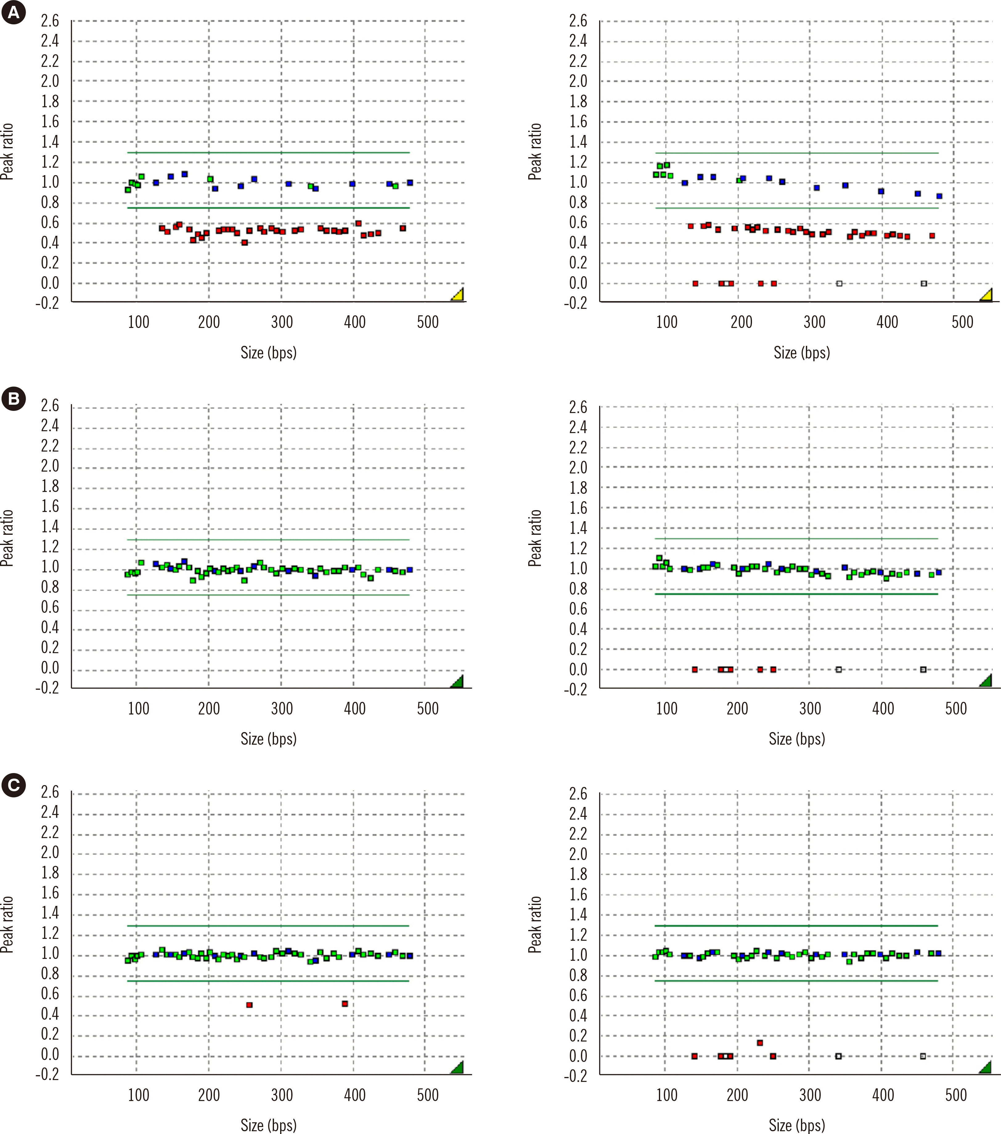
alm-42-1-79-f1.tif
Fig. 2
MS-MLPA results of AS patients. (A) Deletion type of AS. (B) Non-deletion type of AS. (C) IC deletion of AS. (Left panels: undigested, right panels: digested).
Abbreviations: AS, Angelman syndrome; IC, imprinting center; MS-MLPA, methylation-specific multiplex ligation-dependent probe amplification.
alm-42-1-79-f2.tif
Fig. 3
Distribution of molecular mechanisms in (A) PWS and (B) AS patients.
Abbreviations: AS, Angelman syndrome; IC, imprinting center; ID, imprinting defect; PWS, Prader–Willi syndrome; UPD, uniparental disomy.
alm-42-1-79-f3.tif
Table 1
Frequency of PWS Holm diagnostic criteria in 45 PWS patients
Diagnostic criteria Patients, N (%)
Major
1. Neonatal and infantile central hypotonia with poor suck, gradually improving with age 36 (80.0)
2. Feeding problems in infancy with need for special feeding techniques and poor weight gain/failure to thrive 31 (68.9)
3. Excessive or rapid weight gain on weight-for-length chart after 12 months and before the age of six years, central obesity in the absence of intervention 6 (13.3)
4. Characteristic facial features with dolichocephaly in infancy, narrow face or bifrontal diameter, almond-shaped eyes, small-appearing mouth with thin upper lip, downturned corners of the mouth 19 (42.2)
5. Hypogonadism 22 (48.9)
6. Global developmental delay in a child younger than six years, mild to moderate mental retardation or learning problems in older children 17 (37.8)
7. Hyperphagia/food foraging/obsession with food 3 (6.7)
8. Deletion of 15q11-q13 or other appropriate molecular abnormality in this chromosome region, including maternal disomy 45 (100.0)
Minor
1. Decreased fetal movement or infantile lethargy or weak cry in infancy, improving with age 25 (55.6)
2. Characteristic behavior problems, temper tantrums, violent outbursts, and obsessive/compulsive behavior 2 (4.4)
3. Sleep disturbance or sleep apnea 0 (0.0)
4. Short stature for genetic background by 15 years of age 2 (4.4)
5. Hypopigmentation-fair skin and hair compared with other family members 12 (26.7)
6. Small hands ( < 25th percentile) and/or feet ( < 10th percentile) for height age 4 (8.9)
7. Narrow hands with straight ulnar border 0 (0.0)
8. Eye abnormalities 0 (0.0)
9. Thick, viscous saliva with crusting at corners of the mouth 0 (0.0)
10. Speech articulation defects 0 (0.0)
11. Skin picking 0 (0.0)
Total 45

Adopted from “Prader–Willi syndrome: consensus diagnostic criteria,” by Holm VA, et al. 1993 [5].

Abbreviation: PWS, Prader–Willi syndrome.

Table 2
Frequency of AS diagnostic criteria in 24 AS patients
Diagnostic criteria Patients, N (%)
1. Normal prenatal and birth history with normal head circumference and absence of major birth defects 4 (16.7)
2. Developmental delay evident by 6–12 months of age 8 (33.3)
3. Delayed but forward progression of development 0 (0.0)
4. Normal metabolic, hematologic and chemical laboratory profiles 2 (8.3)
5. Structurally normal brain using MRI or CT 6 (25.0)
A. Consistent
1. Functionally severe developmental delay 14 (58.3)
2. Movement or balance disorder, usually ataxia of gait and/or tremulous movement of limbs 6 (25.0)
3. Behavioral uniqueness 3 (12.5)
4. Speech impairment, none or minimal use of words 3 (12.5)
B. Frequent
1. Delayed, disproportionate growth in head circumference, usually resulting in microcephaly by two years of age 3 (12.5)
2. Seizures, onset usually before three years of age 6 (25.0)
3. Abnormal EEG, characteristic pattern with large amplitude slow-spike waves, facilitated by eye closure 3 (12.5)
C. Associated
1. Flat occiput 0 (0.0)
2. Occipital groove 0 (0.0)
3. Protruding tongue 0 (0.0)
4. Tongue thrusting, suck/swallowing disorders 0 (0.0)
5. Feeding problems during infancy 0 (0.0)
6. Prognathia 0 (0.0)
7. Wide mouth, wide-spaced teeth 0 (0.0)
8. Frequent drooling 0 (0.0)
9. Excessive chewing/mouthing behaviors 0 (0.0)
10. Strabismus 1 (4.2)
11. Hypopigmented skin, light hair and eye color 2 (8.3)
12. Hyperactive lower extremity deep tendon reflexes 0 (0.0)
13. Uplifted, flexed arm position especially during ambulation 0 (0.0)
14. Increased sensitivity to heat 0 (0.0)
15. Sleep disturbance 3 (12.5)
16. Attraction to/fascination with water 0 (0.0)
Total 24

Adopted from “Angelman syndrome: consensus for diagnostic criteria,” Williams CA, et al. 1995 [6].

Abbreviations: AS, Angelman syndrome; CT, computed tomography; EEG, electroencephalogram; MRI, magnetic resonance imaging.

Table 3
Clinical features and molecular mechanisms in PWS and AS patients
Case number Sex Age at diagnosis (month) Birth weight (kg) MS-PCR MS-MLPA Clinical features
1 F 13 3.4 PWS Deletion (Type II) Neonatal hypotonia
2 M 2 2.6 PWS Deletion (Type II) Neonatal hypotonia
3 M 2 3.1 PWS Deletion (Type II) Neonatal hypotonia
4 M 40 2.4 PWS Deletion (Type I) Developmental delay
5 F 6 3.0 PWS Deletion (Type II) Developmental delay
6 F 1 2.8 PWS Deletion (Type II) Altered mentality
7 F 1 2.6 PWS Deletion (Type II) Neonatal hypotonia
8 F 25 3.3 PWS UPD/ID Developmental delay
9 M 35 1.9 PWS UPD/ID Developmental delay
10 M 87 2.0 PWS Deletion (Type II) Waddling gait
11 F 4 2.9 PWS Deletion (Type I) Developmental delay
12 M 1 3.1 PWS UPD/ID Neonatal hypotonia
13 M 3 2.2 PWS UPD/ID Neonatal hypotonia
14 M 6 2.7 PWS Deletion (Type II) Neonatal hypotonia
15 M 11 2.8 PWS UPD/ID Neonatal hypotonia
16 M 1 2.5 PWS Deletion (Type II) Poor sucking
17 F 1 2.8 PWS Deletion (Type I) Poor sucking
18 M 123 2.2 PWS UPD/ID Developmental delay
19 M 4 2.8 PWS Deletion (Type II) Neonatal hypotonia
20 F 5 3.2 PWS UPD/ID Neonatal hypotonia
21 F 2 2.6 PWS Deletion (Type I) Developmental delay
22 M 187 2.2 PWS UPD/ID Developmental delay
23 M 1 2.9 PWS Deletion (Type I) Neonatal hypotonia
24 F 5 2.3 PWS UPD/ID Developmental delay
25 M 4 3.1 PWS UPD/ID Fever
26 F 1 2.6 PWS Deletion (Type II) Neonatal hypotonia
27 M 1 2.6 PWS Deletion (Type I) Neonatal hypotonia
28 M 26 2.3 PWS UPD/ID Developmental delay
29 F 114 2.2 PWS UPD/ID Obesity
30 M 144 2.8 PWS Deletion (Type II) Obesity
31 M 50 0.7 PWS UPD/ID Failure to thrive
32 F 4 2.8 PWS Deletion (Type II) Developmental delay
33 M 2 2.4 PWS Deletion (Type II) Neonatal hypotonia
34 F 5 1.5 PWS UPD/ID Developmental delay
35 M 2 3.0 PWS UPD/ID Neonatal hypotonia
36 M 2 2.2 PWS Deletion (Type II) Neonatal hypotonia
37 F 1 2.7 PWS Deletion (Type I) Neonatal hypotonia
38 F 10 3.2 PWS Deletion (Type II) Developmental delay
39 M 150 3.6 PWS Deletion (Atypical) Lymphadenopathy
40 F 3 1.9 PWS UPD/ID Neonatal hypotonia
41 M 1 2.4 PWS Deletion (Type I) Neonatal hypotonia
42 F 5 2.6 PWS UPD/ID Neonatal hypotonia
43 F 155 2.3 PWS UPD/ID Headache
44 F 7 2.6 PWS UPD/ID Developmental delay
45 M 2 2.8 PWS Deletion (Type II) Neonatal hypotonia
46 M 21 3.4 AS Deletion (Type II) Developmental delay
47 M 79 3.0 AS Deletion (Type I) Developmental delay
48 F 36 N/A AS UPD/ID Developmental delay
49 F 56 2.5 AS Deletion (Type I) Developmental delay
50 M 14 2.9 AS Deletion (Type I) Developmental delay
51 F 34 N/A AS UPD/ID Developmental delay
52 M 63 2.7 AS UPD/ID Seizures
53 F 25 2.4 AS Deletion (Type I) Developmental delay
54 F 12 3.0 AS Deletion (Type I) Developmental delay
55 F 16 N/A AS Deletion (Type I) Developmental delay
56 F 11 2.6 AS Deletion (Type II) Developmental delay
57 F 17 2.6 AS Deletion (Type II) Developmental delay
58 F 9 2.5 AS Deletion (Type II) Torticollis
59 F 17 2.6 AS Deletion (Type II) Developmental delay
60 F 13 4.0 AS Deletion (Type II) Developmental delay
61 F 29 2.3 AS Deletion (Type II) Developmental delay
62 F 19 3.2 AS Deletion (Type I) Developmental delay
63 F 19 3.1 AS Deletion (Type II) Developmental delay
64 M 95 3.7 AS UPD/ID Developmental delay
65 F 31 3.3 AS UPD/ID Developmental delay
66 M 38 3.0 AS IC deletion Developmental delay
67 M 76 4.6 AS UPD/ID Developmental delay
68 M 55 3.2 AS UPD/ID Developmental delay
69 M 24 N/A AS Deletion (Type II) Developmental delay

Abbreviations: AS, Angelman syndrome; F, female; IC, imprinting center; ID, imprinting defect; M, male; MS-MLPA, methylation-specific multiplex ligation-dependent probe amplification; MS-PCR, methylation-specific PCR; N/A, not available; PWS, Prader–Willi syndrome; UPD, uniparental disomy.

TOOLS
Similar articles