

 PDF
PDF Citation
 Print
Print


INTRODUCTION
Antinuclear antibody (ANA) is a useful biomarker for the diagnosis of ANA-associated rheumatic diseases (AARDs), such as systemic lupus erythematosus (SLE), systemic sclerosis (SSc), mixed connective tissue disease (MCTD), primary Sjögren’s syndrome (SjS), and polymyositis/dermatomyositis (PM/DM) [1-4]. The most recent European League Against Rheumatism/American College of Rheumatology classification criteria require at least one positive ANA assay result to diagnose SLE [5]. ANA screening is less useful for the diagnosis of other autoimmune rheumatic diseases, such as rheumatoid arthritis [1-3]. ANA can be detected in various other diseases, including liver diseases, thyroid diseases, infectious diseases, and malignancies, and even in apparently healthy individuals [2, 3, 6].
The indirect immunofluorescence assay (IIFA), which was introduced in 1950, is still the gold-standard method for ANA screening [7]. The IIFA uses human epidermoid laryngeal carcinoma cells (HEp-2 or HEp-2000 cells), which serve as substrates presenting more than 100 autoantibodies [3, 4]. The overall sensitivity of the IIFA varies depending on the AARD: it is high for SLE and SSc, but relatively low for SjS and PM/DM [1]. The IIFA exhibits high false positivity in healthy individuals and patients with non-rheumatic diseases [4, 7]. Further, it is labor-intensive, and the determination of results is subjective, making standardization difficult [3]. In a recent survey, the American Association of Medical Laboratory Immunologists investigated several IIFA patterns that laboratory professionals found difficult to read [8]. With the development of novel technologies, automated ANA screening has become possible, and some of the limitations of IIFA have been addressed [9-11].
Recently, two fully automated immunoassays for ANA screening were introduced: EliA CTD Screen (Thermo Fisher Scientific, Freiburg, Germany) and QUANTA Flash CTD Screen Plus (Inova Diagnostics, San Diego, USA). Previous studies have compared these automated immunoassays with IIFA alone [3, 4, 12-14]. To our knowledge, no studies have evaluated samples with discrepant automated immunoassay and IIFA results by confirming the presence of anti-extractable nuclear antigen (ENA) antibodies. In this study, we evaluated the clinical performance of the EliA and QUANTA Flash for ANA screening compared with the reference method, IIFA, and samples with discrepant results were analyzed.
MATERIALS AND METHODS
Study population
We assayed serum samples obtained at Konkuk University Medical Center (KUMC), Seoul, Korea, between December 2018 and January 2019. The study protocol was approved by the KUMC Institutional Review Board (KUH1200079). Informed consent was not required as the study used residual samples left over after requested assays. In total, 406 samples were collected. Routine samples (N=206; 83 females and 123 males; median age [range], 51 years [17–79 years]) were obtained from patients who visited KUMC for a routine medical check-up. Rheumatology clinic samples (N=200; 168 females and 32 males; 48 years [17–82 years]) were obtained from patients for whom an ANA assay was requested. Serum samples were prepared from whole blood samples and were stored at –70°C until use. The data were analyzed anonymously. The study population is presented in Table 1.
Assays
All the samples were assayed using EliA and QUANTA Flash. EliA is a fluorescence enzyme immunoassay (FEIA) run on the Phadia 250 system (Thermo Fisher Scientific). EliA detects autoantibodies against 14 antigens: centromere (CENP-B), double-stranded (ds)DNA, Jo-1, Mi-2, proliferating cell nuclear antigen (PCNA), polymyositis (PM)-Scl, ribosomal-P, RNA Pol III, Scl-70, Sm, Sjögren’s syndrome antigen A (SS-A)/Ro (Ro52 and Ro60), SS-B/La, U1-RNP (RNP-70, A, and C), and fibrillarin. The results of EliA were interpreted as positive, equivocal, or negative according to the cut-off values specified by the manufacturer (>1.0 ratio, positive; 0.7–1.0 ratio, equivocal; <0.7 ratio, negative) [15]. QUANTA Flash is a chemiluminescence immunoassay (CIA) that is run on the BIO-FLASH system (Inova Diagnostics). QUANTA Flash detects autoantibodies against 15 antigens: centromere (CENP-A and -B), dsDNA, Jo-1, Mi-2, PCNA, PM-Scl, ribosomal-P, RNA Pol III, Scl-70, Sm, SS-A/Ro (Ro52 and Ro60), SS-B/La, U1-RNP (RNP-70, A, and C), Ku, and Th/To. QUANTA Flash results were interpreted as positive or negative according to the cut-off values specified by the manufacturer (≥20.0 chemiluminescent units [CU], positive; <20.0 CU, negative) [12]. The units of EliA and QUANTA Flash cannot be compared because these assays have different principles and unit conversion calculations [12, 15].
IIFA was performed for the rheumatology clinic samples using the Kallestad HEp-2 Cell Line Substrate (Bio-Rad Laboratories, Hercules, CA, USA) on a PhD Ix system (Bio-Rad Laboratories). A trained and experienced immunology expert interpreted the IIFA results using an immunofluorescence microscope at 200×. IIFA results were interpreted by dividing the grades of fluorescence intensity at serum dilutions of 1:40 and additional dilutions according to the manufacturer’s instructions. We interpreted ≥1:80 as positive and <1:80 as negative [5].
For confirmation, an anti-ENA antibody assay was performed using EUROLINE ANA Profile 3 (EUROIMMUN, Lübeck, Germany) for samples that were positive in any of the three ANA screening assays. EUROLINE is a line immunoassay (LIA) that is run on the EUROBlotMaster system (EUROIMMUN). EUROLINE detects autoantibodies against 14 antigens: AMA M2, CENP-B, dsDNA, histones, Jo-1, nRNP, nucleosomes, PCNA, PM-Scl, ribosomal-P, Scl-70, Sm, SS-A/Ro (Ro52 and Ro60), and SS-B/La. EUROLINE results were interpreted according to the signal intensity specified by the manufacturer(>50, strong positive; 11–50, positive; 6–10, borderline; 0–5, negative) [16]. All assays were performed according to the manufacturers’ instructions.
Statistical analysis
The positivity of EliA and QUANTA Flash in the routine and rheumatology clinic samples was compared using McNemar’s Chi-squared test for paired proportions. The concordance and agreement among ANA screening assays were evaluated in total samples as well as in routine and rheumatology clinic samples separately. The concordance was calculated by dividing the total number of samples with the same results in multiple assays by the total number of samples [17]. The agreement was calculated using Cohen’s kappa (κ) with 95% confidence intervals (CIs), which was interpreted as follows: ≤0.20, none; 0.21–0.39, minimal; 0.40–0.59, weak; 0.60–0.79, moderate; 0.80–0.90, strong; and >0.90, nearly perfect [18].
The clinical performance of the three ANA screening assays for diagnosing AARDs, which was evaluated based on the clinical diagnosis determined by medical chart review, was compared using ROC curves from the rheumatology clinic samples. Samples from patients diagnosed as having AARDs, including SLE, SSc, MCTD, SjS, and PM/DM, were classified as the AARD group, and samples with any other diagnosis were classified as the non-AARD group. Additionally, the clinical performance of EliA and QUANTA Flash in diagnosing AARDs was evaluated in all samples. The areas under the ROC curve (AUCs) with 95% CIs were calculated and interpreted as follows: AUC=0.5, non-informative; AUC=0.5–0.7, less accurate; AUC=0.7–0.9, moderately accurate; AUC=0.9–1.0, highly accurate; and AUC=1.0, perfect [19]. The sensitivity and specificity of manufacturers’ cut-off values were evaluated, and adjusted cut-off values for EliA and QUANTA Flash were calculated using the Youden index to optimize combined sensitivity and specificity. P<0.05 was considered statistically significant. Statistical analyses were performed using MedCalc Statistical Software version 19.5.1 (MedCalc Software, Ostend, Belgium).
RESULTS
The positivity of EliA and QUANTA Flash was 4.9% and 8.3%, respectively, in the routine samples and 48.5% and 52.0%, respectively, in the rheumatology clinic samples. The positivity of IIFA was 59.5% in the rheumatology clinic samples. In the rheumatology clinic samples, the positivity of EliA, QUANTA Flash, and IIFA was higher in the AARD group than in the non-AARD group (Table 1). QUANTA Flash showed slightly, albeit not significantly, higher positivity than EliA in both the routine and rheumatology clinic samples (P=0.165, routine samples; P=0.485, rheumatology clinic samples). The concordance and agreement between EliA and QUANTA Flash were 92.6% and strong (κ=0.82; 95% CI, 0.76–0.88), respectively, in total samples, and 93.7% and weak (κ=0.49; 95% CI, 0.25–0.72), respectively, in the routine samples. In the rheumatology samples, the concordance and agreement were 91.5% and strong (κ=0.83; 95% CI, 0.75–0.91) between EliA and QUANTA Flash, 79.0% and weak (κ=0.58; 95% CI, 0.47–0.69) between EliA and IIFA, and 80.5% and moderate (κ=0.61; 95% CI, 0.50–0.72) between QUANTA Flash and IIFA, respectively.
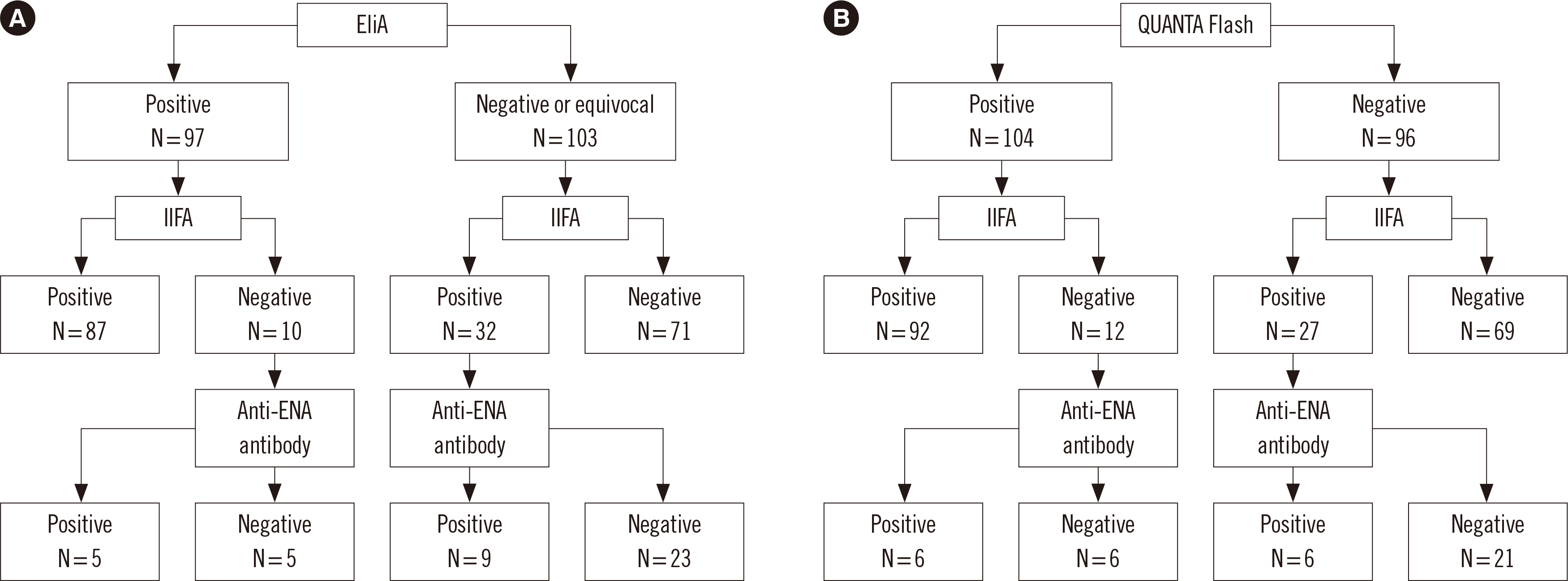
One hundred fifty-four samples tested positive on at least one ANA screening assay. There were 42 samples with discrepant EliA and IIFA results, and 39 samples with discrepant QUANTA Flash and IIFA results (Fig. 1). The positivity of the anti-ENA antibody assay was 50.0% in automated immunoassay-positive, IIFA-negative samples (5/10 EliA-positive, IIFA-negative samples; 6/12 QUANTA Flash-positive, IIFA-negative samples). The positivity of the anti-ENA antibody assay was 28.1% (9/32) in EliA-negative, IIFA-positive samples (Fig. 1A), and 22.2% (6/27) in QUANTA Flash-negative, IIFA-positive samples (Fig. 1B). The results of IIFA and anti-ENA antibody assay for samples with discrepant results are presented in Table 2. The anti-ENA antibodies detected in the EliA- or QUANTA Flash-positive, IIFA-negative samples were all anti-SS-A/Ro (Ro60) antibodies (N=6). The anti-ENA antibodies detected in EliA-negative, IIFA-positive samples were antibodies against histones, Jo-1, nRNP, nucleosomes, Scl-70, and SS-A/Ro (Ro60), whereas those detected in QUANTA Flash-negative, IIFA-positive samples were antibodies against dsDNA, histones, Jo-1, nRNP, nucleosomes, ribosomal-P, Scl-70, and Sm. Except for one patient (detection of anti-Jo-1 antibody) who was diagnosed as having SSc, all patients with EliA- or QUANTA Flash-negative, IIFA-positive results were diagnosed as having SLE.
EliA and QUANTA Flash showed a high accuracy (AUC=0.917 and 0.911, respectively), and IIFA showed a moderate accuracy (AUC=0.862) in diagnosing AARDs using rheumatology clinic samples (Table 3). The difference in AUCs between EliA and IIFA was statistically significant (P=0.033); however, the differences in AUCs between EliA and QUANTA Flash and between QUANTA Flash and IIFA were not significant (P=0.714 and P=
0.058, respectively). Using manufacturers’ cut-off values, the sensitivities of EliA, QUANTA Flash, and IIFA were 72.4%, 77.2%, and 84.6%, respectively. Using adjusted cut-off values for EliA and QUANTA Flash, their sensitivities were improved (82.9% and 87.8%, respectively). EliA and QUANTA Flash showed similar sensitivity and specificity in total samples (Table 3). The difference in AUCs between EliA and QUANTA Flash was not significant (P=0.308).
DISCUSSION
We evaluated the clinical performance of EliA and QUANTA Flash and analyzed samples with discrepant automated immunoassay and IIFA results for the first time. The present study provided baseline data for the clinical application of EliA, QUANTA Flash, and IIFA in patients undergoing routine checkups and in rheumatology clinic patients. The concordance and agreement between QUANTA Flash and IIFA were similar to those between EliA and IIFA. In a previous study, opposite results were obtained [3]. Therefore, our results alone do not suggest that QUANTA Flash is more suitable than EliA as a replacement for IIFA.
Automated immunoassays reportedly are less sensitive than IIFA for detecting ANA [20]. In this study, however, the anti-ENA antibody assay positivity was higher in automated immunoassay-positive, IIFA-negative samples than in automated immunoassay-negative, IIFA-positive samples. Moreover, the anti-ENA antibodies detected by automated immunoassays and IIFA differed. All automated immunoassay-positive, IIFA-negative samples were anti-SS-A/Ro (Ro60) antibody-positive. This indicates that automated immunoassays can detect the anti-SS-A/Ro antibody, which may be missed by the IIFA. These results are in line with those of previous studies in which solid phase assays, such as FEIA and LIA, detected the anti-SS-A/Ro antibody, which was not detected by IIFA [21, 22]. According to international recommendations for ANA screening, patients should be assayed for specific anti-ENA antibodies, such as SS-A/Ro, even if the IIFA result is negative, in cases of high clinical suspicion [7]. EliA and QUANTA Flash may be suitable for ANA screening of patients with clinically suspected anti-SS-A/Ro antibody.
The anti-ENA antibodies detected in automated immunoassay-negative, IIFA-positive samples were variable. These antibodies have been reported to be associated with SLE [23-25]. In the present study, all patients with automated immunoassay-negative, IIFA-positive results were diagnosed as having SLE, except for one patient. The ANA titer reported in IIFA is not consistently associated with disease severity; but higher ANA titers are more clinically significant, as healthy individuals generally have low ANA titers [26]. For these reasons, automated immunoassays should be used in combination with the IIFA in patients who are clinically suspected of having AARDs, especially, SLE.
Both EliA and QUANTA Flash were highly accurate in diagnosing AARDs. Although the AUCs of EliA and QUANTA Flash were as high as that of IIFA in this study, the manufacturers’ cut-off values showed lower sensitivity than the adjusted cut-off values. The cut-off values can be adjusted to optimize combined sensitivity and specificity according to the assay purpose. Enzyme immunoassay pre-screening followed by IIFA reportedly reduces the overall time compared with screening by IIFA alone [27]. For ANA screening, cut-off values with high sensitivity, adjusted to nearly half of the manufacturers’ cut-off values in this study, can be used. This study showed a tendency toward higher specificity for EliA and higher sensitivity for QUANTA Flash; however, as in previous studies, the differences between EliA and QUANTA Flash were not statistically significant [3, 4].
This study had several limitations. First, although we included the rheumatology clinic samples, the number of abnormal samples from patients diagnosed as having AARDs was relatively small; further studies with large numbers of abnormal samples are needed. Second, there were differences in antigen composition between EliA, QUANTA Flash, and the anti-ENA antibody assay. We did not perform further confirmatory assays for specific antibodies that were missed by EUROLINE. Third, we did not perform separate ROC curve analyses for individual diseases, while previous studies have evaluated the clinical performance for not only AARDs, but also individual diseases [3, 4]. Disease-specific performance of the two automated immunoassays would be required to identify clinically suspected patients. Last, in patients with SLE, ANA can be detected several years before clinical symptoms appear [28]. As ANA screening using EliA and QUANTA Flash was performed without patient identification, it was not possible to follow up patients with ANA screening among those undergoing routine checkups.
In conclusion, EliA and QUANTA Flash showed reliable performance compared with the IIFA, and adjusting the cut-off values improved their sensitivities. These automated immunoassays may be used in combination with the IIFA in clinical immunology laboratories with a high ANA screening workload. The clinical cut-off values can be adjusted according to the workflow in each laboratory.
 XML Download