

 PDF
PDF Citation
 Print
Print


INTRODUCTION
Intrauterine contraceptive devices (IUDs) are widely used for contraception in South Korea. However, several complications of IUDs have been reported, including inflammation, obstruction, perforation, and fistula.1-3 IUD perforation is the rarest of these complications but is also severe. IUD perforation has an incidence rate between 0.05/1,000 and 13/1,000.4-6 Migrated IUDs can be retrieved through endoscopy, laparoscopy, or laparotomy.7 We report an atypical case of incomplete removal of an IUD perforating the sigmoid colon through laparoscopic surgery.
Go to : 
CASE REPORT
A 54-year-old woman (gravida 3, para 3) was admitted to the emergency department after a colonoscopy conducted during a medical check-up at a local clinic identified a foreign body in the sigmoid colon. The colonoscopy showed a yellow tube penetrating the sigmoid colon with edematous mucosal change and pus (Fig. 1A). The woman had received the T-shaped Mirena IUD (levonorgestrel-releasing intrauterine system, 52 mg) after her second vaginal delivery 22 years ago. However, she became pregnant one month after IUD insertion. Two months after the third delivery, transvaginal sonography did not indicate the presence of the IUD in the uterine cavity. The woman had suffered lower abdominal pain on two occasions that year but had not visited the hospital. She had not experienced gastrointestinal bleeding, including hematochezia and melena. She also denied experiencing other symptoms including vaginal bleeding, constipation, diarrhea, changes in bowel habits, and fever.
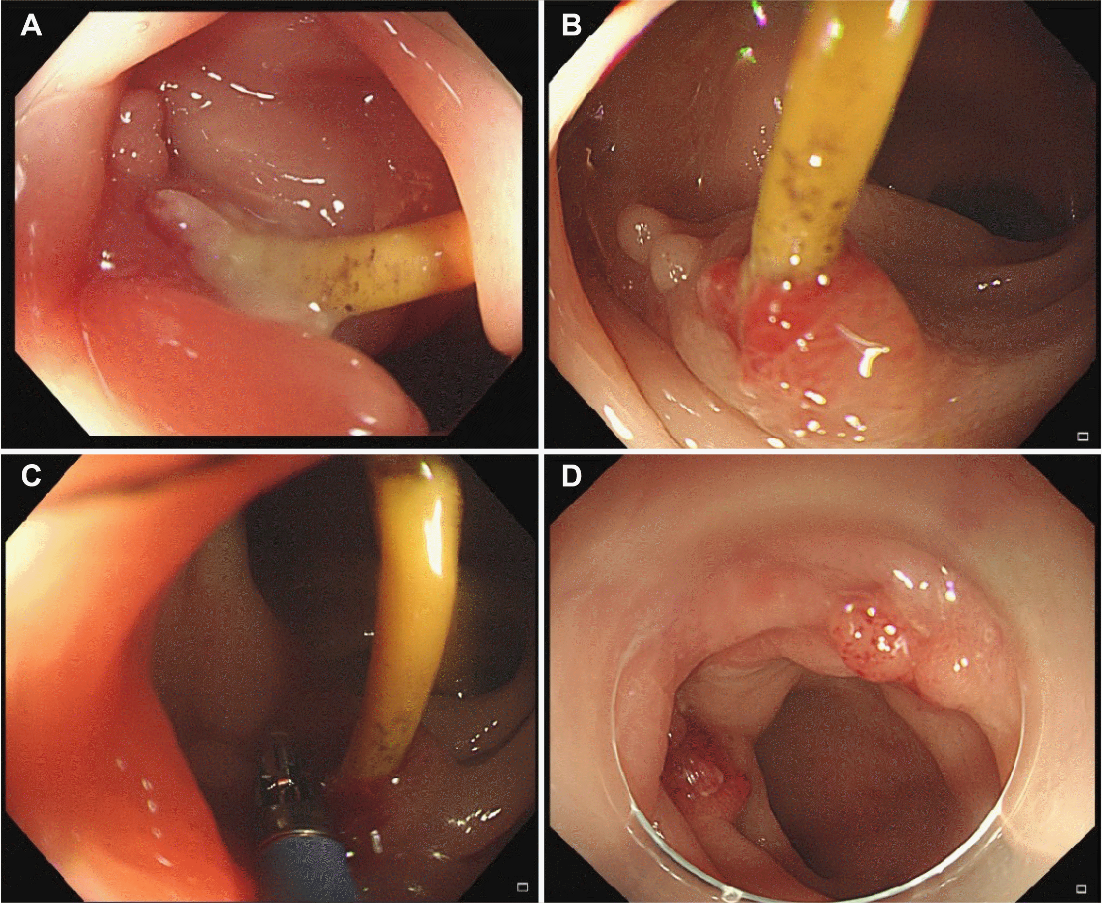 | Fig. 1Colonoscopy findings: (A) a yellow tube penetrating sigmoid colon with edematous mucosal change and pus, (B) Intrauterine device (IUD) had penetrated the lumen of the distal sigmoid colon and was surrounded by granulation tissue, (C) attempts to retrieve the IUD using biopsy forceps failed because of severe adhesions, (D) the missing part of the IUD was absent from the colon lumen.
|
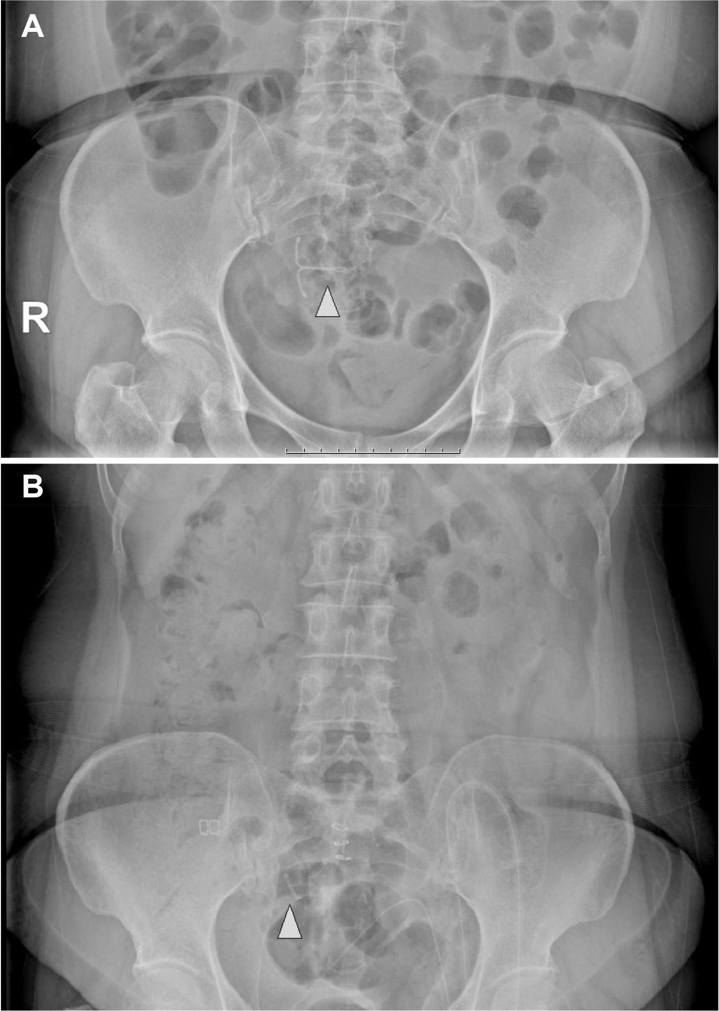
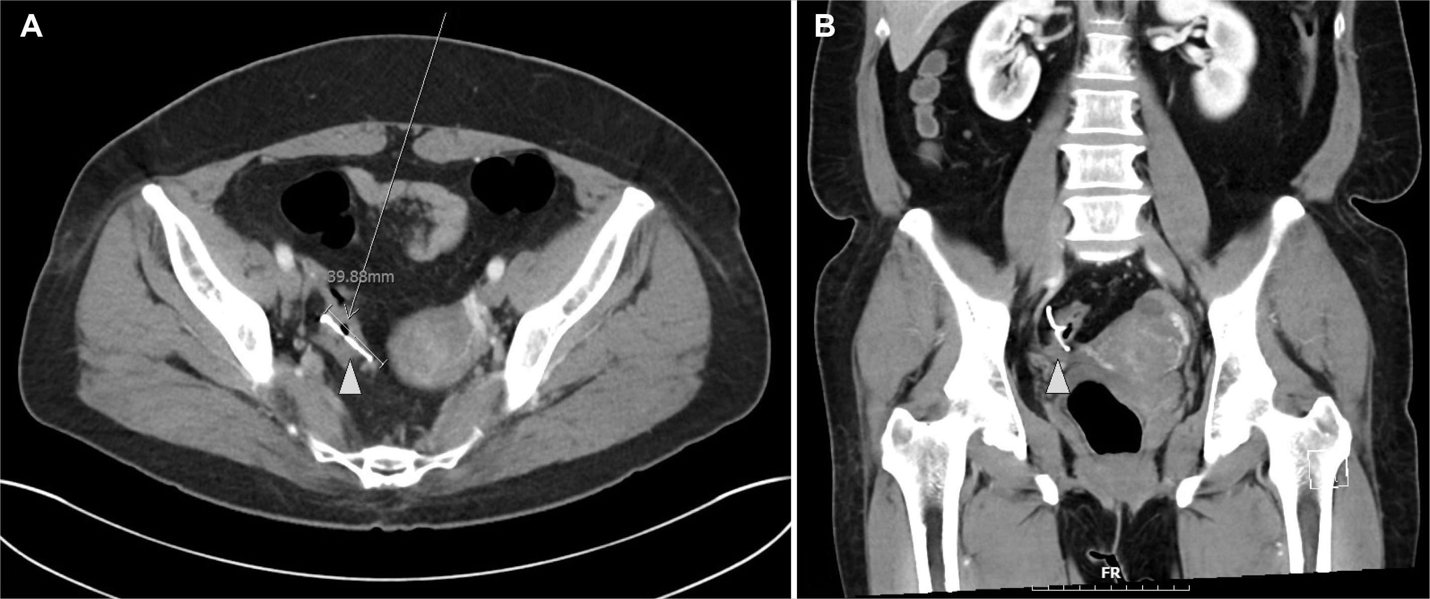
The woman was hemodynamically stable at presentation. The initial laboratory findings were within the normal range (white blood cell count 4.70×10⁹/L, segmented neutrophils 57.9%, hemoglobin 13.8 g/dL, CRP 0.09 mg/dL, erythrocyte sedimentation rate 5 mm/hour, PT/INR 1.10, aPTT test 30.2 sec). The initial abdominal X-ray revealed an IUD in the pelvis (Fig. 2A). A CT scan of the abdomen was performed and revealed a 4-cm sized IUD penetrating the wall of the sigmoid colon (Fig. 3). A colonoscopy showed that the IUD had penetrated the lumen of the distal sigmoid colon and was surrounded by granulation tissue. However, attempts to retrieve the IUD through endoscopy using biopsy forceps failed because of severe adhesions between the IUD and colon wall (Fig. 1B, C).
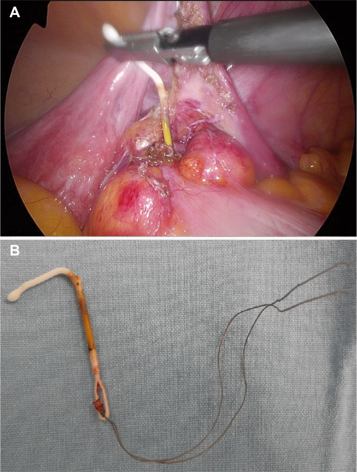
After endoscopic retrieval failed, laparoscopic foreign body removal was performed. An 11-mm laparoscopic port was inserted into the umbilicus and three 5-mm ports were inserted into the right lower quadrant, right upper quadrant, and left lower quadrant under general anesthesia. At first, the migrated IUD was not visible because of the right adnexa adhesion. After the adhered right adnexa had been dissected, the migrated IUD was found on the antimesenteric side of the distal sigmoid colon (25 cm from the anal verge). The IUD was retrieved by the grasper and the perforated colon wall was repaired with V-lock #3-0, after which the four laparoscopic ports were removed and the port insertion sites repaired (Fig. 4A). We had expected to recover a T-shaped IUD, but the IUD was imperfectly removed. The 1-cm right arm of the IUD was suspected to have been left in the lumen of the colon (Fig. 4B).
Post-operative follow-up colonoscopy was performed to check for remnants of the IUD and the missing part of the IUD was not found in the colon lumen (Fig. 1D). However, a post-operative follow-up abdominal X-ray revealed that part of the IUD had remained in the right lower abdomen (Fig. 2B). Due to the small size of the IUD remnant, we believed it may not pose a serious risk for complications and could remain in the bowel wall. Furthermore, the patient was also unwilling to undergo a laparotomy for complete removal of the remnant. The patient was symptom-free at discharge. The patient has not reported any complications or abnormalities in the post-surgical follow-up over the last 4 months since the surgery.
Go to :
DISCUSSION
IUDs have been widely used as a simple, effective form of contraception. However, their use is associated with several complications, including vaginal bleeding, abdominal pain, ectopic pregnancy, infection, and perforation of adjacent organs.1,8 IUD perforation stands out among the complications due to its severity and varied clinical features, including asymptomatic, chronic abdominal pain, lower urinary tract symptoms, unexpected pregnancy, changes in the bowel habits, and penetration of adjacent organs.2
The mechanism and timing of the sigmoid colon penetration are still unclear. However, many IUD perforations occur immediately after insertion.9 Therefore, it is recommended that patients are assessed for symptoms and signs of IUD perforation 6 weeks after insertion.10,11 Abdominal X-rays and transvaginal ultrasonography are recommended initially when IUD migration is suspected. A CT scan may also be needed to determine the exact site of perforation.4,12 Risk factors for IUD perforation include lack of gynecologist experience with IUD insertion, uterine immobilization, and anatomical defects of the uterus.3
Methods of retrieving migrated IUDs include colonoscopy, laparoscopy, and laparotomy. The choice of the IUD retrieval method depends on the location of the IUD and the degree of involvement with adjacent organs. IUD removal via colonoscopy is recommended, especially in cases where the IUD is located within the wall of the colon. This is the safest and easiest method and does not cause peritonitis. Consequently, retrieval by colonoscopy has gradually increased in popularity. Furthermore, an experienced endoscopist would be able to remove the IUD by incision with an endoscopic needle knife. However, if the IUD is firmly embedded within an adjacent organ, removal by colonoscopy is not advisable.7,13
If IUD removal by colonoscopy is unsuccessful, laparoscopic surgery is recommended as the next step. However, laparoscopic retrieval may fail in cases involving severe adhesions, severe perforations, or non-visible IUDs located in the lumen of the colon or retroperitoneum. In these cases, surgical laparotomy is required.14,15 Surgical laparotomy is also recommended in cases of acute abdomen, peritonitis, abscess formation, and fistula.1 Although surgical laparotomy is more invasive than endoscopic or laparoscopic retrieval, it is the most effective method in the case of severe adhesions.
In short, the endoscopic removal of the IUD is the best option for an IUD placed in the wall of the colon without free perforation. Surgical laparoscopy is the next option for IUD removal when the IUD is firmly embedded within the adjacent organs. Finally, surgical laparotomy can be useful in complicated cases, such as failed laparoscopic attempts due to a non-visible location of the IUD or patient instability.
In the present case, we initially attempted to remove the IUD via colonoscopy but found that the IUD was firmly embedded in an adjacent organ. Thus, the IUD was retrieved incompletely through laparoscopic surgery. The embedded IUD was fragile because it was a deteriorating, 22-year-old device. We then had the option of completely removing the remnants of the IUD through laparotomy. However, the patient refused laparotomy due to the absence of any significant symptoms associated with the remnant, including abdominal pain and discomfort. The surgeons agreed not to perform the laparotomy because the remnant was small in size and could remain embedded in the colon wall without any increased risk of severe complications. The patient wanted to undergo surgery only if and when symptoms occurred. Therefore, the removal of the IUD perforating the sigmoid colon was deferred.
In this case, colonoscopy showed that the IUD penetrated the sigmoid colon and both arms of the IUD were firmly embedded in the colon wall. Therefore, removal of the IUD using a colonoscopy could not succeed. In addition, the IUD was incompletely removed by laparoscopic surgery. Because we ignored the fragility of the firmly embedded 22-year-old IUD, we had to perform the laparotomy or attempt to use the endoscopic needle knife instead of laparoscopic surgery as the appropriate choice.
In conclusion, proper patient selection and access to treatment for IUD perforation are very important. Initially, endoscopy can be attempted as a simple method for confirming IUD perforation and removing the IUD. In some cases, if endoscopy fails, surgical retrieval should be performed as the next step. Laparoscopy can be attempted prior to laparotomy to avoid unnecessary extended surgery. However, there are many complex cases where laparotomy is more appropriate than laparoscopy. This rare case report underlines the importance of proper patient selection and access to treatment for IUD perforation for improving prognosis and therapy.
Go to :
 XML Download