

 PDF
PDF Citation
 Print
Print


INTRODUCTION
Gingival recession (GR) refers to exposure of the surfaces of tooth roots due to apical displacement of the keratinized gingiva in relation to the cementoenamel junction. Bone dehiscence is an anatomical prerequisite for GR development.1-4 Periodontal disease, mechanical trauma, a thin gingival biotype, traumatic occlusion, and aging are possible etiological factors for GR.3,4
Orthodontic movements without buccal or lingual control increase the risk of bone dehiscence, which can cause GR.5-9 Evaluation of the alveolar bone condition before orthodontic treatment and adequate biomechanical control of tooth movement are factors contributing to the prevention of alveolar defects.9-11 In this regard, cone-beam computed tomography is a gold-standard diagnostic modality used to evaluate the periodontal condition as well as osseous and tegumentary tissues, particularly in adult patients.9,12
In humans, orthodontic tooth movement toward the center of the alveolar process with the use of conventional appliances has reportedly resulted in complete or partial regeneration of the buccal bone plate and a consequent reduction in GR.8,13-15 Over recent years, a clear aligner system called the Invisalign® system (Align Technology, San Jose, CA, USA) became an orthodontic treatment option for cases of mild and moderate malocclusions. This system is not only esthetical, comfortable and easy to clean, but also uses biomechanical principles that allows three-dimensional tooth movement.16-19 Moreover, light forces allow a favorable periodontal response associated with better oral health.20 In this report, we demonstrate the effectiveness of the Invisalign® system in the treatment of severe GR and bone dehiscence through torque, translation, and intrusion movements in a young woman.
Go to : 
DIAGNOSIS AND ETIOLOGY
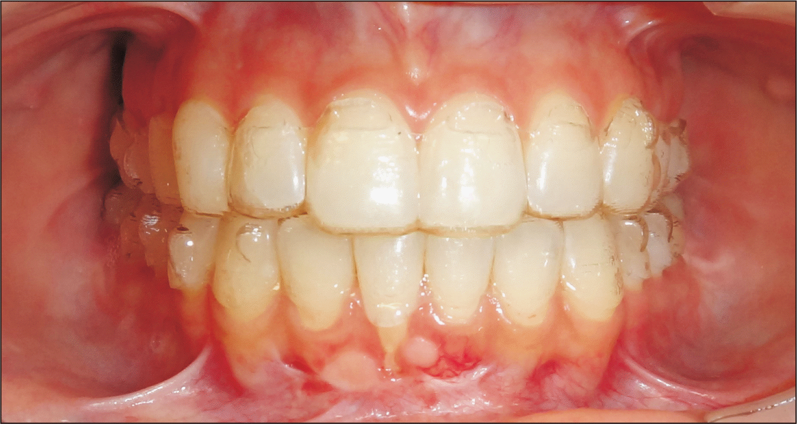
A 25-year-old woman visited an orthodontic clinic for evaluation in May 2016. Her chief complaint was severe GR on the buccal surface of the mandibular right central incisor (#41). During clinical evaluation, she mentioned that the recession developed 4 years after orthodontic treatment with a fixed appliance. At that time, two periodontal surgeries involving gingival grafting were performed for root coverage and esthetic improvement; however, they were unsuccessful. The 4-mm GR was clinically graded as Class II recession according to Miller’s classification.21 It extended beyond the mucogingival junction without bone loss and tegumentary tissue in the interdental area (Figure 1A). The patient had no history of chronic diseases or smoking.
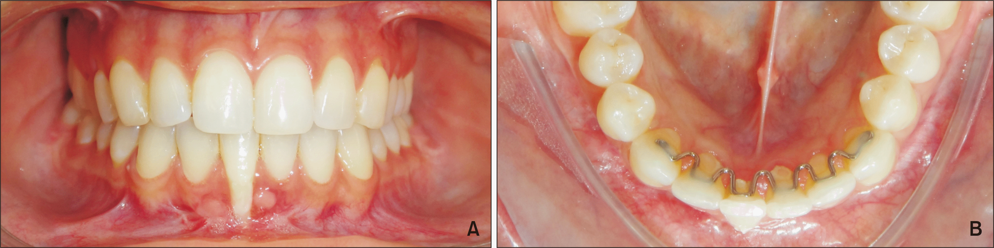
A V-loop lingual retainer22 was bonded in the region of tooth #41, with less resin relative to the amount used for the adjacent teeth (Figure 1B). We suspected that failure during fixed retainer placement and the lack of post-treatment follow-up were the primary etiological factors for the development of bone dehiscence and GR in this case.14,15,23 According to the literature, fixed retainers bonded to the six mandibular anterior teeth are adequate to maintain their alignment, although poor placement and mastication of hard food after the treatment can generate forces that can move the teeth to improper positions and result in periodontal problems, mainly when flexible wires are used.14,23 We observed that the affected mandibular incisor was also in traumatic contact; however, a vitality test showed a positive result. The other mandibular teeth showed no abnormalities. The patient’s gingival biotype was not favorable; her gingiva in both arches was thin with little resistance.
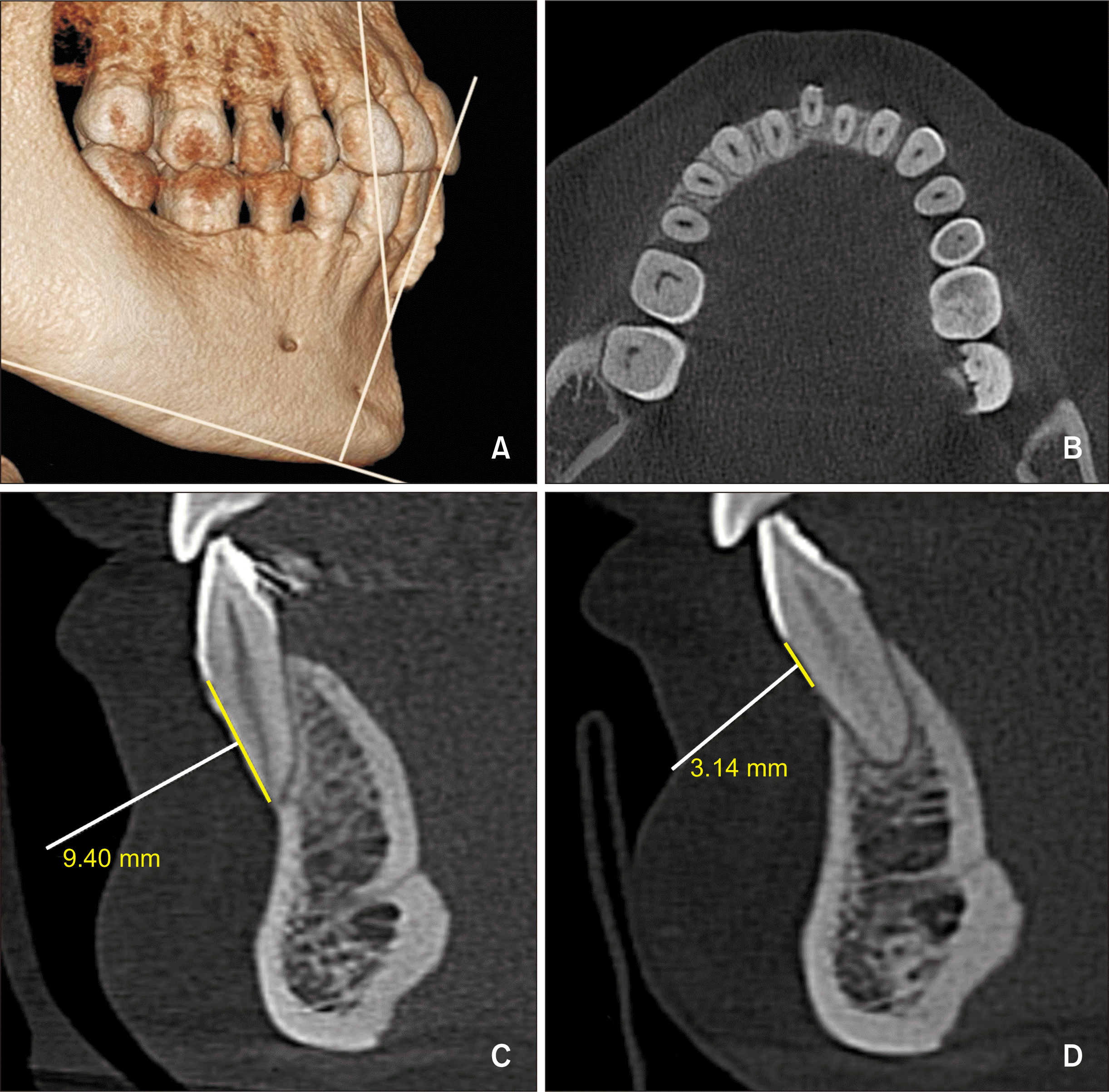
Cone-beam computed tomography was ordered to aid in diagnosis and treatment planning. Cephalometric analysis showed a Class I skeletal-sagittal relationship, a balanced vertical pattern, slight lingual inclination of the maxillary incisors and buccal inclination of the mandibular incisors (#31, #32, and #42) (Figure 2). On the other hand, the root of tooth #41 showed excessive buccal displacement, and it was vertically positioned out of the bone plate with bone dehiscence of 9.40 mm (Figure 3A–3C). The initial IMPA (the angle formed by the long axis of the lower incisor and the mandibular plane) was 90°. Model analysis showed Bolton’s discrepancy in the mandibular arch, with an anterior excess of 1.85 mm. The shape of the arch was normal, with no dental crowding.
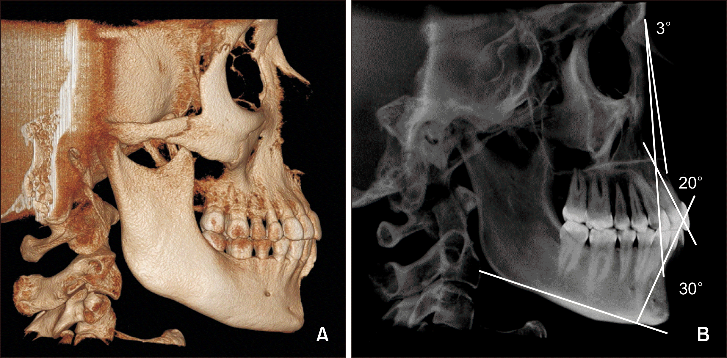 | Figure 2
A, Facial cone-beam computed tomography. B, ANB angle = 3°, 1.NA for tooth #21 = 20°; 1.NB for tooth #31= 30°.
ANB, the angle formed by the lines Nasion to point A and Nasion to point B; 1.NA, the angle formed by the long axis of the upper incisor and the line Nasion to point A; 1.NB, the angle formed by the long axis of the lower incisor and the line Nasion to point B.
|
Go to :
TREATMENT OBJECTIVES
The therapeutic objectives were determined as follows:
To orthodontically move the root of tooth #41 toward the center of the alveolar bone, in association with root lingual torque, by using clear aligners
To increase the thickness of the buccal bone plate and decrease the bone dehiscence
To reduce GR
To eliminate the traumatic contact for tooth #41
Go to :
TREATMENT ALTERNATIVES
As an alternative treatment, we considered extraction of tooth #41, which would be followed by distribution of the remaining teeth in the created space with the help of aligners. The crowns of the mandibular incisors would be resized using compound resin. The treatment plan would be interdisciplinary, involving orthodontics and restorative dentistry so that both esthetic and functional goals could be met after the treatment.
Orthodontic treatment using metallic or esthetic brackets was also offered as an option to the patient.
Go to :
TREATMENT PROGRESS
Treatment with aligners was chosen as the treatment method because the patient had already undergone orthodontic treatment with a fixed appliance. The Vivera® (Align Technology) retainer was selected for treatment. The patient consented to the treatment plan although she was aware that tooth #41 could be lost if the treatment failed. The lower fixed retainer was removed, and the patient was referred for periodontal treatment.
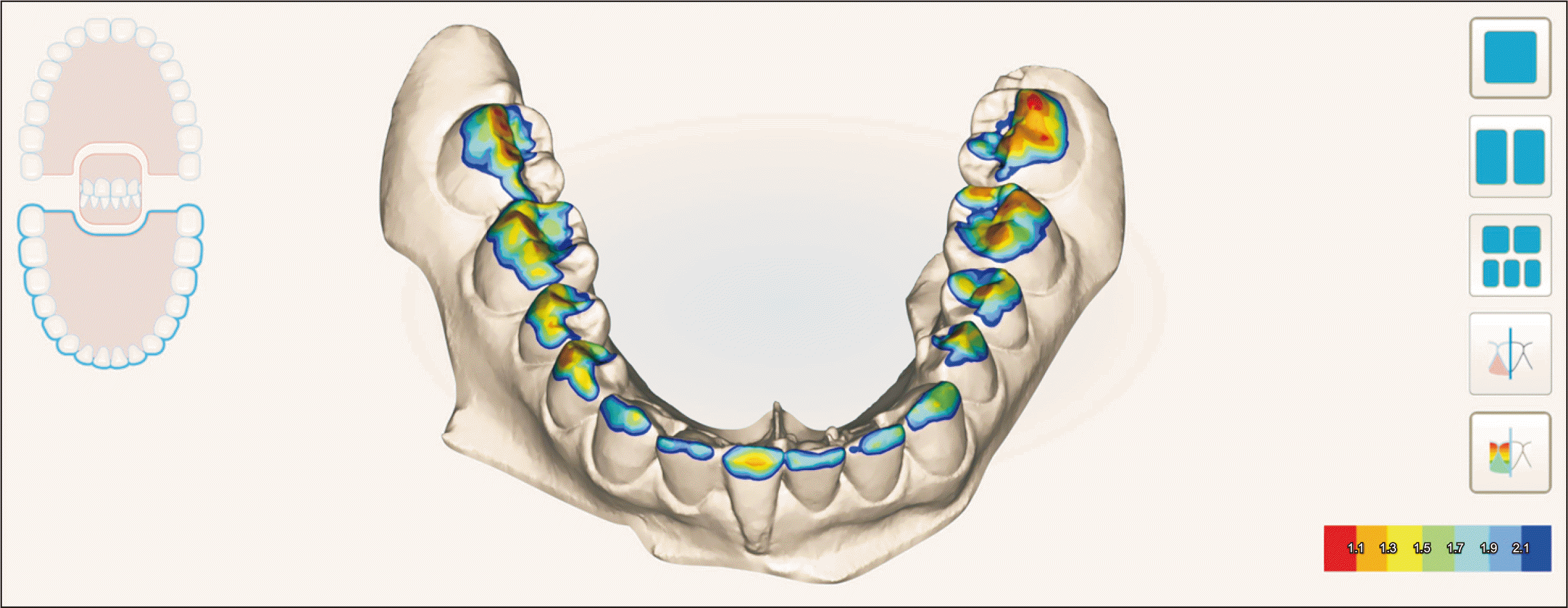
Traumatic contact and grade II mobility of tooth #41 were observed during clinical examination. Analysis of three-dimensional models using the scanner’s occlusogram tool (iTero Element®; Align Technology) also revealed premature contact (Figure 4).
Orthodontic treatment involved the use of 28 active aligners for the mandibular arch and 24 active and four passive aligners for the maxillary arch. Power ridges® (pressure lines close to the gingival margin to increase contact pressure on the tooth surface)19 were used for the application of torque forces on the maxillary incisors and tooth #41 throughout the treatment. Interproximal enamel reduction of 0.4 and 0.2 mm between the canine and first premolar (#43 and #44) and the lateral incisor and canine (#42 and #43), respectively, was planned. The aim was to allow lingual movement of tooth #41. The patient changed the aligners #1 to #19 every 14 days and the other aligners every 7 days. The total treatment time was 10 months, with a reduction in GR observed after 5 months (Figure 5). Periodontal treatment involving scaling and polishing was performed every 3 months during the orthodontic treatment.
Go to :
RESULTS
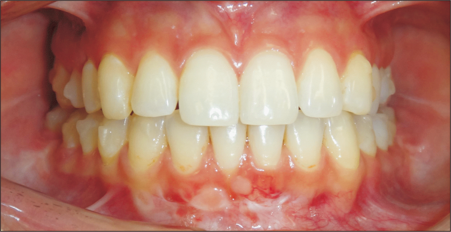
The final result of orthodontic treatment with clear aligners showed a significant decrease in GR of tooth #41 (Figure 6), whose root was lingually inclined by 11.7°, lingually moved by 3.23 mm into the alveolus, and intruded by 0.41 mm (Figure 7). The final IMPA for tooth #41 was 91°. Bone dehiscence was reduced by 6.26 mm (Figure 3D). In the post-treatment phase, the clinical outcome was esthetically and periodontally maintained, with no mobility of tooth #41.
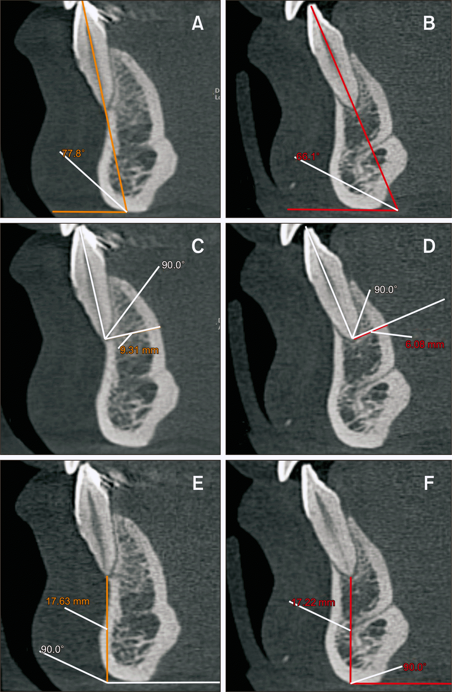 | Figure 7Angle formed by the tooth’s long axis and symphysis base. A, Initial examination, angle = 77.8°. B, Final examination, angle = 66.1°; Distance from the tooth apex to the posterior limit of the symphysis. C, Initial examination, distance = 9.31 mm. D, Final examination, distance = 6.08 mm. Distance from the tooth apex to the lower limit of the symphysis. E, Initial examination, distance = 17.63 mm. F, Final examination, distance = 17.22 mm.
|
Go to :
DISCUSSION
There are several triggering factors for GR involving the incisors.24 One of them is inappropriate dental root position which is out of biological limits due to inadequate orthodontic movement and/or fixed retainer bonding.3,8,9,13-15,23 Renkema et al.25 and Katsaros et al.26 estimate that 2.7% to 5.0% patients wearing fixed retainers with flexible wires develop adverse dental changes that may result in periodontal problems during the post-treatment phase. Nevertheless, rigid wires can also be related to bonding failure and improper tooth movements, thus contributing to the development of alveolar defects and GR, as seen in the present case.23 The patient was wearing a rigid wire retainer bonded to the six mandibular anterior teeth and presented severe GR of tooth #41. Scientifically and clinically,23 it is thought that failure may have occurred during placement of the retainer, thus resulting in force exertion on the tooth over time with buccal displacement of the root. For this reason, proper placement of retainers with passive adaptation to the lingual surface of the incisors and regular clinical control by the practitioner are indispensable in the post-treatment phase.
Other causes such as periodontal disease, traumatic occlusion, a thin gingival biotype, mechanical trauma (e.g., tooth brushing), upper labial frenum insertion, smoking, improper restoration, and aging are also associated with the development of GR.2,8 In the present clinical case, biofilm accumulation and dental calculus on the lingual face of the teeth, a thin gingival biotype, and traumatic contact of tooth #41 were factors leading to the periodontal problems. Before orthodontic therapy, the patient was referred to a periodontist, who treated the inflammatory condition and periodontal disease and educated her about the importance of proper hygiene during the treatment period. The interdisciplinary treatment not only improved the supportive and protective periodontium but also resulted in esthetic improvements, functional occlusion, and oral health and well-being.
Proper repositioning of the tooth inside the alveolus by means of orthodontic movement has been reported to have beneficial effects on the periodontium1,8,14,15 and, consequently, bone regeneration.14 Laursen et al.13 presented a series of 12 patients with GR on the buccal or lingual surfaces of the mandibular incisors, which showed poorly positioned roots. Segmented mechanics with torque control consistently reduced the recession. In the present case, proper root movement favored the alveolar bone architecture of tooth #41. At the start of treatment, the root was buccally displaced and there was no bone plate. During treatment, the root was moved toward the center of the alveolar process, which improved the patient’s periodontal health.
Although the literature reports that orthodontic treatment with corrective appliances is correlated with favorable occlusive and periodontal effects, regardless of the technique used, the findings from the present case showed that clear aligners were effective in correcting the root position which improved the periodontal condition. Power ridges®19 were used for the aligner placed on tooth #41 from the beginning to the end of the treatment; this helped in achieving the desired torque movement and proper root repositioning by mild force application. Figure 7A and 7B shows the change of 11.7° in the angle formed between the tooth’s long axis and symphysis base, which differed from the virtually planned change (19.3° with root movement). This suggests the loss of force and need of overcorrection in cases of aligners, in agreement with the literature.27,28 With regard to the buccal-lingual translation, orthodontic movement with aligners guided the roots lingually, decreasing the distance from the root apex to the posterior limit of the symphysis by 3.23 mm; this also differed from the virtually planned change (6.5 mm) (Figure 7C and 7D). In the virtual plane, intrusive movement of 1.2 mm resulted in a final movement of 0.41 mm (Figure 7E and 7F, Table 1). These results suggest that practitioners should consider possible tooth movements beyond the estimated amounts, with application of overcorrections during treatment planning.27,28 The adjacent incisors in our patient showed no side effects because no force was applied on them. These clinical results confirm that SmartForce® (Align Technology) features are needed for proper control of tooth movements and promotion of an optimal biological response. Moreover, when indicated and used with the patient’s co-operation, clear aligners have advantages over fixed appliances, such as easy oral hygiene, comfort, esthetics and reduced chair time.17,18,29,30 Tepedino et al.31 concluded that the Nuvola® aligner system (GEO S.r.l., Rome, Italy) generally resulted in achievement of the movements predicted in the digital configuration. Tooth movement out of the alveolar process poses a risk of bone dehiscence, which can be accompanied by GR. On the other hand, repositioning of the root into the alveolar process by means of orthodontic appliances can result in a better marginal bone level and a spontaneous reduction in GR.1,8,13-15 The tomographic images of our patient showed the beneficial effects of the clear aligners on her periodontal health. Before treatment initiation, the patient was aware of the poor positioning of tooth #41 as well as the failure of the gingival graft surgeries previously performed for root coverage. After considering the offered treatments, she opted for the repositioning of the tooth using clear aligners, despite knowing the possible complications. Therefore, as a protocol, periodontal follow-up was indispensable to maintain the periodontal health and achieve therapeutic success. In the final phase of the treatment, gingival grafting was not performed because of the positive results.
Table 1
Root movement of the mandibular right central incisor (tooth #41) before (T0) and after (T1) the treatment of gingival recession and bone dehiscence using Invisalign® aligners
| Tooth #41 | T0 | T1 |
|---|---|---|
| Tooth long axis - symphysis base | 77.8° | 66.1° |
|
Root apex - posterior limit symphysis |
9.31 mm | 6.08 mm |
| Root apex (90°) - symphysis base | 17.63 mm | 17.22 mm |
![]()
However, this report described only a single case, and a well-designed clinical trial with a large sample size is required to further validate the findings. Another limitation of this report was the lack of availability of the initial periodontal records, including the periodontal charting. Nevertheless, a clear reduction in GR with the use of clear aligners was demonstrated through computed tomography images, clinical photographs, and periodontal evaluation.
Go to :
CONCLUSION
The findings from this case suggest that repositioning of the root into the alveolar process using Invisalign® aligners allows bone neoformation and a reduction in GR. Adequate diagnosis and treatment planning, application of mild forces, and periodontal follow-up for treating inflammatory conditions contributed to the success of treatment in the present clinical case.
Go to :
 XML Download