

 PDF
PDF Citation
 Print
Print


INTRODUCTION
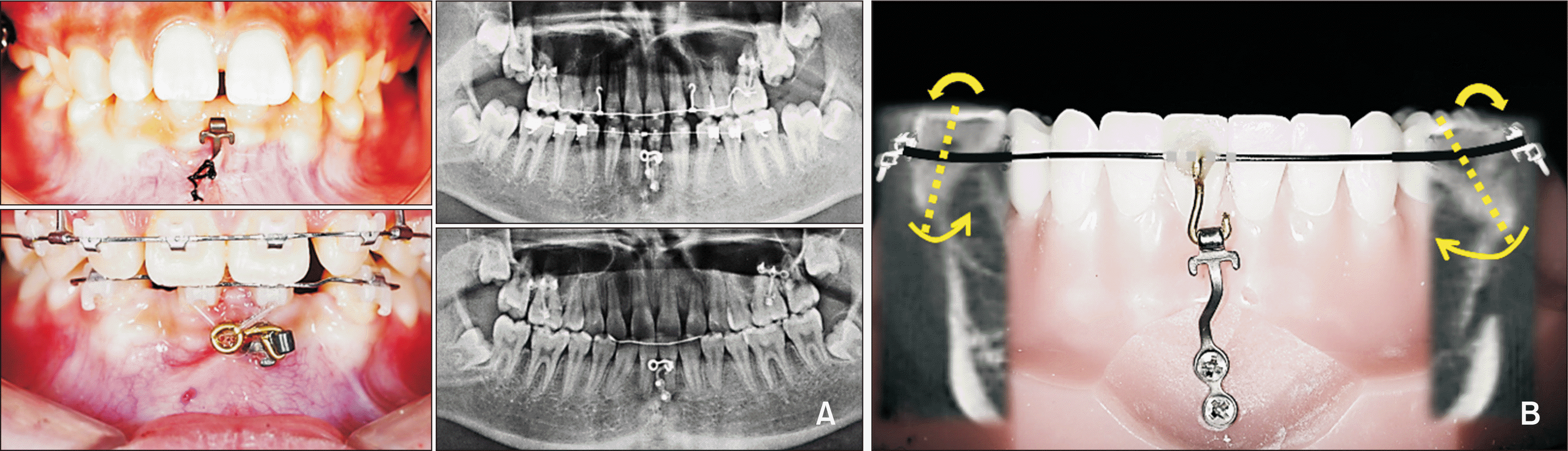 | Figure 1Clinical application of the orthodontic miniplate (OMP) used in this study. A, OMP for lower intrusion and molar protraction. Intraoral photographs and panoramic radiographs show multidirectional tooth movement achieved using a bendable OMP. B, Schematic illustration of an OMP combined with a reverse curve NiTi application for lower posterior uprighting with intrusion.
|
MATERIALS AND METHODS
Patients
OMP placement protocol
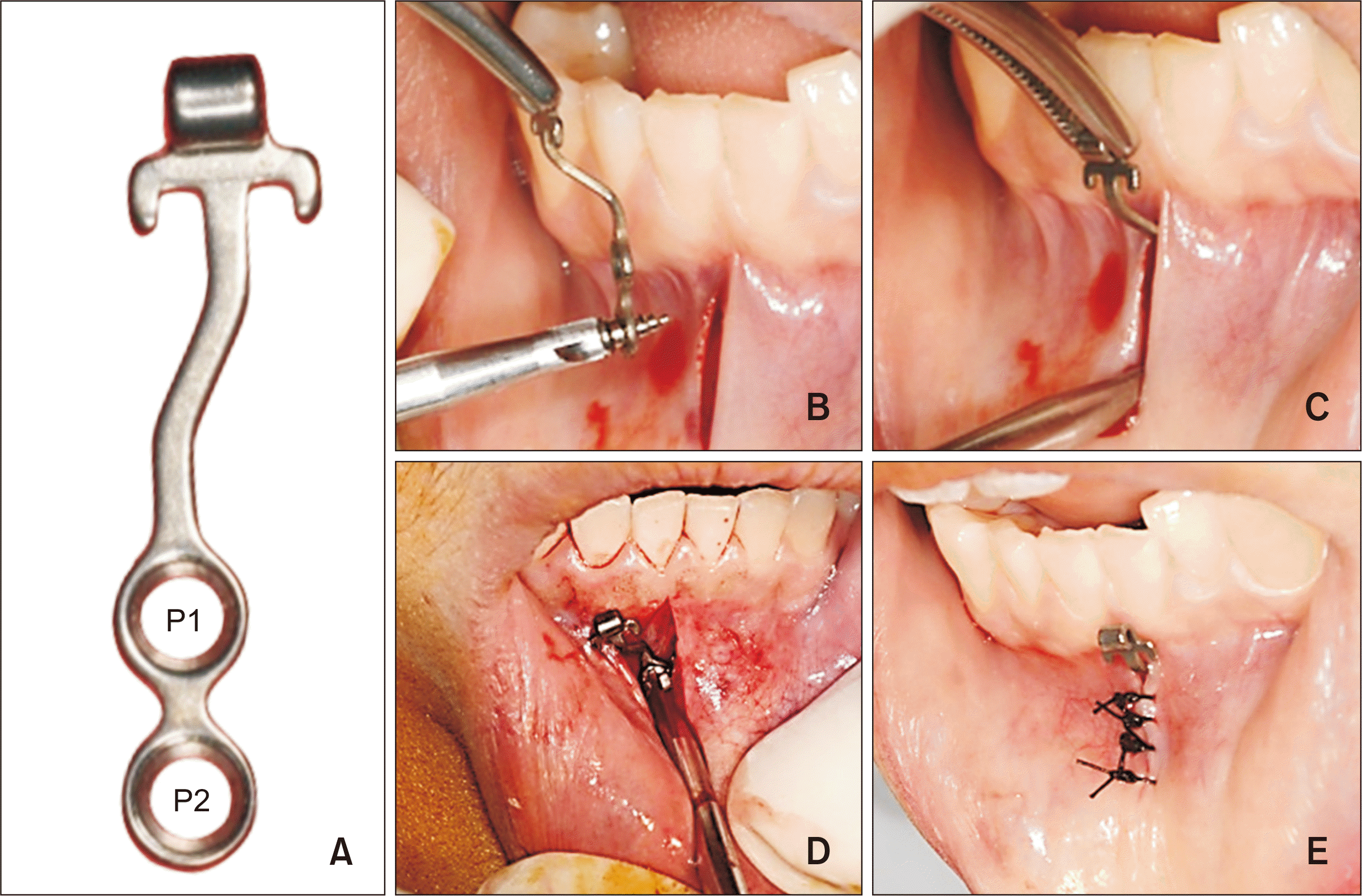 | Figure 2Placement protocol of the orthodontic miniplate (OMP). A, The I-type OMP with two holes and a tube-shaped head. B, The OMP is placed with self-drilling miniplate anchoring screws (MPASs). C, The OMP is placed in the incised area. D, MPASs are fixed to the cortical bone under the mucosa by using a manual screwdriver. E, The incised area is sutured with 4-0 silk.
P1, position 1; P2, position 2.
|
CBCT protocol and measurement
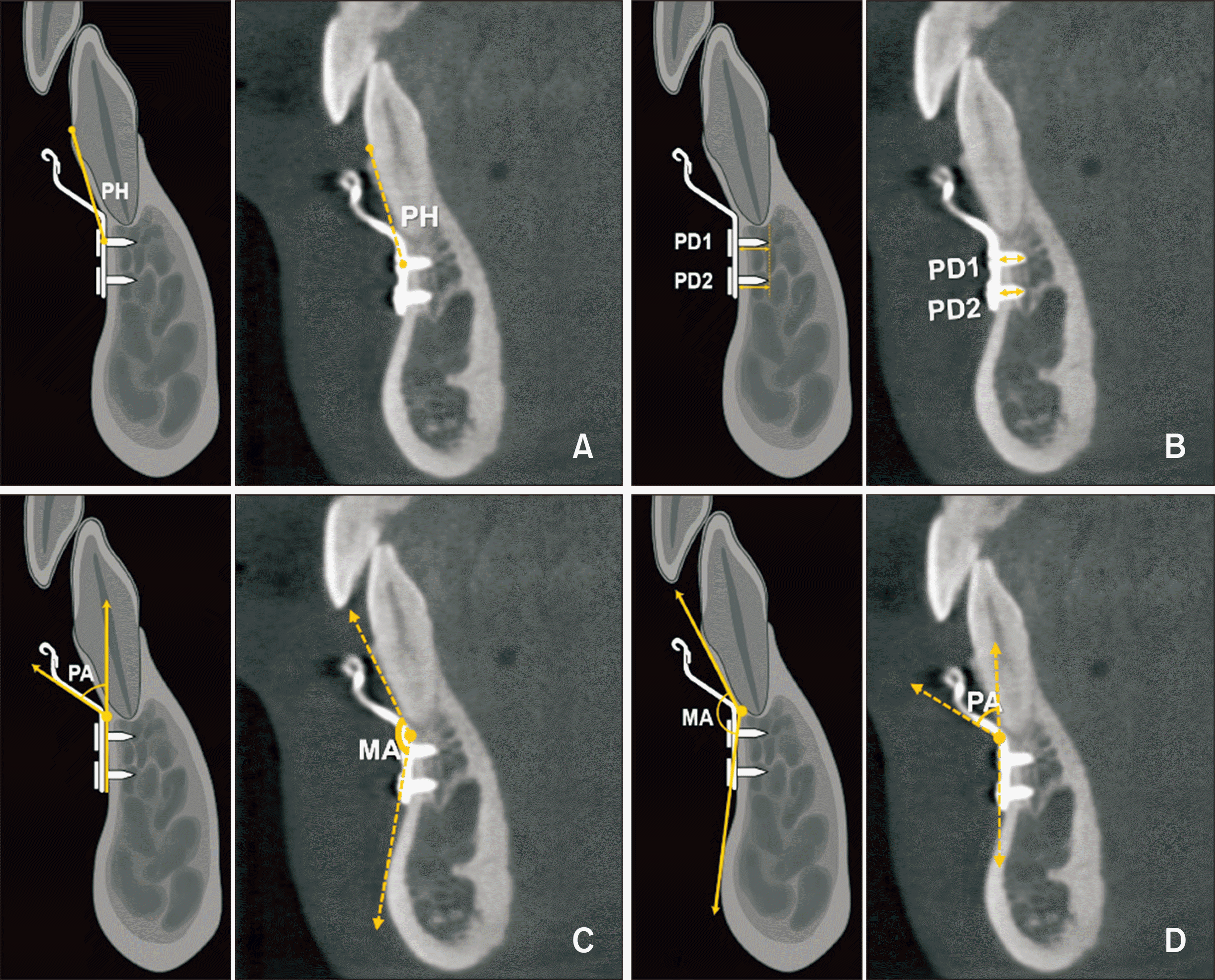 | Figure 3Schematic illustration and actual cone-beam computed tomographic image of the orthodontic miniplate (OMP) in the anterior mandible. A, The vertical distance from the cementoenamel junction to the center of the miniplate anchoring screw (MPAS) at position 1 (P1) (placement height, PH). B, Distance from the outer surface of the labial cortical bone to the tip of the MPASs at P1 (PD1) and P2 (PD2). C, The angle of the step-up bend on the OMP that was made by an orthodontist based on the soft- and hard-tissue anatomic features of the incision site (plate angle, PA). D, The angle made by the two tangents on the sagittal plane, with one from the most prominent midpoint of the chin and the other from the most prominent point on the labial root surface (mental fossa angle, MA).
|
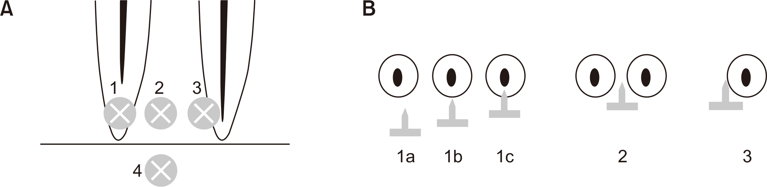 | Figure 4Schematic illustration showing the classification of root proximity according to the location of the miniplate anchoring screw (MPAS) at position 1. A, Frontal view: group 1, frontal overlap (no contact [1a], frontal contact [1b, the MPAS contacts the labial surface of the mandibular incisor], and root perforation [1c]); group 2, interdental space; group 3, lateral contact; and group 4, no-root area. B, Axial view: 1a, no contact; 1b, frontal contact; 1c, root perforation; 2, interdental space; 3, lateral contact.
|
Statistical analysis
RESULTS
Table 1
| Variable | Value |
|---|---|
| PH (mm) | 11.42 ± 2.05 |
| PD1 (mm) | 2.01 ± 0.43a |
| PD2 (mm) | 2.23 ± 0.38b |
| PA (°) | 54.16 ± 11.55 |
| MA (°) | 140.77 ± 8.41 |
PH, vertical distance from the cementoenamel junction to the center of the miniplate anchoring screw (MPAS) at position 1; PD, distance from the outer surface of the labial cortical bone to the tip of the MPASs at positions 1 (PD1) and 2 (PD2); PA, angle of the miniplate placed on the mandibular symphysis; MA, angle made by the two tangents, with one from the most prominent midpoint of the chin and the other from the prominent point on the buccal root surface.
![]()
Table 2
| Variable | Skeletal classification | p-value | ||
|---|---|---|---|---|
| Class I (n = 33) | Class II (n = 50) | Class III (n = 14) | ||
| PH (mm) | 11.32 ± 2.05 | 11.41 ± 2.15 | 11.69 ± 1.80 | 0.8841 |
| PD1 (mm) | 1.97 ± 0.33 | 2.04 ± 0.48 | 2.02 ± 0.46 | 0.7208 |
| PD2 (mm) | 2.22 ± 0.32 | 2.28 ± 0.42 | 2.09 ± 0.36 | 0.2567 |
| PA (°) | 50.57 ± 9.25b | 54.24 ± 12.59b | 63.32 ± 8.55a | 0.0039 |
| MA (°) | 142.08 ± 7.12 | 139.25 ± 9.40 | 143.15 ± 6.74 | 0.4629 |
PH, vertical distance from the cementoenamel junction to the center of the miniplate anchoring screw (MPAS) at position 1; PD, distance from the outer surface of the labial cortical bone to the tip of the MPASs at positions 1 (PD1) and 2 (PD2); PA, angle of the miniplate placed on the mandibular symphysis; MA, angle made by the two tangents, with one from the most prominent midpoint of the chin and the other from the prominent point on the buccal root surface.
![]()
Table 3
| Independent variable | Dependent variable | Spearman’s correlation | Multiple generalized linear model | |||||
|---|---|---|---|---|---|---|---|---|
|
Correlation coefficient |
p-value | Estimate | 95% CI | p-value | ||||
| MA | PH | 0.28 | 0.0062** | 0.78 | 0.31 | 1.25 | 0.001** | |
| MA | PA | 0.09 | 0.3994 | −0.01 | −2.85 | 2.83 | 0.996 | |
| MA | PD1 | 0.09 | 0.4045 | 0.05 | −0.06 | 0.16 | 0.346 | |
| MA | PD2 | 0.12 | 0.2497 | 0.06 | −0.04 | 0.15 | 0.243 | |
| PA | PH | −0.24 | 0.0189* | −0.43 | −0.78 | −0.09 | 0.015* | |
| PA | PD1 | −0.21 | 0.0407* | −0.08 | −0.16 | −0.01 | 0.035* | |
| PA | PD2 | 0.06 | 0.5314 | 0.03 | −0.04 | 0.10 | 0.354 | |
| PH | PD1 | 0.23 | 0.0222* | 0.05 | 0.01 | 0.09 | 0.021* | |
| PH | PD2 | 0.08 | 0.4209 | 0.01 | −0.03 | 0.05 | 0.538 | |
PH, vertical distance from the cementoenamel junction to the center of the miniplate anchoring screw (MPAS) at position 1; PD, distance from the outer surface of the labial cortical bone to the tip of the MPASs at positions 1 (PD1) and 2 (PD2); PA, angle of the miniplate placed on the mandibular symphysis; MA, angle made by the two tangents, with one from the most prominent midpoint of the chin and the other from the prominent point on the buccal root surface; CI, confidence interval.
![]()
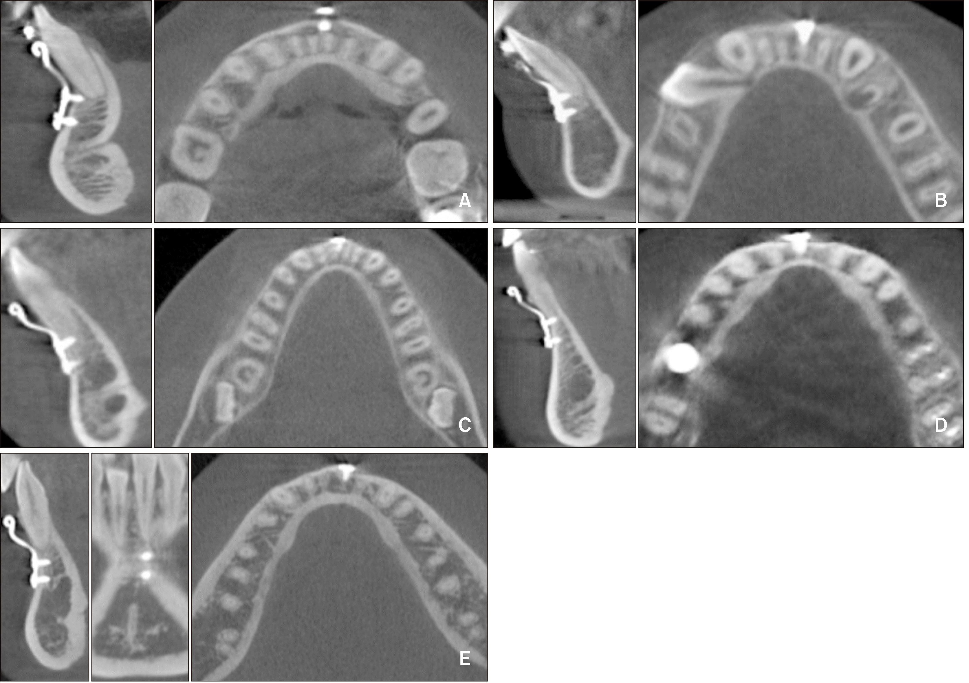 | Figure 5Cone-beam computed tomographic image of the orthodontic miniplate according to the classification of root proximity. A, Group 1 with subdivision a, no contact. B, Group 1 with subdivision b, frontal contact. C, Group 3, lateral contact. D, Group 2, interdental space. E, Group 4, no-root area.
|
Table 4
| Variable | Root proximity | p-value | |||
|---|---|---|---|---|---|
| No contact (n = 11) | Contact (n = 19) | Interdental space (n = 18) | No-root area (n = 49) | ||
| PH (mm) | 10.88 ± 1.98b | 9.75 ± 1.69b | 9.96 ± 1.66b | 12.73 ± 1.36a | < 0.0001 |
| PD1 (mm) | 1.81 ± 0.44 | 1.95 ± 0.42 | 2.01 ± 0.42 | 2.09 ± 0.43 | 0.0968 |
| PD2 (mm) | 2.29 ± 0.58 | 2.26 ± 0.47 | 2.25 ± 0.40 | 2.20 ± 0.28 | 0.8296 |
| PA (mm) | 51.59 ± 11.40 | 49.39 ± 9.54 | 52.56 ± 10.01 | 57.18 ± 12.22 | 0.0823 |
| MA (mm) | 138.35 ± 6.47 | 142.19 ± 7.81 | 139.99 ± 9.88 | 141.06 ± 8.54 | 0.4169 |
PH, vertical distance from the cementoenamel junction to the center of the miniplate anchoring screw (MPAS) at position 1; PD, distance from the outer surface of the labial cortical bone to the tip of the MPASs at positions 1 (PD1) and 2 (PD2); PA, angle of the miniplate placed on the mandibular symphysis; MA, angle made by the two tangents, with one from the most prominent midpoint of the chin and the other from the prominent point on the buccal root surface.
![]()
 XML Download