

 PDF
PDF Citation
 Print
Print


Go to : 
INTRODUCTION
Piggyback hepatectomy technique in liver transplantation (LT) has a potential disadvantage in patients with hepatocellular carcinoma (HCC) because tumor cells can be left at the inferior vena cava (IVC) margins or tumor cells can spread through the short hepatic vein branches. Manipulation of the HCC-bearing liver during piggyback hepatectomy may increase the risk of HCC spread [1]. However, piggyback hepatectomy is inevitable when performing living donor liver transplantation (LDLT). We have previously reported a technique of no-touch en bloc recipient hepatectomy with IVC replacement in a patient with HCCs close to the retrohepatic IVC [2], in which the extent of hepatectomy is compatible to that of deceased donor liver transplantation (DDLT) with classical IVC replacement. If some distance exists between HCCs and the retrohepatic IVC, the possibility of direct tumor cell invasion into the IVC wall may be relatively low. Considering that IVC replacement with a prosthetic graft cannot be a routine procedure of LDLT for HCC [2,3], a compromising method between the conventional piggyback IVC preservation and IVC replacement can be attempted to prevent iatrogenic tumor cell spread during liver manipulation. The basic technique of this method is similar to that of total hepatic vascular exclusion (THVE) [4,5]. Iatrogenic tumor cell spread can be theoretically prevented if piggyback recipient hepatectomy is performed under THVE. The recipient liver is destined to be removed, thus there is no time limit for the duration of THVE [6,7]. However, considering that the majority of patients undergoing LDLT for HCC have less advanced liver cirrhosis and poor development of portal collaterals, active venovenous bypass can be combined to prevent prolonged THVE-associated hemodynamic instability and splanchnic venous congestion. We herein present our experience of recipient hepatectomy under THVE and active venovenous bypass for LDLT in a patient with multiple HCCs closely located to the retrohepatic IVC.
Go to :
CASE REPORT
This study was approved by the Institutional Review Board of Asan Medical Center (IRB No. 2020-0822). The requirement for informed consent from the patient was waived due to the retrospective nature of this study.
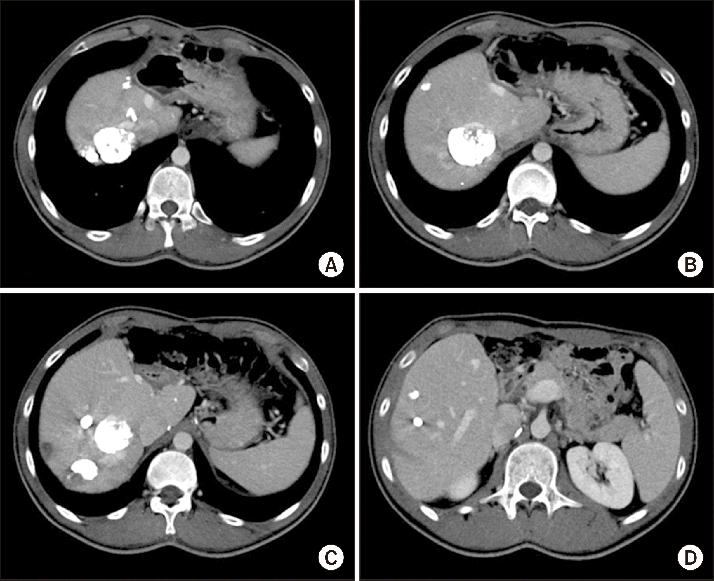
A 19-year-old Mongolian male patient diagnosed with hepatitis B virus (HBV)-associated HCC was admitted to our institution for LDLT. He had been diagnosed with HBV at 8 months after birth. This patient underwent left lateral sectionectomy 14 months before and also received 4 sessions of transarterial chemoembolization (TACE) due to post-hepatectomy tumor recurrence. Liver dynamic computed tomography (CT) scan showed lipiodol uptake of multiple viable HCCs (Fig. 1). Pretransplant work-up studies revealed no evidence of extrahepatic metastasis. There were three small nodules at the right lower lobe and the left upper lobe of the lung with showing no changes for 2 months on chest CT follow-up. These nodules were highly like to be inflammatory nodules. HCC tumor markers were decreased after the last session of TACE, with serum α-fetoprotein (AFP) level at 1,637.7 ng/mL and des-γ-carboxyprothrombin (DCP; protein induced by vitamin K antagonists or absence-II [PIVKA-II]) at 67 mAU/mL. These clinical sequences after HCC resection implicated a high risk of posttransplant HCC recurrence. However, his family members eagerly wanted him to undergo LDLT with expectation of a prolonged survival. Thus, LDLT was performed after observation for two months following the last session of TACE. The donor was a 24-year-old brother of the patient. A modified right liver graft from this donor weighed 750 g, making a graft-to-recipient weight ratio of 1.19%. The donor recovered uneventfully from donor operation. He was discharged at 10 days after operation.
During recipient operation, the hepatoduodenal ligament was meticulously dissected. The right hepatic artery and bile duct were then transected (Fig. 2A). The supra- and infra-hepatic portions of the retrohepatic IVC were encircled with vascular tourniquets under very gentle handling of the liver (Fig. 2B). The right portal vein was transected and portal flow was diverted through an active venovenous bypass connected to the internal jugular vein pathway. Under THVE and portal vein bypass, the Spigelian lobe and paracaval portion of the caudate lobe were meticulously dissected from the retrohepatic IVC (Fig. 2C). After complete detachment of the caudate lobe from the IVC, the right liver was fully mobilized (Fig. 2D). The duration of THVE and portal vein bypass for recipient hepatectomy took approximately 70 minutes.
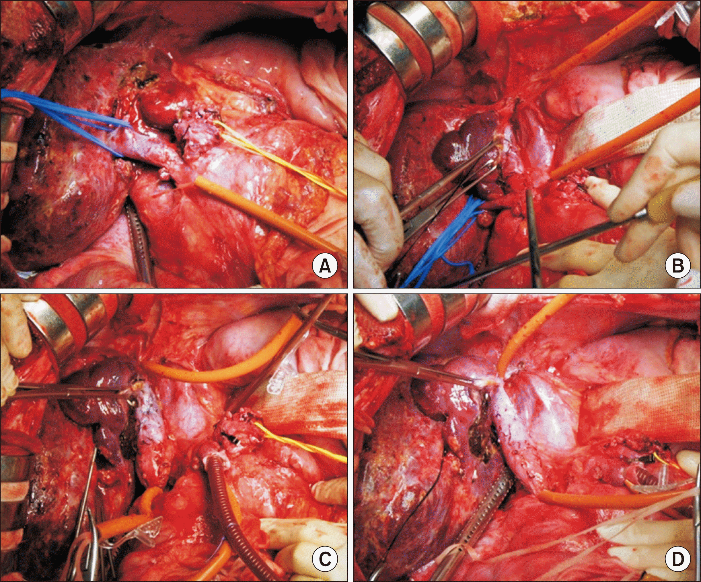 | Fig. 2Intraoperative photographs of recipient hepatectomy. (A) The hepatoduodenal ligament is meticulously dissected, and then the right hepatic artery and the bile duct are transected. (B) The supra- and infra-hepatic portions of the retrohepatic inferior vena cava (IVC) are encircled with vascular tourniquets. (C) The right portal vein is transected and portal flow is diverted through the active venovenous bypass connected to the internal jugular vein pathway. (D) Under total hepatic vascular exclusion and portal vein bypass, the caudate lobe is meticulously dissected from the retrohepatic IVC and the right liver is fully mobilized.
|
Immediately after recipient hepatectomy, the isolated retrohepatic IVC was vigorously flushed with heparinized saline to remove the stagnated blood. A modified right liver graft was implanted according to standard procedures of LDLT. The graft right hepatic vein and interposed middle hepatic vein conduit were separately reconstructed to the recipient’s right hepatic vein and left-middle hepatic vein stumps, respectively. The recipient portal bifurcation was used for anastomosis with the graft portal vein. A single right hepatic artery was reconstructed with the corresponding recipient right hepatic artery under surgical microscopy. Biliary reconstruction was performed as a duct-to-duct anastomosis.
The pathology report of the explant liver showed three viable HCCs. The largest mass was 4.5×3.5×3.0 cm in size, having Edmondson-Steiner grade III/II, 50% necrosis and microvascular invasion. Each of 3 cm- and 1.5-cm-sized masses showed 30% necrosis (Fig. 3). The liver showed HBV-associated septal fibrosis. Other multiple small tumors of total necrosis were also present.

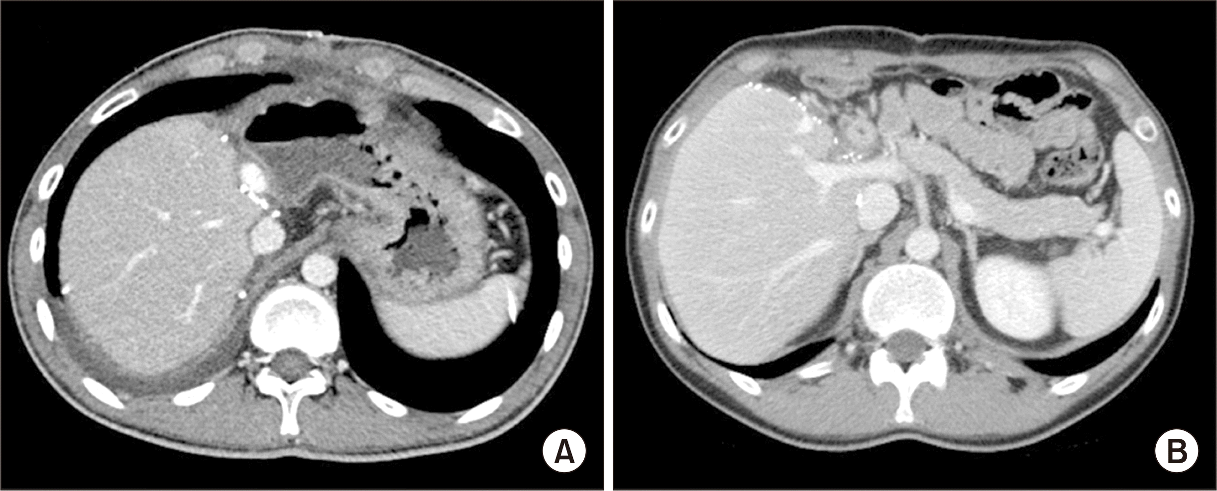
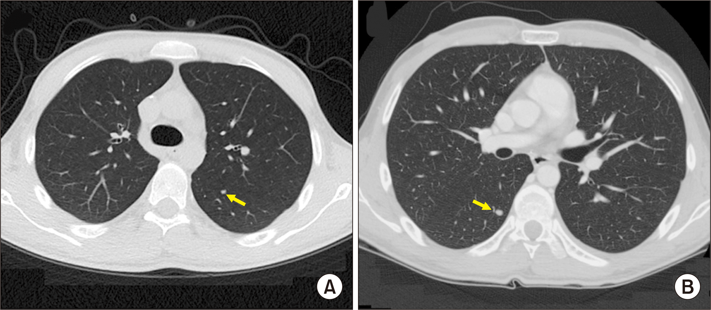
The patient recovered uneventfully from the LDLT operation (Fig. 4), and was discharge 21 days after LDLT. Reduced-dose tacrolimus combined with mycophenolate mofetil was used for the primary immunosuppressants. However, chest CT taken 4 months after the LDLT showed multiple small nodules (Fig. 5A), indicating high probability of multiple pulmonary metastasis. There was no noticeable increase in serum AFP and PIVKA-II levels. The immunosuppressive regimen was changed to combination therapy with everolimus and low-dose tacrolimus. Sorafenib was administered for 5 months, but it was stopped due to disease progression (Fig. 5B). The patient is currently administering lenvatinib for 3 months. Despite the occurrence of slowly growing pulmonary metastasis, the patient has been doing well for 12 months after LDLT.
Go to :
DISCUSSION
The present case demonstrated the surgical technique of recipient hepatectomy under THVE and venovenous bypass to prevent iatrogenic tumor cell spread. The primary reason why we present this technique is that there are no established procedures for this method in the literature although it might have been performed previously.
Tumor cells released from the HCC-bearing liver during recipient hepatectomy could be entrapped in the lung as the first filtering system, which can be the background reason why the lung is the most common site of distant metastasis following LT or hepatic resection for advanced HCC [8]. For surgical resection of large HCCs at the right liver, hepatic parenchymal transection with anterior approach can achieve longer survival outcome than the conventional approach because the former requires less tumor handling, thus having less chance of tumor cell spread [9].
We have performed no-touch en bloc recipient hepatectomy with IVC replacement for patients with HCCs close to the retrohepatic IVC [2] because the extent of hepatectomy is compatible to that of classical DDLT. LDLT for HCC satisfying the Milan criteria does not compromise patient survival or increase HCC recurrence compared to DDLT [10]. On the contrary, in patients with HCC exceeding the Milan criteria, it was occasionally presented that LDLT might lead to worse outcomes than DDLT [11]. IVC replacement with resection of the IVC during LDLT is a feasible technical option with an acceptable risk of morbidity and a low risk of thrombus formation [12]. However, it is still regarded as an unfamiliar procedure. Our current method with THVE and venovenous bypass is a compromising method between the conventional piggyback IVC preservation and IVC replacement. It does not appear to be demanding because THVE and active venovenous bypass are familiar to LT surgeons.
We also previously presented the technique of no-touch hepatectomy with left approach for LDLT and multiple or large HCCs in the right liver lobe [13], in which the basic concept is similar to that of the present case. The technique included hilum dissection without liver mobilization; temporary portocaval shunt is required if the recipient does not have large porto-systemic collaterals to prevent bowel edema caused by splanchnic congestion; clamping and splitting of the middle-left hepatic vein common trunk after mobilization of the left liver lobe, and ligating and transecting short hepatic veins from the left side of the IVC; clamping of the right hepatic vein before right liver mobilization; and finally, the right liver is mobilized and the native liver is removed [13].
Besides THVE, we have applied prolonged hepatic inflow occlusion technique to reduce bleeding and prevent tumor cell spread during recipient hepatectomy in patients with HCC beyond the Milan criteria [6,7]. We have previously reported the mean duration of hepatic inflow occlusion was 68.2±19.1 minutes [6]. Such prolonged occlusion of the hepatoduodenal ligament can be applied during right liver mobilization with intention to minimize hematogenous spread of HCC cells during LT, although its beneficial effect has not been objectively proven yet.
However, unlike patients with advanced liver cirrhosis, the majority of patients undergoing LDLT for HCC have less advanced liver cirrhosis and subsequently poor development of portal venous collaterals. A prolonged hepatic inflow occlusion can induce splanchnic venous congestion and prolonged THVE can result in hemodynamic instability. Prolonged prehepatic portal venous congestion or sinistral portal hypertension is also a potential risk factor of acute pancreatitis after LT [14]. Active venovenous bypass is an effective solution to solve these problems. Thus, we think that there is no reason to hesitate to perform venovenous bypass if indicated. A review study has revealed that the venovenous bypass has the following disadvantages: (1) it does not guarantee normal perfusion of abdominal organs and lower limbs or improved outcome; (2) it may worsen postreperfusion syndrome; (3) there is no evidence that it can reduce or prevent the occurrence of postoperative renal failure; (4) it may worsen cerebral edema following reperfusion of the graft; (5) it may potentiate bleeding by causing hemolysis and platelet depletion; and (6) morbidity and mortality are also associated with its use. Its advantages were as follows: (1) it can reduce hemodynamic instability during anhepatic phase; (2) it is useful in patients with pulmonary hypertension and cardiomyopathy who tolerate anhepatic period poorly; (3) it can maintain intraoperative renal function; (4) it can maintain cerebral perfusion pressure in patients with acute fulminant failure by avoiding rapid swings in blood pressure; and (5) it can facilitate difficult surgery and reduce blood loss [15].
As shown in the posttransplant clinical sequences of the present case, pulmonary metastasis occurred a few months after LDLT. Pretransplant sequences of HCC treatments and responses implicated that this patient would have a high risk of posttransplant HCC recurrence. We recently presented that the independent risk factors of salvage LDLT are beyond the Milan criteria and the ADV score (multiplication of AFP, PIVKA-II [DCP] and tumor volume) greater than 4log [8]. The pretransplant extent of HCC exceeded the Milan criteria and the explant ADV score was calculated to be 6.9log, thus high probability of early HCC recurrence after LDLT was anticipated. In principle, this patient was not indicated for salvage LDLT in principle in our institution. However, considering the patient age of only 19 years at LT, it is difficult to define it as a futile LDLT, although pulmonary metastasis is slowly progressing. Although post-recurrence patient survival following salvage LDLT is known to be inferior to that of primary LDLT, a small number of patients showing post-transplant HCC recurrence survived over a few years [8]. We presumed that the early occurrence of pulmonary metastasis in the present case was associated with advanced nature of HCC per se rather than ineffectiveness of THVE and venovenous bypass. The tumor extent appeared to exceed the range of protective coverage via THVE and venovenous bypass.
In conclusion, we suggest that recipient hepatectomy under THVE and venovenous bypass is a feasible technical option to cope with the risk of iatrogenic tumor cell spread during LDLT in patients with advanced HCCs located close to the retrohepatic IVC. The real-world role of this technique should be assessed through further validation studies.
Go to :
 XML Download