

 PDF
PDF Citation
 Print
Print


Introduction
Industrial production, food preservation and packaging are on the increase in response to world population growth and rapidly changing environment. In all of these processes, there are some essential chemicals frequently used, one of these is bisphenol-A (BPA). Like most chemicals, it is not without some health risks [1, 2]. In spite of compelling evidence that showed disturbing adverse effects in humans and animal experiments, few food safety agencies certified BPA to pose no risk in foods packed with BPA-made paper glass and plastic, BPA–coated tin, and BPA-lamented carton [1-3]. These include, epigenetically-inducing type 2 diabetes and lipids deregulation in mice [4, 5], predisposing to a host of life threatening diseases such as cardiovascular diseases [6, 7], obesity [8], and thyroid dysfunction [9] in humans.
In consideration of the possible health challenges that may result from accidental or unintentional exposures to BPA, it is enlisted in the global list of emerging environmental toxicants [10-13]. More than the threat of exposures through industrial food containers is the evidence of BPA accumulation in plants from contaminated environment, raising more concern of un-aware toxicity in the herbivorous population who rely on plants as source of nutrients. Chemical profiling in selected plants shows that every parts of the plants, from flowers, seeds, leaves, stems to roots contained a biologically significant amount of buildup of BPA [14].
Despite the various media of possible exposure to BPA, free forms of BPA are detectable in many vital body tissues, with the evidence of successive increase in unconjugated BPA concentrations in breast milk, brain, liver and the adipose tissue [15]. In addition, age seems to affect serum concentration of free forms of BPA. While concentration ranges from 2.3 to 2.4 ng/ml in adults, it is 4.3 ng/ml in children and 2.8 ng/ml in adolescents [16]. The increased concentration in children may not be unrelated to the BPA in baby food packs combined with in-utero and breastfeeding exposures, and this have been reported to predispose to defective brain development and behavior in humans and animals [17-19].
Some of the BPA-induced toxicity has been reported due to its hormone disrupting properties in the hypothalamic-pituitary axis, with evidences of variation in cerebral sexual differentiation, increase gonadotropin-releasing hormone and prompt early puberty [20]. Aside the hormone disrupting effects, one of the very important mechanisms of BPA induced toxicity is oxidative damages, commonly manifested as reactive oxygen species (ROS) and/or reactive nitrogen species (RNS), over generation, impaired antioxidant systems, mitochondrial dysfunction, and other molecular signaling related to oxidative functions [21, 22]. Understanding that with the genomic and non-genomic mediated interferences by BPA in the biological systems is the induced oxidative damages and impaired anti-oxidant functions, exploring novel antioxidants like melatonin (MEL) with reproductive related effects in BPA toxicity is thus essential, more so that many antioxidants are reported efficacious against BPA toxicity [23-31].
MEL is known to have physiological roles in various survival functions in the body, and its antioxidant and mitigative efficacies against reproductive toxicity have been previously reported [26, 32-34]. Thus, this study, assessed the oxidative implications, genetical manifestations and morphological modifications following PBA exposure in the ovarian tissues of adolescent female Wistar rats.
Go to : 
Materials and Methods
Ethical clearance
The animals used were carefully treated as stipulated by the National Institutes of Health guide for the care and use of laboratory animals. The experimental design used was reviewed and acknowledged by the Ethical Review Committee (ERC) of University of Ilorin, Ilorin, Nigeria, with approval number UERC/ASN/2018/1154. The College of Health Sciences’ animal holding was used in carrying out the research. All institutional and national guidelines provided for animal care and use of laboratory animals were duly adhered to.
Acquisition of experimental materials
BPA was procured from Sigma® (CAS-No: 80-05-7; Munich, Germany), while absolute ethanol, corn oil and MEL were procured from a standard laboratory in Ilorin, Nigeria.
Grouping and breeding of animals
Mature female rats and male rats weighing 150±10 g and 200±20 g respectively were procured from an experimental rodent breeding farm in Oyo State, Nigeria. The rats were kept in a healthy and controlled environment at a temperature of 22°C±2°C and at least 14 hours of light from 6:00 a.m. to 20:00 p.m. daily. The rats also had a constant access to pelletized rat feed that was purchased from a standard feed-mill in Ilorin, Nigeria. They also had access to clean water throughout the study.
Sexually matured male rats were introduced to adult female rats in the first stage of their estrous cycle (the proestrus phase) in a ratio of 2 female rats to 1 male rat. Subsequently, vaginal smears were done to check for sperm in the female rats, and the day when sperm was seen in the vaginal washings was labelled as the 1st day of gestation and the pregnant females were isolated and kept in separate cages until the day of parturition.
After parturition, 42 female litters were identified, randomly divided into 7 groups I to VII (n=6), and were administered as follows: group I, control; group II, vehicle control (corn oil); group III, 10 mg/kg MEL; group IV, 25 mg/kg BPA; group V, 25 mg/kg BPA+10 mg/kg MEL; group VI, 50 mg/kg BPA; group VII, 50 mg/kg BPA+10 mg/kg MEL.
BPA is first dissolved in ethanol, then mixed with corn oil and administered according to the doses per kg/bw. The administration started on postnatal day (PND) 19, daily for 7 weeks (PND, 19–68 days) via oral gavage. All the groups were left until day 120±4 days before sacrifice, which was carried out in their proestrus phase of cycles. The ±4 days was due to different animals being in proestrus at different days.
Sample collection and processing
The rats were euthanized using 20 mg/kg body weight of ketamine intraperitoneally. Blood samples for hormonal analysis were obtained from the apex of the heart with the aid of a 5 ml syringe from all rats employed in the study. The serum was separated by centrifugation at 3,000 rpm for 15 minutes. The brains were then excised with isolation of the hypothalamus and the pituitary, which were transferred into RNA later, then snapped frozen in liquid nitrogen prior to storing in –80°C freezer before analysis. One ovary from each animal was then fixed in 4% paraformaldehyde for histological analysis while the second ovary was used as homogenate for enzyme studies of oxidative stress markers.
Histomorphometric analysis
The histomorphometry (i.e., corpus counts, defective, primary, preantral and antral follicular counts) were from photomicrographs of the ovary taken using Olympus microscope (SC50; Olympus, Tokyo, Japan) at 40× magnification.
Biochemical assay
The blood samples were collected from the apex of the heart and centrifuged for 15 minutes. The collected blood was used for serum anti-mullerian hormone (AMH), follicle stimulating hormone (FSH), luteinizing hormone (LH), estrogen, progesterone, and testosterone. The oxidative stress markers (superoxide dismutase [SOD], Glutathione Peroxidase [GPx], Nitric Oxide Synthase [NOS], and uridine 5’-diphospho [UDP]) were assessed by an enzyme-linked immunosorbent assay kit (IB79174; IBL-America, Minneapolis, MN, USA) using the ovarian tissue homogenate.
Quantitative real time polymerase chain reaction procedure
Total RNA was prepared with Euro Gold Tri-Fast solution (Euro Clone, Brussels, Belgium). The tissue/RNA extraction solution was then pulverized with a tissue homogenizer. DNase treatment was performed on total RNA samples extracted in order to eliminate genomic DNA contamination from total RNA preparation. RNA was purified through acid phenol chloroform, precipitated and suspended in distilled water (dH2O).
Reverse transcription of total RNA was performed using M-MLV reverse transcriptase (Invitrogen, Carlsbad, CA, USA). A total of 1 µg RNA were retrotranscribed for quantification of mRNA expression in experiments. Samples were mixed by gentle up and down pipetting and incubated at 37°C. M-MLV reverse transcriptase was inactivated for 15 minutes at 70°C. cDNA was kept at –20°C.
cDNA and RNA concentrations were measured by NanoDropTM 1000 spectrophotometer (Life Technologies, Carlsbad, CA, USA). Ratios of absorbance at 260/280 nm and 260/230 nm were used as indication of nucleic acids quality. Quantitative real time-polymerase chain reaction (qRT-PCR) was used to measure relative amounts of transcript of a specific gene via two step procedure using Sybr green supermix (Biorad, Hercules, CA, USA)—the amplification reaction and subsequent generation of melting curves of the amplicons. The PCR product was run on 2% agarose gel to verify melting curve data. The efficiency of primers was tested in reactions with six serial 1:10 dilutions of cDNA as a template to perform a calibration curve. The primers sequence using Rattus norvegicus is as shown in Table 1.
Table 1
Primers sequence (Rattus norvegicus)
![]()
Data analysis
The data obtained was analyzed using two-way analysis of variance (ANOVA) and subsequently with Turkey’s honestly significant difference multiple comparison test using GraphPad Prism v.6 (GraphPad Software Inc., La Jolla, CA, USA). Data were presented as means±standard error of mean (SEM), and statistical significance was considered at P-values less than 0.05.
Go to :
Results
Bisphenol A-induced hormonal disruptions and melatonin intervention
Being a known hormonal disruptor, effects of BPA on reproductive hormones were investigated in adolescent rats (7 weeks). First, we measured the plasma levels of AMH to extrapolate the status of transiting resting primordial follicles to growing follicles. The results showed a decrease in plasma AMH in lower BPA-treated rats compared to normal saline (NS), vehicle control (VC), and MEL only treated animals. This decrease was reversed with MEL treatment in the group that received 25 mg BPA+MEL. Specifically, there was an increase AMH level in 25 mg/kg BPA (LBP-A)+MEL-treated rats compared to LBP-A only-treated animals (Fig. 1). Although there was a marked decrease in the AMH level of higher dose BPA group (50 mg) when compared to NS-treated group, there is no observable reversal with MEL administration in this group (Fig. 1A).
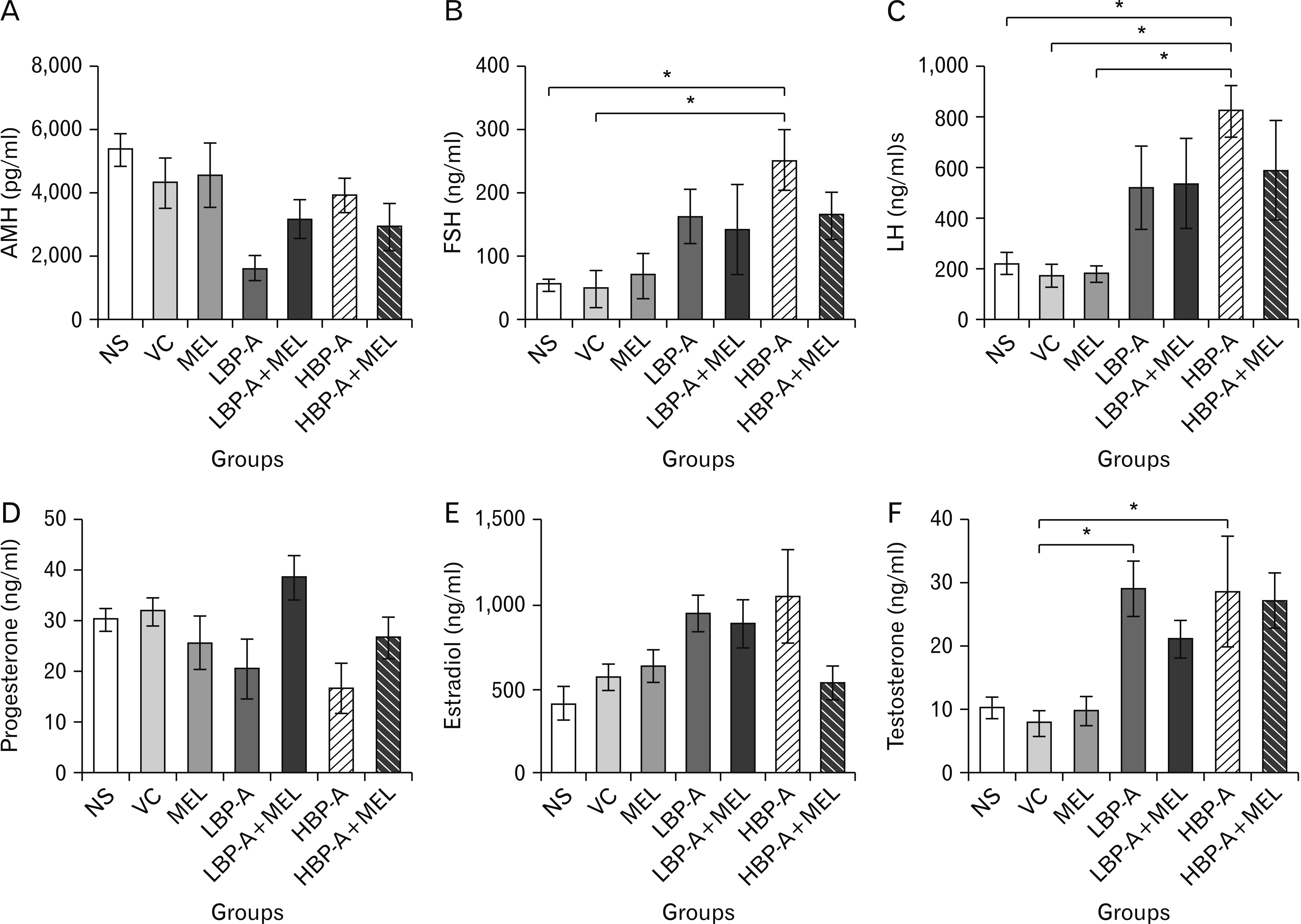 | Fig. 1Showing the plasma levels of anti-mullerian hormone (AMH) (A), follicle stimulating hormone (FSH) (B), luteinizing hormone (LH) (C), progesterone (D), estradiol (E), and testosterone (F) following prenatal exposures to normal saline (NS), vehicle control (VC), 10 mg/kg melatonin (MEL), 25 mg/kg bisphenol-A (BPA) (LBP-A), 25 mg/kg BPA+10 mg/kg melatonin (LBP-A+MEL), 50 mg/kg BPA (HBP-A) and 50 mg/kg BPA+10 mg/kg melatonin (HBP-A+MEL). Values are presented as mean±standard error of mean. Asterisk indicates significant increase from all or specific groups (*P<0.05).
|
Next, we analyzed FSH, which is the earliest hormone to be recruited during antral stage, and LH, a hormone that is involved in ovulation and supports FSH in promoting growth and secretory activity of the follicle. Estradiol level, a naturally occurring type of estrogen hormone known to govern various aspects of healthy reproductive system in female animals was also studied. There were significant increases in the FSH, LH and estradiol levels in BPA only treated groups compared to controls, while there was a trend of lowering these levels in groups co-treated with MEL (Fig. 1B, C, E).
Then, we assessed the plasma concentration of progesterone, as this hormone is known to work in synergy with estradiol. There was an observable trend in the decrease in progesterone levels in rats treated with the two dosages of BPA when compared to NS-, VC- and MEL-treated rats. Like in estradiol, MEL treatments significantly increased progesterone levels in LBP-A+MEL and HBP-A+MEL respectively when compared with the BPA only treated rats (Fig. 1E). Finally, testosterone, a substrate in the metabolic pathway for estrogen synthesis was then measured. There was a significant increase in testosterone level in low and high BP-treated rats compared to animals treated with NS, VC, and MEL. MEL cotreatment similarly reversed the observed disruption in testosterone levels, by reducing the testosterone levels (Fig. 1F).
Differential effects of BPA on anti-oxidants and metabolic activities
Evaluation of BPA involvement in oxidative stress showed increased activities of SOD and NOS in all BPA-treated groups. In specific term, there was a significant increase in activities of SOD and NOS in BPA treated groups compared to NS-, VC- and MEL-treated groups, indicating NO and O as the main players in this oxidative stress. Attempt with co-administration of BPA and MEL had no significant effect on the activities of the two enzymes, SOD and NOS (Fig. 2A, B), although there was a mild trend observed in lowering these levels by MEL. Similar results were obtained when activities of GPx and UDP were evaluated. Activities of GPx and UDP were significantly decreased in all groups that had BPA administered compared to NS-, VC-, and MEL-treated group (Fig. 2C, D), while a similar effect of mild increasing trend was observed with coadministration of MEL.
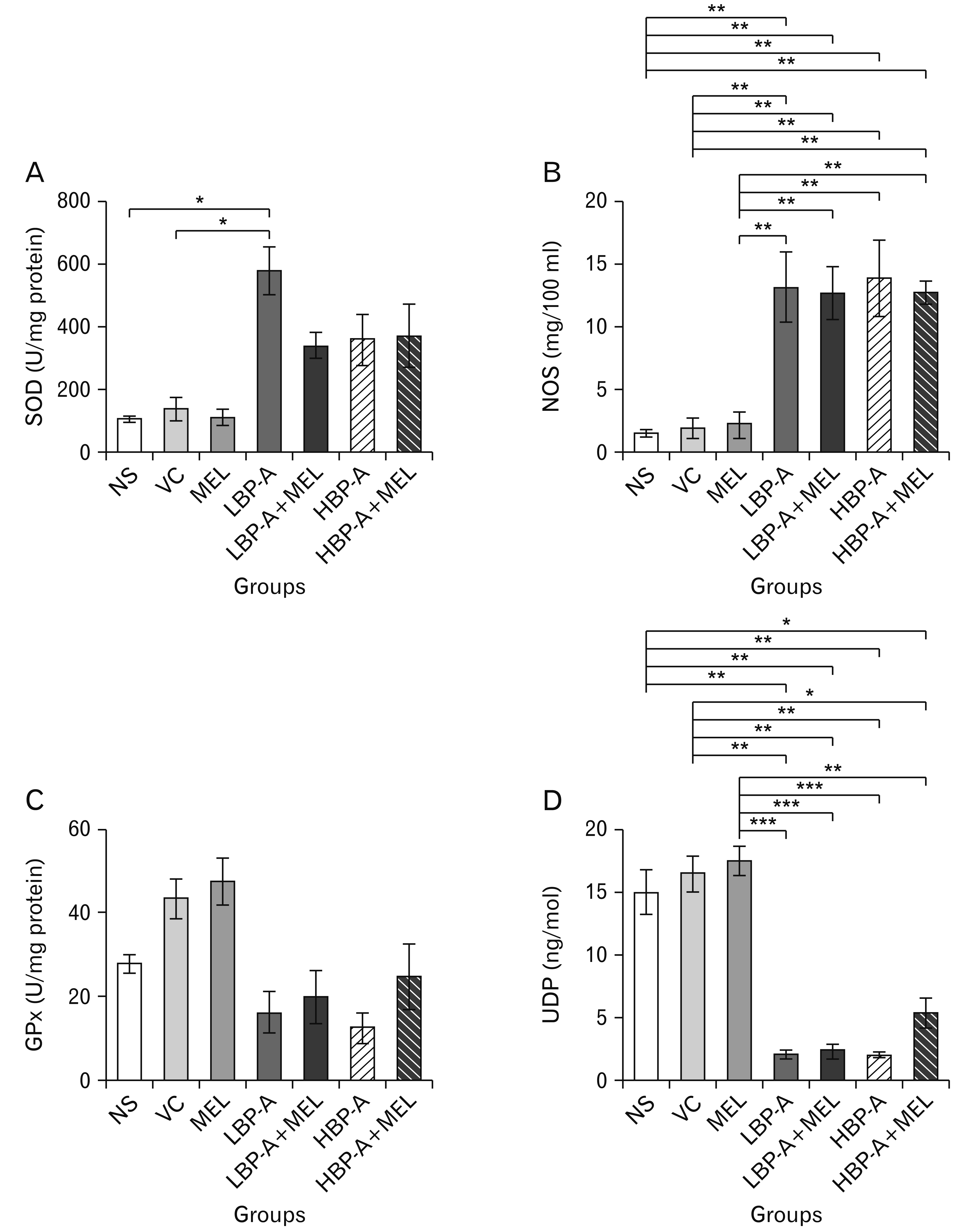 | Fig. 2Showing the ovarian levels of (A) superoxide dismutase (SOD), (B) nitric oxide synthase (NOS), (C) glutathione peroxidase (GPx), and (D) uridine 5’-diphospho (UDP) following exposures to normal saline (NS), vehicle control (VC), 10 mg/kg melatonin (MEL), 25 mg/kg bisphenol-A (BPA) (LBP-A), 25 mg/kg BPA+10 mg/kg melatonin (LBP-A+MEL), 50 mg/kg BPA (HBP-A), and 50 mg/kg BPA+10 mg/kg melatonin (HBP-A+MEL). Values are presented as mean±standard error of mean. Asterisks indicate significant differences at *P<0.05, **P<0.01, and ***P<0.001, respectively from all or specific groups.
|
Bisphenol-A interference with protein receptors expression
Expression of relevant receptor proteins were investigated with qPCR. There was a significant increase in expression of Kiss 1, GnRH receptor (GnRHr) and GnRH mRNA in rats treated with LBP-A, HBP-A, LBP-A+MEL and HBP-A+MEL compared to NS-, VC-, and MEL-treated animals. The density of expressed Kiss 1, GnRHr and GnRH mRNA in lower or higher dose BPA-treated animals was not statistically different with MEL intervention, and its coadministration groups (Fig. 3A-C). BPA significantly downstream the estradiol receptor expression but was reversed with MEL co-treatments. Specially, there were significant decrease in density of expressed estrogen receptor (EsR) in BPA only groups compared to NS-, VC-, and MEL-treated groups. When treated with MEL, there was a significant upstream expression of EsR in 50 mg BPA+MEL-treated rats compared to its corresponding BPA only administered group (Fig. 3D). Unlike the obtained result on EsR expression, androgen receptor expression was not significantly different among all the groups (Fig. 3E). Also, protein expression for AMH was not affected by BPA treatments. There was no significant difference in expressed AMH protein in all BPA treated rats when compared to NS- and VC-treated animals. Interestingly, AMH expression was significantly increased in MEL-treated rats compared to other groups (Fig. 3F).
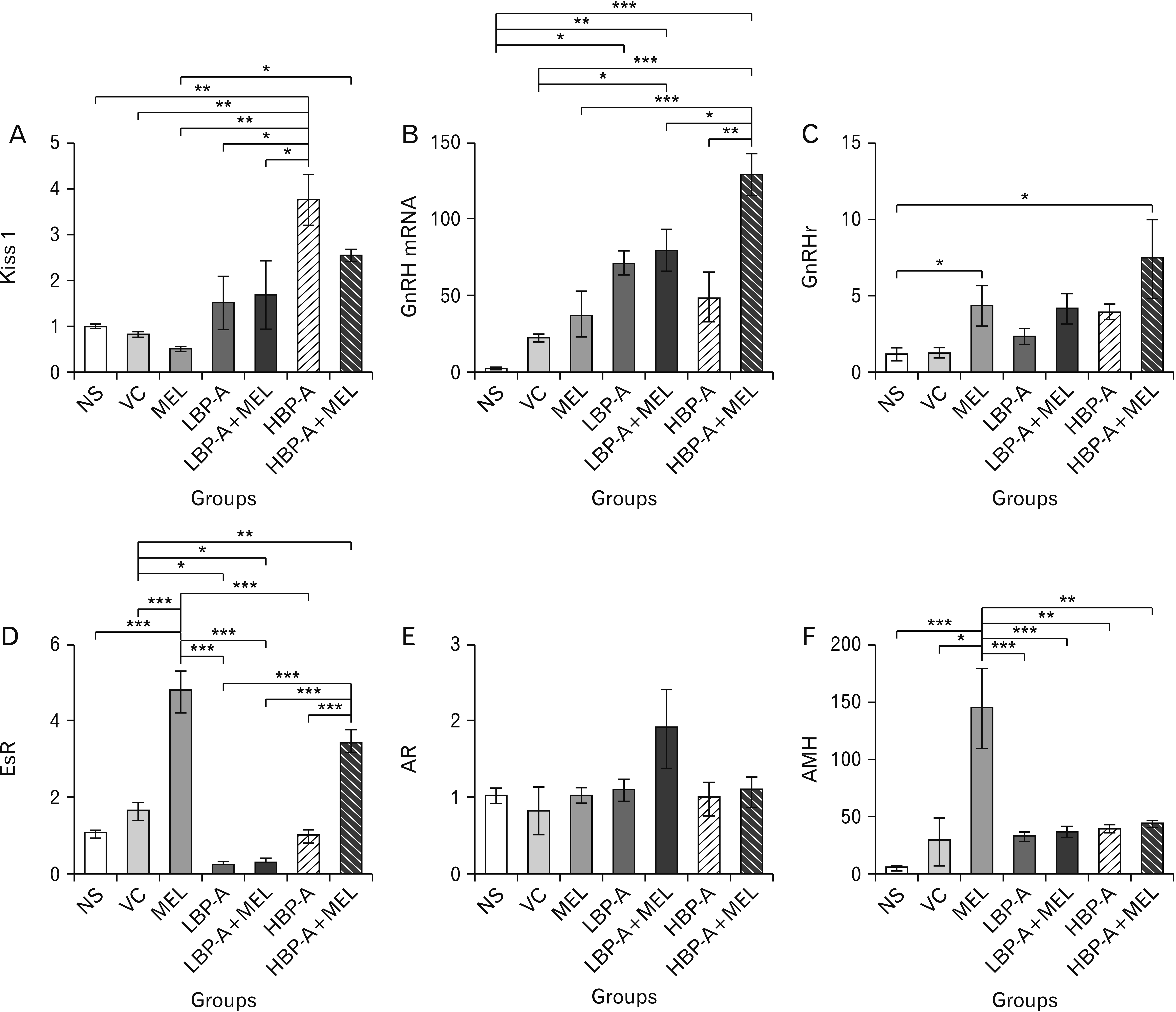 | Fig. 3Showing the expression of Kiss 1 (A), GnRH mRNA (B), GnRH receptor (GnRHr) (C), estrogen receptor (EsR) (D), androgen receptor (AR) (E), and anti-mullerian hormone (AMH) (F) following exposures to normal saline (NS), vehicle control (VC), 10 mg/kg melatonin (MEL), 25 mg/kg bisphenol-A (BPA) (LBP-A), 25 mg/kg BPA+10 mg/kg melatonin (LBP-A+MEL), 50 mg/kg BPA (HBP-A), and 50 mg/kg BPA+10 mg/kg melatonin (HBP-A+MEL). Values are presented as mean±standard error of mean. Asterisks indicate significant differences at *P<0.05, **P<0.01, and ***P<0.001, respectively from all or specific groups.
|
Bisphenol A-induced histological variations
Structural evidence showed that all doses of BPA used in this study significantly decreased follicular count in rats compared to NS- and VC-treated animals. MEL coadministration with BPA was shown to have some increase in this follicular count, especially in the high dose group. Similarly, MEL co-treatment was associated with increase in primary and preantral follicles compared to BPA treated only groups. There was however increase in defective follicles in both BPA and MEL treated groups when compared to control groups (Figs. 4, 5).
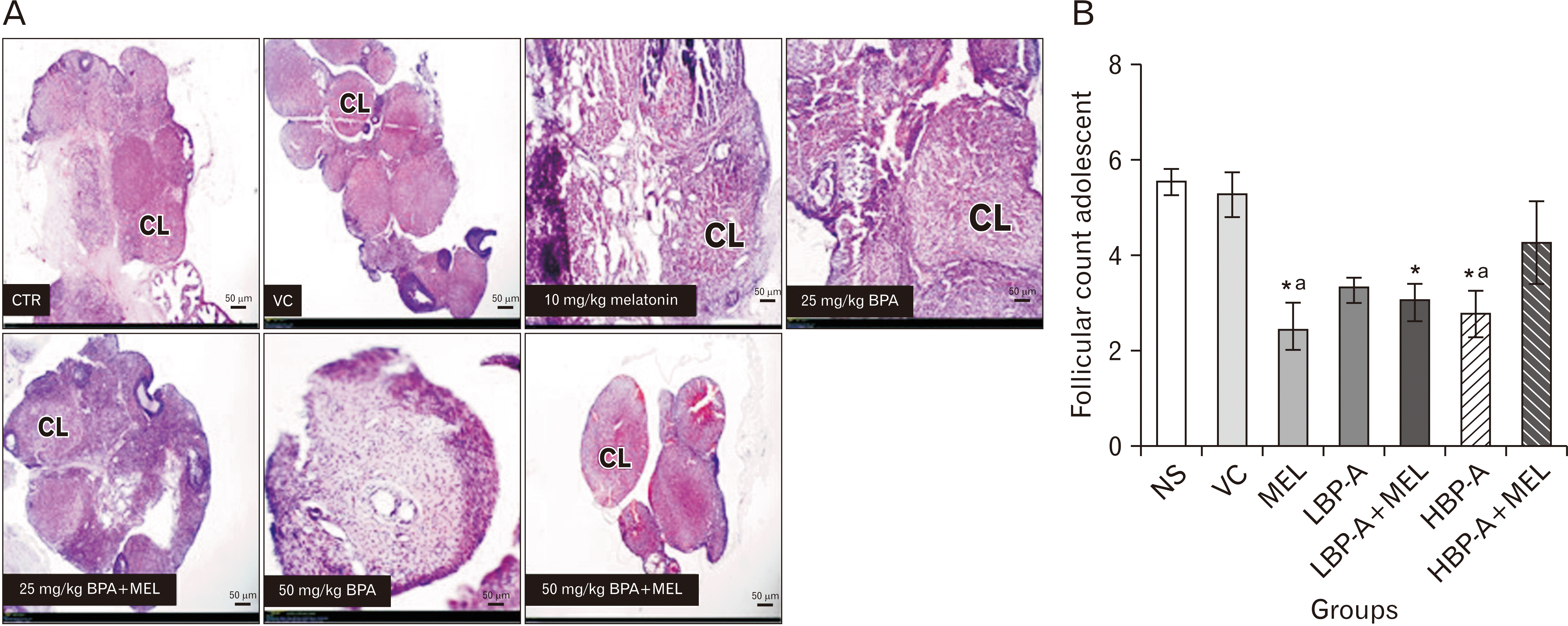 | Fig. 4Showing (A) representative photomicrographs of the ovaries (H&E, ×40) and (B) follicular count following prenatal exposures to normal saline (NS), vehicle control (VC), 10 mg/kg melatonin (MEL), 25 mg/kg bisphenol-A (BPA) (LBP-A), 25 mg/kg BPA+10 mg/kg melatonin (LBP-A+MEL), 50 mg/kg BPA (HBP-A), and 50 mg/kg BPA+10 mg/kg melatonin (HBP-A+MEL). Values are presented as mean±standard error of mean. *Significant decrease from the control; aSignificant decrease from the vehicle control group. CL, corpus luteum; CTR, control.
|
 | Fig. 5Showing the (A) ovarian histoarchitecture (H&E, ×100) and (B) follicle count in adolescent rats exposed to normal saline (NS) is the control group (CTR), vehicle control (VC), 10 mg/kg melatonin (MEL), 25 mg/kg bisphenol-A (BPA), 25 mg/kg BPA+MEL, 50 mg/kg BPA, and 50 mg/kg BPA+MEL. AF, antral follicles; AT, atretic follicles; CL, corpus luteum; FD, follicular degeneration; PA, preantral; PF, primary follicle.
|
Go to :
Discussion
Adolescent stage is a vulnerable phase where slight hormonal alteration could elicit proteins programming that might adversely affect reproductive life in adulthood. In the present study, we profiled the impact of BPA exposure on female rats in their adolescent age via probing the hypothalamo-pituitary-ovarian axis. The question we attempted to answer borders on whether adolescent-related disparity in genetic disposition and physiological handling exist in BPA-exposed adolescent rats. Knowledge on this would improve our understanding on potential stage of target for therapeutics. Although, it was recommended that the Lowest Observed Adverse Effect Level (LOAEL) in animal studies is 50 mg/kg [35-39], however, even at this LOAEL, uncertainties as to the safety of BPA still abound. Therefore, the recommended LOAEL for rodent animals was used, and a lower dose of 25 mg/kg to observe for any possible adverse effects even when exposed to a lower dose. To achieve the set objectives, two doses of BPA, 25 mg/kg and 50 mg/kg were used, and both doses impacted adversely on the animals’ reproductive biology in a none dose-dependent pattern. First, we assessed BPA interference on plasma levels of reproductive hormones. Similar to what was previously reported in adult rodents [40], in the present study, BPA caused hormonal alteration in the adolescent rats used, evidenced in the marked increase in the plasma levels of FSH, LH, estradiol and testosterone in the exposed rats. BPA exposure has been described to encourage the onset of PCOS-like (poly cystic ovarian syndrome-like) abnormalities, which is characterized by elevated gonadotrophs (FSH, LH) as well as testosterone and estradiol [41]. Similarly, our findings are corroborated by some other studies, who have also described these steroid abnormalities, that leads to distortion of these reproductive hormones following exposure to BPA [42, 43].
There were no statistically significant changes observed with coadministration of MEL. There was however a trend of MEL lowering the heightened hormones (FSH, LH, testosterone). This is similar to the findings noted by Anderson et al. [44], where they also found no marked difference when MEL was administered. These suggest that the antioxidant effect of MEL did not have much effect on the restoration of the reproductive hormonal milieu.
Further strengthening the reproductive affective activities of BPA, the plasma anti-mullerian and progesterone hormones were depleted. Exposure to BPA results in antagonism of estradiol effect on progesterone. By decreasing expression in progesterone receptor, BPA decreases the ability of progesterone to inhibit estradiol action. Therefore, this affects the balance between estrogen and progesterone, leading to an increased estrogen response and a marked decrease in progesterone level when compared to controls [45].
Based on previous studies that showed existence of high free BPA in exposed adolescence animals compared to adult [16]. In contrast, to our findings [46], reported increased LH and testosterone levels following MEL administration in young male rats [46].
The physiological relevance of the hormonal disruptions above could at immediate or later in the life of the animal cause appreciable deterioration in reproductive functions [40], and in combination with the persistent decrease in anti-mullerian and progesterone could lead to induced-infertility and lower probabilities of having live births respectively [47]. Thus, our observation on BPA-induced hormonal imbalance at adolescent age seems capable of initiating programming for later life reproductive challenges in the BPA-exposed animals.
Like any other essential chemicals with risks of toxicity, BPA exposure had been implicated to worsen oxidative stress [48]. Thus, MEL intervention was introduced to ameliorate oxidative stress and scavenge radicals’ outburst in BPA exposure in the present study. Our findings here of the over production of NOS and depletion of GPx and UDP suggesting induced oxidative stress following BPA exposure, can be strengthened with previous reports of increased ROS, lipid peroxidation by increased MDA, depletion of catalase, glutathione and glutathione-s-transferase, decreased antioxidant enzyme activity and induced DNA damage, leading to organ toxicity, including reproductive toxicity [49-51].
SOD protects oocytes from oxidative stress. This explains the increased levels of SOD following BPA administration. As BPA may bind to the SOD sites, thereby inhibiting the enzymatic activity of SOD. Hence, decrease the activities of SOD, thereby leading to increase concentration of SOD [52].
Elevated androgen concentration leads to down regulation of the UDP-glucuronosyltransferase activity, and this result in a decrease in detoxification and clearance of BPA [53].
In contrast to the observed oxidative events in BPA toxicity above, MEL alone and its interventions in BPA exposed improved anti-oxidant capacity by reducing NOS, and increasing GPx and UDP productions in the ovaries. This finding confirms the previously reported MEL’s anti-oxidant efficacy [32, 34, 54], and other antioxidants such as vitamins [23, 55], quercetin [24], Garlic acid [25], Ginseng [56], honey [26], and others. It is thus tempting to speculate that the antioxidant capacity of MEL could be used as a potential defence and/or treatment regime against BPA induced toxicity.
Transcript abundance was assessed using qPCR for relevant mRNA (protein) associated with hypothalamo-pituitary-ovarian axis. Amazingly, both doses of BPA and same in combination with MEL caused increased expression of GPR54 in the hypothalamus. With this result, a quick assumption would be that there is high possibility that BPA-exposed adolescent rats could attain sexual maturation as GPR54 plays essential roles in initiation and conservation of mammalian fertility [57]. However, at the adolescence stage of life, abundant expression of GPR54 could induce precocious activation of gonadotropic axis leading to surge in serum LH and estrogen, increased uterus weight and induced ovulation [58]. The gonadotropin regulators, more specifically the mRNAs for gonadotropin releasing hormone and its receptor were then investigated since it acts as direct inputs from the hypothalamus to the pituitary gland.
The reported high expression of the transcriptome for GnRH in the hypothalamus and GnRH receptor on pituitary gonadotropin cells in all the tested groups is in support of our submission on GPR54. Abundant mRNA for GnRH and its receptors showed that protein synthesis capacity of hypothalamus and pituitary gland might not be compromised but its regulation may be perturbed. The regulation for these genes expression is believed to be affected due to the increased transcriptome in the treated groups. Once the gonadotropin cells (via GnRH receptor) are stimulated, the gonadotropin hormones (LH and FSH) are released to act on the gonads. The increase in genes expression recorded also commensurate increased FSH and LH levels with BPA exposures.
Importantly, the released gonadotropins are known to stimulate steroidogenesis in the gonads leading to the synthesis of steroid hormones-estrogen, androgen and progesterone. Our analysis of the androgen hormone (testosterone) showed an appreciable increase in all the BPA-tested groups, while a decline in the mRNA for estrogen receptors in form of the EsR gene in all BPA-treated groups, meanwhile there is no observable difference in the expression of androgen receptor gene. This might be that BPA diminished the expression of ER gene or there might be down-regulation of ER due to excessive secretion of estrogen hormone as alluded to above. Actions of estrogen are carried out via its receptors, thus the decreased ER gene could indirectly translate to affected repair, replace and formation of new ER, which may interfere with the development and regulation of reproductive system as well as secondary sex features.
Our findings show that Androgen receptors were not affected by BPA administration. However, testosterone levels were significantly increased. This is in keeping with previous studies [42]. A blockage of androgen binding sites, alters the feedback control mechanisms. Therefore, this leads to an elevation of circulating testosterone level [42, 43]. Another explanation for this increase in testosterone level is that BPA increase testosterone concentration by stimulating the ovaries to produce testosterone, and it also inhibits T-hydroxylase activity [53].
Lastly, BPA did not pose any observable interference on the mRNA for AMH in all exposed groups, indicating the presence of viable follicles and some preserved ovarian follicular functions. Nonetheless, in contrary to the inference from the AMH gene expression, an histological examination of the ovarian tissue revealed a remarkable reduction in corpus luteum count and noticeable increase in defective follicles following BPA exposures, pointing to the detrimental effects phases of the follicular development. This is corroborated by the observed decrease in the AMH, which is suggestive of a decrease in residual ovarian function compared to the controls.
As the present study demonstrates the effects of BPA exposure on the ovary, which include decrease follicular counts, especially decrease in preantral as well as antral follicles. There has also been report of such effects in rodents that were exposed to different doses of BPA [59]. We examined the effect of long-term MEL treatment on the ovaries. Studies have demonstrated that MEL does not improve maturation rate in oocytes from sheep ovaries [60]. Maganhin et al. [61] also did not find any significant difference in follicle numbers between controls and group treated with MEL. This suggests that MEL does not necessarily affect the follicular counts, as also documented in this study. There has been report that MEL improves ovarian function by increasing the number of secondary and Graaffian follicles as well as corpus luteum and also protects oocytes and granulosa cells from nicotine damage [62], which is contrary to our findings, whereas vitamin C, which is also an antioxidant, has been associated with an improvement in ovarian follicular numbers, though its administration was for a much longer duration than that of MEL in this study vitamin C restores ovarian follicular reservation in a mouse model of aging [63]. MEL not showing reparative numbers may have been due to the short administration compared to that of the vitamin C study, which may not have been long enough for obvious reparative effects on the follicular counts in the ovary, following the prolonged BPA induced effects. We found that different doses of BPA treated animals have morphological changes in the ovaries, having decreased follicle numbers. This is in accordance with the AMH result, which is a hormone that is used in determining ovarian reserve level. We can conclude that in female rats, early exposure to BPA, causes histological alternations in ovary, disrupts folliculogenesis resulting in degenerative changes of ovarian follicles, however, MEL administration is not shown to have obvious histological reparative effects on these follicular counts.
In conclusion, BPA exposure in adolescent rats favoured increase in expression of GRP54, GnRH, and GnRH receptor gene which in turn encourages increase steroidogenesis that lead to production of excessive FSH, LH, Estradiol and testosterone; and eventually down-regulation of the ER. Down-regulated ER synergizes with the induced generation of oxidative radicals to diminishing proper female reproductive development and increased defective follicular cells. Interestingly, although MEL may not have completely been effective which may likely be due to the dosages employed in this study, as well as it being only co-administered, its antioxidant effects gives some promising outturns against BPA induced toxicities.
Go to :
 XML Download