

 PDF
PDF Citation
 Print
Print


Introduction
The external carotid plexus (Figs. 1 and 2) encapsulates the external carotid artery and its branches to innervate the majority of the mid and lower face [1]. The superior pole of the superior cervical ganglion gives rise to postganglionic sympathetic fibers of which some of these branches form connections along the external carotid artery and combine as the external carotid plexus [2, 3]. Generally, the anterior communicating branch of the superior cervical ganglion to the external carotid artery travels directly to the medial side of the external carotid artery [2, 4]. Whereas another branch, the interior communicating branch of the superior cervical ganglion to the superior laryngeal nerve, travels inferiorly and wraps around the superior thyroid artery (STA) before returning superiorly to anastomose with the prior branch to the external carotid artery [2, 5] Branches of the plexus partially continue onto the branches of the external carotid artery. As a result, implications to the plexus or nearby artery may result in neural abnormalities to facial muscles and sweat glands [3]. As this plexus is poorly described in the literature, the present paper aims to review the anatomy, function, and clinical applications of the external carotid plexus for a better understanding among anatomists and clinicians alike.
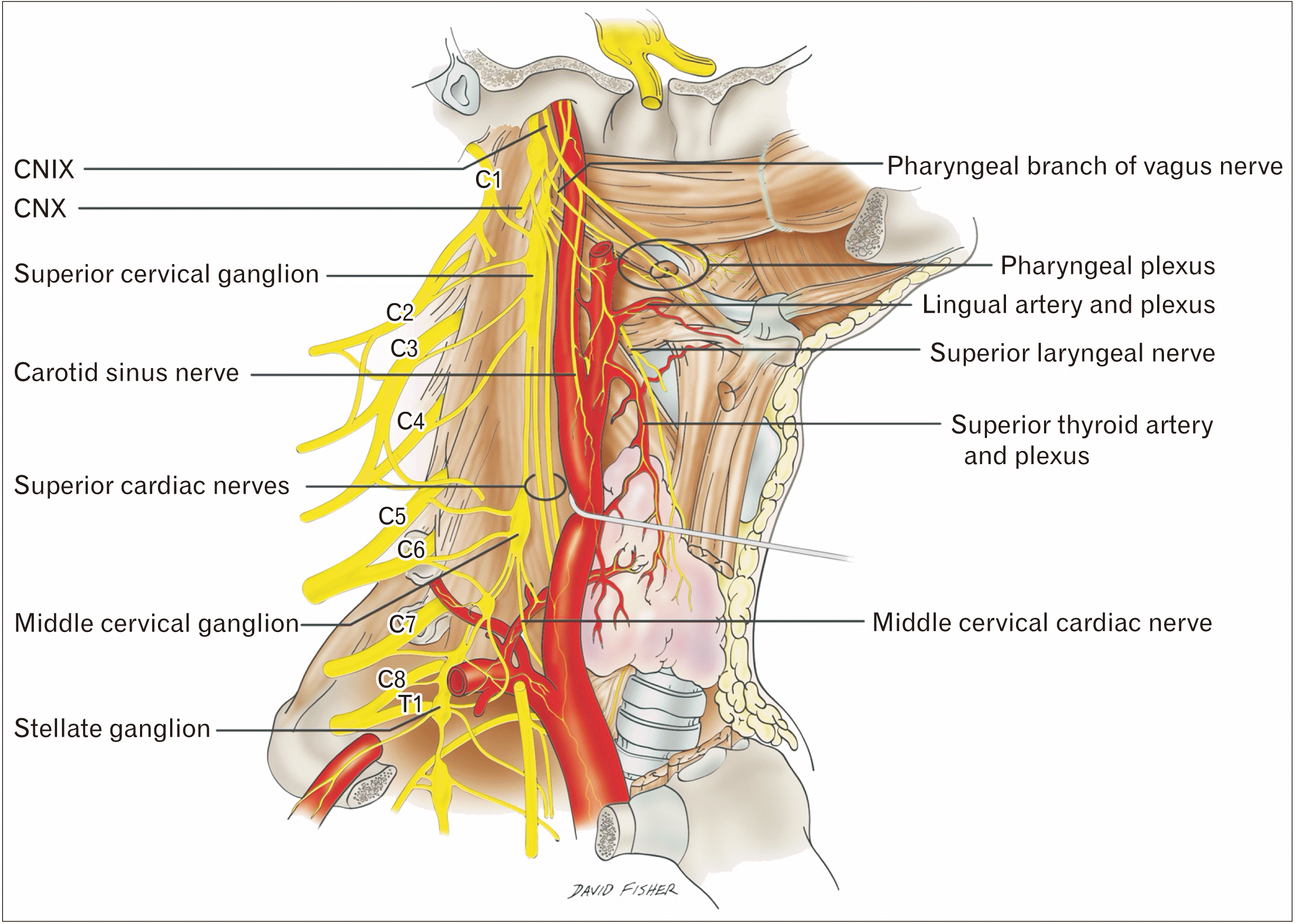 | Fig. 1Schematic drawing illustrating the external carotid plexus. Note the nervous plexus lying on the external carotid artery and its branches e.g., lingual artery and superior thyroid artery.
|
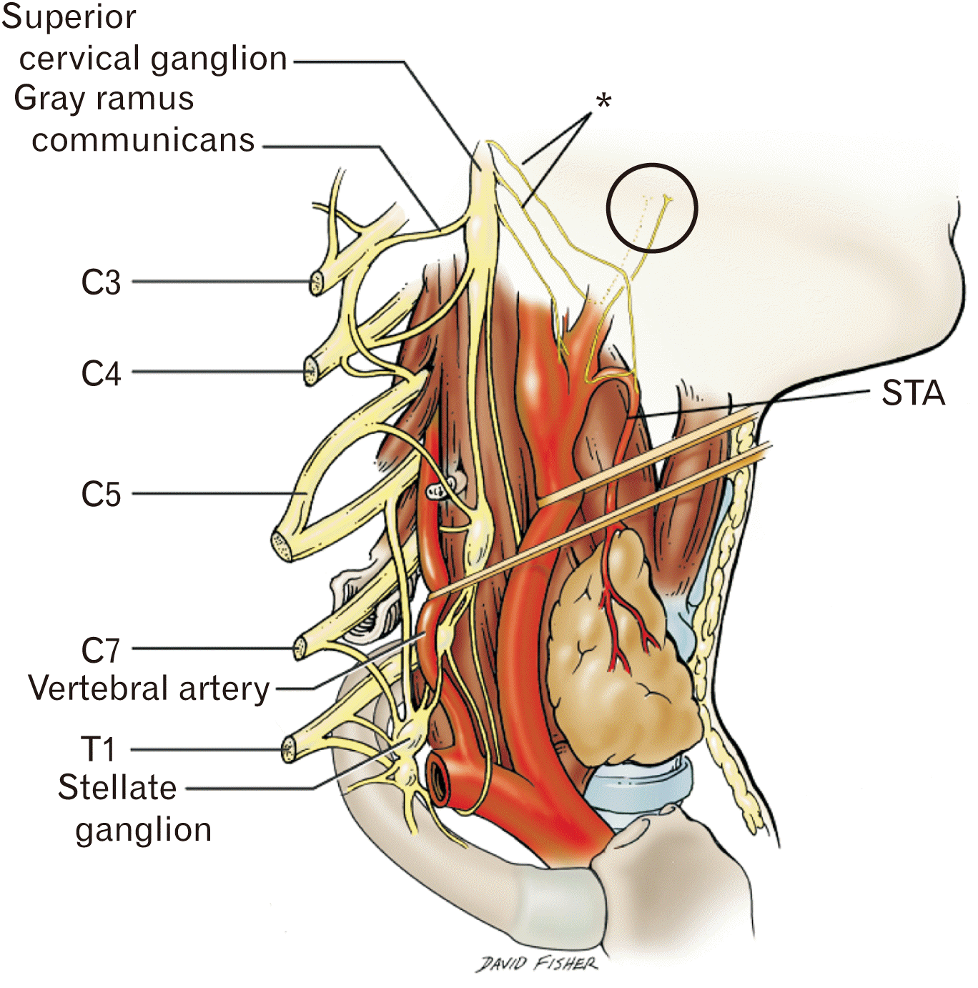 | Fig. 2Schematic of the source of the external carotid plexus on the right side. The superior medial two branches (*) of the superior sympathetic ganglion are shown as the main contributions to the plexus which at its source is seen at the circle with contributions from a branch of the superior cervical ganglion that travels medial (dotted lines) to the external carotid artery and a branch of the ganglion that travels first along the STA then recurs up along the lateral surface of the external carotid artery. A third and unlabeled medial branch from the superior cervical ganglion travels to contribute to the intercarotid plexus between the origin of the external and internal carotid arteries at their origin from the common carotid artery. The combined upper two branches of the superior cervical ganglion will thus travel along the branches of the external carotid artery onto the face. STA, superior thyroid artery.
|
Go to : 
Anatomy
The external carotid plexus originates from the superior pole of the superior cervical ganglion [2]. Typically, this pole either produces one branch that splits into several branches or may immediately produce an anastomosing plexus from small fascicles that later splits into three to four branches [2]. Three branches are commonly identified from the superior pole of the superior cervical ganglion. The first branch, the inferior branch of the superior cervical ganglion, is involved with the formation of the intercarotid plexus [2, 6]. The second branch, the anterior communicating branch of the superior cervical ganglion to the external carotid artery, follows the external carotid artery medially and is involved with the formation of the external carotid plexus [2, 4]. The third branch, the interior communicating branch of the superior cervical ganglion to the superior laryngeal nerve, travels posteriorly and medially down the external carotid artery until it arrives at the STA [2, 5]. This branch anastomoses with the superior laryngeal branch of the vagus nerve [2, 5]. Then, this branch travels around the STA or the infrahyoid branch of the STA and returns anterior to the external carotid artery until it anastomoses with the second branch [2]. Upon the combination of the second and third branches, the external carotid plexus forms within the connective tissue surrounding the external carotid artery [2].
The external carotid plexus runs partially into the branches of the external carotid artery. This plexus travels down the external carotid artery and continues onto the common carotid artery, connecting with the common carotid plexus [7]. The communicating branch of the superior cervical ganglion to the superior laryngeal nerve gives off fine filaments that supply the STA with the vagus nerve, resulting in a secondary plexus known as the superior thyroid plexus [2, 7]. Additionally, this branch supplies twigs to the lingual artery, which form a secondary plexus named the lingual plexus [7]. Small branches of the external carotid plexus travel with the occipital artery and the ascending pharyngeal artery [2]. The external carotid plexus continues to travel superiorly with fine rami traveling shortly with the superficial temporal artery [2]. Many more branches travel with the facial artery to form an additional facial artery plexus [7]. The external maxillary plexus then intertwines perpendicularly with branches of the facial nerve near the mandible [2]. Along the maxillary artery, the plexus divides into small filaments within dense connective tissue including few filaments from the internal maxillary plexus that travel with the middle meningeal artery to form the meningeal plexus [2, 7]. The meningeal plexus gives off additional filaments to the otic ganglion, which provides sympathetic innervation to the parotid gland, and occasionally to the external superficial petrosal nerve [8]. Few filaments continue with the inferior alveolar artery [2]. Moreover, small filaments join blood vessels into the submandibular gland and ganglion [2].
The majority of the external carotid plexus originates from the interior communicating branch of the superior cervical ganglion to the superior laryngeal nerve [2]. This branch remains fairly constant [2]. Occasionally, the communicating branch of the superior cervical ganglion to the superior laryngeal nerve may instead travel around the lingual artery and provide a separate branch that travels to the STA [2]. Additionally, this plexus may develop a ganglion enlargement, also known as the temporal ganglion, near the origin of the posterior auricular artery [9]. Previous cases have also identified communications between this plexus and other nerves. For example, cutaneous branches of the external carotid plexus in the cervical region were found intermingling with the supernumerary branch of the glossopharyngeal nerve as well as branches that originated from the vagus nerve [10, 11].
Research in other animals, such as Bos grunniens or yaks, identified their external carotid nerve to consist of two nerve branches [12]. Of those two branches, the branch originating from the caudoventral margin of the cranial cervical ganglion travelled ventrally on the external pharynx and ultimately joined the external carotid plexus near the carotid sinus [12].
Go to :
Function
The external carotid plexus is composed of postganglionic sympathetic fibers originating from the superior cervical ganglion [3]. Since the external carotid plexus encompasses the external carotid artery and partially continues onto the branches of the external carotid artery, the external carotid plexus is involved with innervating the majority of the mid and lower face [1]. Ligature to arteries, such as the superior thyroid artery, may disrupt the innervations of the external carotid plexus and its branches [2]. Branches of the external carotid plexus also periodically dissociate from the external carotid artery’s blood vessels to join peripheral nerves as they enter into target tissues [1]. Therefore, the external carotid plexus is involved in many innervations to separate parts of the face.
The pterygopalatine fossa receives its sympathetic input through the external carotid plexus, combined with the internal carotid plexus, the vidian nerve, and the plexus of the maxillary nerve [13]. Additionally, the external carotid plexus is involved in innervating facial sweat glands [14]. Previous research suggest that the external carotid plexus innervates sweat glands on the lateral side of the forehead, whereas the internal carotid plexus innervates the medial forehead’s sweat glands [15]. The external carotid plexus passes filaments to the carotid body and thyroid glands [7]. Furthermore, the external carotid plexus innervates the parotid gland [16]. Activation of all facial glands does not likely occur upon solely stimulating the external carotid nerves. Specifically, research on rat superior cervical ganglia has discovered that though the cervical sympathetic nerve may excite both the external and internal carotid nerves, stimulation of either the internal or external carotid nerves only initiates antidromic effects on the cervical sympathetic nerve [3]. Transmission of action potentials is not typically transferred from the external carotid nerve to the internal carotid nerve or vice versa [3]. Additionally, nerve fibers travel from the cervical sympathetic nerve through the superior cervical ganglion and to the external carotid nerve with no synaptic relay [3]. Consequently, the effects of the external carotid plexus do not usually interact with the internal carotid plexus unless their branches are combined at a single target tissue.
Go to :
Pathology
Damage to sympathetic fibers at any location along the cervical sympathetic trunk oftentimes lead to symptoms of Horner’s syndrome [17]. Symptoms of Horner’s syndrome include miosis, ptosis, anhidrosis, enophthalmos, and facial flushing. For example, retracting the carotid sheath or longus colli muscle using an anterior cervical surgical approach runs the risk of causing tension on the sympathetic trunk and may result in Horner’s syndrome [17]. Because of the external carotid plexus’ relationship to the sympathetic trunk, traction on the plexus may also lead to temporary symptoms or partial Horner’s syndrome. Harlequin syndrome, unilateral facial flushing that often occurs with Horner’s syndrome, may also result from injury or tension on the external carotid plexus as the nerves supply the cheeks and other facial features [18]. Additionally, the branches of the external carotid artery and its surrounding plexus may likely be involved in other neurological conditions such as Sluder’s neuralgia, cluster headache and sympathetic neuralgia in the face [13]. With these neurological conditions, anesthesia using specific or non-specific adrenergic blocks were used to relieve the pain [13].
Carotid sympathetic plexus schwannoma, benign brain tumors, are rare and have been documented in only three cases [14]. In these three cases, the schwannoma was present in either the cavernous sinus part of the plexus or the petrous part of the carotid canal [14]. Therefore, these schwannomas typically arise from the internal carotid nerve [14]. Removal of the schwannoma from the cavernous plexus also resulted in partial Horner syndrome [14].
Go to :
Clinical Applications
The external carotid plexus is an intricate and important neural aspect when considering surgical procedures of the head and neck. Symptomatic internal carotid artery occlusion has been treated successfully using an external carotid artery endarterectomy [19]. This method has been able to increase cerebral perfusion and improve cognitive function [19]. In a study of 195 external carotid artery endarterectomies, 83% of patients had resolution of their symptoms, 3% of patients had perioperative mortality rates most commonly due to neurologic deaths, and 5% of patients experienced an overall neurologic complication rate [20]. Therefore, in most cases, the external carotid plexus was likely able to remobilize and develop new connections to maintain neurologic functioning. Atypical facial neuralgia and buccal neuralgia are also involved in pain typically in areas supplied by the external carotid artery.2 Consequently, identifying and denervating the external carotid plexus may provide some relief to the pain without severe complications such as Horner’s syndrome [2].
The branches of the external carotid artery also provide methods for chemoembolization of head and neck tumors [21]. Common feeding vessels to these tumors include the STA and the lingual artery [21]. Nevertheless, with excision of these tumors, functional and aesthetic disabilities may occur such as abnormal anastomosis between the STA and the lingual artery [21]. Various flaps have been developed to improve coverage over these excisions, yet anastomosis issues may damage the flap with necrosis or hemorrhage [21]. Damage to the external carotid plexus may occur with surgical excision of these tumors. Consequently, further neurologic complications from injury to the external carotid plexus may occur in the face and neck, such as pain, abnormal facial sweating, and abnormal facial flushing. Yet with proper implantation of these flaps, new branches may form within the plexus to promote improved neural stimulation around the surgical areas.
During aesthetic and plastic surgery procedures, perforators from the transverse facial artery provide one of the main arterial sources [22]. The transverse facial artery is a branch of the superficial temporal artery, originating from the external carotid artery. This artery supplies the lateral face, parotid gland, parotid duct, masseter muscle, facial nerve, and the integument [22]. Face lift flap procedures may risk hematoma, facial nerve palsy, and facial skin sloughing and necrosis [22]. Surgical procedures involving the ligation, resection or mobilization of the external carotid artery and its branches, such as face lift flap procedures and carotid endarterectomies, increase the risk of injury towards the external carotid plexus with, for example, postoperative complications such as facial flushing [16]. Damage to branches of the external carotid plexus or abnormal postoperative anastomoses of the branches of the plexus during these procedures may then promote the nerve palsy or reduce innervation to the lateral face. Four cases have documented patients’ loss of sympathetic innervation to their parotid gland after a carotid endarterectomy [16]. As a result, the patients developed first bite syndrome, a sudden and intense pain near the parotid region [16]. Therefore, identifying and limiting contact with the external carotid plexus during general, aesthetic and plastic surgery procedures may provide an important marker to decrease abnormal postoperative complications.
Go to :
 XML Download