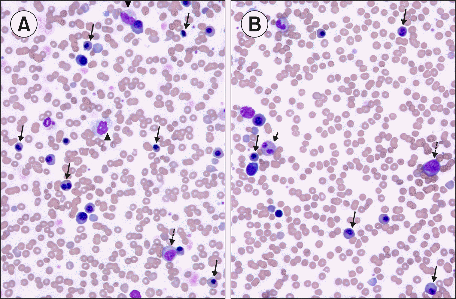

A 15-year-old, previously healthy male with low-risk obesity and recent SARS-CoV-2 exposure presented with severe hyperglycemic hyperosmolar syndrome, metabolic encephalopathy, and hypovolemic shock necessitating mechanical ventilation. Biochemical tests showed elevated ferritin (3,542 mg/L), triglycerides (10.5 mmol/L), and glucose (32.2 mmol/L). His CBC showed mildly elevated leukocytes (13,900/mL) and neutrophils (11,920/mL). An ultrasound showed hepatomegaly. He rapidly developed anemia (Hb 7.6 g/dL), thrombocytopenia (platelets, 57,000/mL), and monocytosis (5,900/mL) with continued worsening shock requiring multiple inotropes, dialysis, and increased ventilatory support. After initially improving, he again developed significant lung disease along with a new fever. He was then diagnosed with SARS-CoV-2 via PCR and severe acute respiratory distress syndrome. Ultimately, he was cannulated on to veno-venous extracorporeal life support for refractory hypoxemic respiratory failure. He was also diagnosed with macrophage activation syndrome secondary to the SARS-CoV-2 infection. For this, he was treated with steroids and 6 days of Anakinra. Laboratory testing showed increased ferritin (19,894 mg/L), sCD25 (1,770 U/mL), inflammatory cytokines (IL-6, IL-8, IL-10, IL-18, and CXCL9), and decreased CD107a (4%). A PB smear showed numerous nucleated RBC (352/100 WBC), including 30% dysmorphic nRBCs (long arrows), immature granulocytes (long dotted arrows) (10%), vacuolated monocytes (arrowheads) (22%), and occasional bilobed neutrophils (short arrows) (A and B). Ultimately, he developed intracranial hemorrhagic complications, and support was withdrawn.


 PDF
PDF Citation
 Print
Print


 XML Download