

 PDF
PDF Citation
 Print
Print


INTRODUCTION
Coronavirus disease 2019 (COVID-19) caused by severe acute respiratory syndrome coronavirus 2 (SARS-CoV-2) has become a global pandemic with continued transmission [1, 2]. Since there are currently no effective treatments for COVID-19, considerable efforts are focused on developing vaccines and therapeutic drugs [3]. However, the dynamics of humoral immune responses of COVID-19 patients using different serological assay platforms are largely unclear.
A wide range of SARS-CoV-2 binding antibody assays have been developed with different antigen targets and assay formats [4]. These assays can detect either isotype-specific antibodies (IgG, IgA, IgM) or total antibodies based on different assay principles such as chemiluminescent immunoassay (CLIA), enzyme immunoassay (EIA), lateral flow immunoassay (LFIA), and microsphere-based antibody assay. Most of these assays mainly target either spike (S) protein (the most exposed viral protein) or its receptor-binding domain (RBD), or nucleocapsid (N) protein, which is abundantly expressed. Although the performance and clinical utility of different binding antibody assays continue to be identified, the currently available assays show variable performance, and data on the early immune response and seroconversion are insufficient [4–6]. Many questions have also been raised about the index value of antibody assays for COVID-19 monitoring.
There is great interest in identifying SARS-CoV-2 neutralizing antibodies for measuring immune status and assessing vaccine responses. Neutralizing antibodies against both the viral S and N proteins have been found in COVID-19 patients, pointing to the potential value of SARS-CoV-2 binding antibody assays as a surrogate for neutralizing titers [7–9]. A SARS-CoV-2 surrogate virus neutralization test (sVNT) (GenScript Inc., Leiden, the Netherlands) is available for detecting neutralizing antibodies that block the interaction between angiotensin-converting enzyme 2 (ACE2) receptor protein and the RBD. However, limited data are available correlating the results of commercial assays with the presence of neutralizing antibodies detected by the sVNT.
We compared the serological characteristics and seroconversion ability in serial serum samples from COVID-19 patients using 12 commercial antibody assays: three automated, high-throughput CLIAs [SARS-CoV-2 IgG assay (Abbott, Chicago, IL, USA), Elecsys Anti-SARS-CoV-2 assay (Roche, Basel, Switzerland), and SARS-CoV-2 Total assay (Siemens, Munich, Germany)]; three EIAs [COVID-19 ELISA IgM+IgA and COVID-19 ELISA IgG (Vircell Microbiologists, Granada, Spain), anti-SARS-COV-2 ELISA (IgA) and anti-SARS-COV-2 ELISA (IgG) (Euroimmun AG, Lübeck, Germany), and Platelia SARS-CoV-2 Total Ab (BioRad Laboratories, Hercules, CA, USA)]; five LFIAs [ichroma COVID-19 Ab (Boditech Med Inc., Gangwon-do, Korea), STANDARD Q COVID-19 IgM/IgG Combo assay (SD Biosensor Inc., Gyeonggi-do, Korea), PCL COVID-19 IgG/IgM Rapid Gold (PCL Inc., Seoul, Korea), SGTi-flex COVID-19 IgM/IgG (Sugentech Inc., Daejeon, Korea), and Biocredit COVID-19 IgG+IgM Duo (Rapigen Inc., Gyeonggi-do, Korea)]; and one SARS-CoV-2 sVNT (GenScript Inc., Piscataway, NJ, USA). To our knowledge, this is the first study to compare 12 SARS-CoV-2 antibody assays using various assay platforms for assessing the early antibody response, seroconversion, neutralizing capacity, and association with disease severity during the early infection period in COVID-19 patients.
MATERIALS AND METHODS
Patients and samples
For antibody response assessment, we retrieved 139 serial serum samples from 49 COVID-19 patients. All diagnoses were confirmed by real-time RT-PCR testing between March 2020 and October 2020 at Seoul St. Mary’s Hospital, Eunpyeong St. Mary’s Hospital, or Samkwang Medical Laboratories, Seoul, Korea. We also retrieved 195 serum samples from healthy donors to assess the negative percent agreement (NPA), including 95 serum samples collected before November 2019 (in the pre-COVID-19 period) and 100 serum samples from organ donors who tested negative for SARS-CoV-2 by real-time RT-PCR at Seoul St. Mary’s Hospital. Real-time RT-PCR with nasopharyngeal swabs was performed in the three laboratories using an Allplex 2019-nCoV Real-time PCR kit (Seegene, Seoul, Korea), PowerChek 2019-nCoV kit (KogeneBiotech, Seoul, Korea), or Real-Q 2019-nCoV Real-Time Detection kit (BioSewoom, Seoul, Korea), according to each manufacturer’s instructions. Serum remnants were retrieved from blood samples collected for routine laboratory assays. All serum samples were stored at 4°C for up to two weeks and aliquoted for assessment. Serum aliquots were stored at −80°C before the assays. Due to insufficient sample volumes, only 109 consecutive serum samples from 36 patients were subjected to the EIAs from Euroimmun, Vircell, and BioRad.
To assess the seroconversion response, we retrieved 75 serum samples from 10 COVID-19 patients during hospitalization at Seoul St. Mary’s Hospital. Each patient’s seroconversion sera set consisted of at least five serial samples with an initially SARS-CoV-2 antibody-negative result from any available commercial assay. Clinical data for the day after symptom onset and disease severity were collected retrospectively from electronic medical records. Disease course was classified as mild, severe, or critical, according to a previous definition [1]. The 10 seroconversion cases comprised six mild cases and four severe cases, including one critical case. All 75 samples used for seroconversion evaluation were subjected to all 12 assays.
This study was approved by the Institutional Review Board (IRB) of the respective institutions (XC20SIDI0069, Seoul and Eunpyeong St. Mary’s Hospitals; S-IRB-2020-007-05-15, Samkwang Medical Laboratories). The requirement for written informed consent was waived by the IRBs because of the retrospective study design.
SARS-CoV-2 antibody assays
All samples were assessed using 11 SARS-CoV-2 binding antibody assays and one sVNT (GenScript). Detailed descriptions of the assay kits are shown in Supplemental Data Table S1. All assays were performed at Seoul St. Mary’s Hospital, according to the manufacturers’ instructions. For the eight assays that present unit or index values (Boditech-IgM and -IgG, Vircell-IgM+IgA and -IgG, Euroimmun-IgA and -IgG, BioRad-Total Ig, Abbott-IgG, Siemens-Total IgG, Roche-Total IgG, and GenScript), we compared quantitative antibody responses of seroconversion panels in patients with a severe or mild disease course. The correlation between SARS-CoV-2 binding antibody assay and sVNT results was also analyzed.
Statistical analysis
Agreement between assays was calculated using the Cohen kappa agreement value. Kappa values were categorized as slight (0–0.20), fair (0.21–0.40), moderate (0.41–0.60), substantial (0.61–0.80), and excellent (0.81–1.00) [10]. Pearson correlation coefficients were calculated for correlations between SARS-CoV-2 binding antibody assay and sVNT results, which were defined as strong (0.7–1.0), moderate (0.5–0.7), and weak (0.3–0.5)[11]; Spearman correlation coefficients greater than 0.5 were deemed strong, those below 0.3 were deemed weak, and those between 0.3 and 0.5 were deemed moderate. Graphs were created with Graph Pad Prism 9.0 (GraphPad Software, Inc., San Diego, CA, USA) and Microsoft Excel 2016 (Microsoft Co., Santa Rosa, CA, USA). The positive percent agreement (PPA) for COVID-19 diagnosis was assessed based on days from symptom onset. To demonstrate PPAs, samples were divided into the following eight groups according to days from symptom onset: 2–5 days, 6–8 days, 9–10 days, 11–13 days, 14–16 days, 17–20 days, 21–27 days, and 28–40 days (Supplemental Data Fig. S1). In addition, PPA and NPA of kits were calculated in three groups according to days from symptom onset: 0–7, 8–14, and >14 days (Table 1). PPA, NPA, and correlation coefficients were calculated using MedCalc version 19.6 (MedCalc Software Ltd., Ostend, Belgium). P<0.05 was considered statistically significant.
Table 1
Positive and negative percent agreement of 12 SARS-CoV-2 antibody assays
| Days from symptom onset | CLIA | EIA | LFIA | sVNT | ||||||||||||||||
|---|---|---|---|---|---|---|---|---|---|---|---|---|---|---|---|---|---|---|---|---|
|
|
|
|
|
|||||||||||||||||
| Abbott | Siemens | Roche | Euroimmun | Vircell | BioRad | Boditech | SD biosensor | PCL | Sugentech | Rapigen | GenScript | |||||||||
|
|
|
|
|
|
|
|
|
|
|
|
|
|||||||||
| IgG (%) | Total (%) | Total (%) | IgA (%) | IgG (%) | IgM+IgA (%) | IgG (%) | Total (%) | IgM (%) | IgG (%) | IgM (%) | IgG (%) | IgM (%) | IgG (%) | IgM (%) | IgG (%) | IgM (%) | IgG (%) | IgG (%) | ||
| PPA | < 8* | 40.0 | 24.0 | 40.0 | 21.1 | 15.8 | 47.4 | 42.1 | 47.4 | 40.0 | 32.0 | 44.0 | 40.0 | 40.0 | 24.0 | 56.0 | 32.0 | 36.0 | 36.0 | 40.0 |
| 8–14† | 71.4 | 63.3 | 75.5 | 82.5 | 55.0 | 72.5 | 77.5 | 90.0 | 73.5 | 75.5 | 83.7 | 69.4 | 83.7 | 71.4 | 91.8 | 73.5 | 87.8 | 75.5 | 85.7 | |
| > 14‡ | 92.3 | 87.7 | 89.2 | 90.0 | 90.0 | 94.0 | 84.0 | 92.0 | 86.2 | 96.9 | 96.9 | 92.3 | 92.3 | 93.8 | 98.5 | 90.8 | 93.8 | 89.2 | 98.5 | |
| NPA | NA§ | 100.0 | 100.0 | 100.0 | 94.9 | 100.0 | 97.4 | 100.0 | 99.5 | 98.5 | 99.0 | 99.0 | 100.0 | 99.0 | 98.5 | 96.4 | 100.0 | 99.0 | 97.9 | NT |
*Includes 25 samples from 22 patients for CLIAs, sVNTs, and LFIAs; 19 samples from 16 patients for EIAs. †Includes 49 samples from 34 patients for CLIAs, sVNTs, and LFIAs; 40 samples from 25 patients for EIAs. ‡Includes 65 samples from 25 patients for CLIAs, sVNTs, and LFIAs; 50 samples from 18 patients for EIAs. §N patients=195.
![]()
RESULTS
Positive and negative percent agreement
As shown in Supplemental Data Fig. S1, the two EIAs and five LFIAs detected IgM or IgA isotype antibodies separately from IgG. IgM or IgA antibody assays tended to have a higher detection rate than the IgG assays, especially in the early infection period (<14 days from symptom onset).
The serum samples were then subdivided into three groups according to days from symptom onset: 0–7, 8–14, and >14 days. The PPA of the 12 antibody assays for each group are shown in Table 1. For samples collected >14 days after symptom onset, the PPA ranged from 87.7% to 92.3% for the CLIAs, from 84.0% to 94.0% for the EIAs, from 86.2% to 98.5% for the five LFIAs, and 98.5% for sVNT.
The NPA was evaluated for the 11 SARS-CoV-2 binding antibody assays, except the sVNT, using the 195 COVID-19–negative sera. All the three CLIAs showed no false-positive results with NPAs of 100%. The NPA for the EIAs and LFIAs ranged from 94.9% to 100%. No sample exhibited false-positivity in the majority of assays.
Seroconversion positivity
To compare the time to seropositivity between assays, the numbers of positive bleeds per total number of serial bleeds were calculated (Table 2). Evaluation of the 10 seroconversion panels consisting of 75 serum samples showed that the ratio of positive bleeds was higher in IgM or IgA assays than in IgG assays except for the results from Vircell kits. Differences in the time to seropositivity were detected between assays for each patient. The five LFIA-IgM and -IgG assays detected 76.0%–88.0% and 64.0%–74.7% seropositivity, respectively. Of the 10 patients, eight showed seroconversion up to two weeks after symptom onset using all 12 assays. Patient 3, who had a critical disease course, showed seropositivity between 18–22 days after symptom onset, whereas patient 10, who had a mild disease course, showed no seropositivity until 25 days using all three CLIAs.
Table 2
Comparison of SARS-CoV-2 antibody assays using 10 seroconversion panels
| Seroconversion panel | CLIA | EIA | |||||||||||
|
|
|
||||||||||||
| Abbott | Siemens | Roche | Euroimmun | Vircell | BioRad | ||||||||
|
|
|
|
|
|
|
|
|||||||
| Patient | N | (Days) | IgG | Total | Total | IgA | IgG | IgM+IgA | IgG | Total | |||
| 1 | 9 | (7–30) | 7/9 (11)* | 7/9 (11) | 7/9 (11) | 7/9 (11) | 7/9 (11) | 9/9 (7) | 7/9 (11) | 9/9 (7) | |||
| 2 | 8 | (6–21) | 5/8 (10) | 5/8 (10) | 5/8 (10) | 6/8 (9) | 4/8 (12) | 5/8 (10) | 5/8 (10) | 7/8 (8) | |||
| 3 | 11 | (9–30) | 7/11 (16) | 5/11 (18) | 5/11 (18) | 5/11 (18) | 5/11 (18) | 1/11 (28) | 5/11 (18) | 6/11 (17) | |||
| 4 | 7 | (3–15) | 7/7 (3) | 2/7 (10) | 6/7 (5) | 1/7 (15) | 2/7 (10) | 7/7 (3) | 7/7 (3) | 7/7 (3) | |||
| 5 | 8 | (12–32) | 6/8 (16) | 7/8 (14) | 7/8 (14) | 8/8 (12) | 7/8 (14) | 7/8 (14) | 7/8 (14) | 8/8 (12) | |||
| 6 | 7 | (10–24) | 6/7 (11) | 2/7 (18) | 6/7 (11) | 7/7 (10) | 1/7 (24) | 6/7 (11) | 7/7 (10) | 7/7 (10) | |||
| 7 | 7 | (9–23) | 7/7 (9) | 7/7 (9) | 7/7 (9) | 7/7 (9) | 5/7 (11) | 7/7 (9) | 7/7 (9) | 7/7 (9) | |||
| 8 | 5 | (5–31) | 4/5 (11) | 4/5 (11) | 4/5 (11) | 4/5 (11) | 4/5 (11) | 4/5 (11) | 4/5 (11) | 4/5 (11) | |||
| 9 | 5 | (5–33) | 4/5 (14) | 4/5 (14) | 4/5 (14) | 4/5 (14) | 4/5 (14) | 4/5 (14) | 4/5 (14) | 4/5 (14) | |||
| 10 | 8 | (5–25) | 0/8 ( > 25) | 0/8 ( > 25) | 0/8 ( > 25) | 6/8 (9) | 4/8 (14) | 6/8 (9) | 4/8 (14) | 6/8 (9) | |||
| Total | 75 | 53/75 | 43/75 | 51/75 | 55/75 | 43/75 | 56/75 | 57/75 | 65/75 | ||||
| 70.7% | 57.3% | 68.0% | 73.3% | 57.3% | 74.7% | 76.0% | 86.7% | ||||||
| Seroconversion panel | LFIA | sVNT | |||||||||||
|
|
|
||||||||||||
| Boditech Med | SD biosensor | PCL | Sugentech | Rapigen | GenScript | ||||||||
|
|
|
|
|
|
|
|
|||||||
| Patient | N | (Days) | IgM | IgG | IgM | IgG | IgM | IgG | IgM | IgG | IgM | IgG | IgG |
| 1 | 9 | (7–30) | 8/9 (9) | 7/9 (11) | 9/9 (7) | 6/9 (14) | 8/9 (9) | 7/9 (11) | 9/9 (7) | 6/9 (14) | 8/9 (9) | 7/9 (11) | 7/9 (11) |
| 2 | 8 | (6–21) | 5/8 (10) | 5/8 (10) | 6/8 (9) | 6/8 (9) | 5/8 (10) | 5/8 (10) | 7/8 (8) | 6/8 (9) | 6/8 (9) | 6/8 (9) | 5/8 (10) |
| 3 | 11 | (9–30) | 6/11 (17) | 6/11 (17) | 6/11 (17) | 6/11 (17) | 5/11 (18) | 4/11 (22) | 7/11 (16) | 4/11 (22) | 6/11 (17) | 4/11 (22) | 7/11 (16) |
| 4 | 7 | (3–15) | 7/7 (3) | 6/7 (5) | 6/7 (5) | 6/7 (5) | 6/7 (5) | 1/7 (12) | 7/7 (3) | 6/7 (5) | 6/7 (5) | 6/7 (5) | 5/7 (6) |
| 5 | 8 | (12–32) | 8/8 (12) | 8/8 (12) | 8/8 (12) | 6/8 (16) | 8/8 (12) | 7/8 (14) | 8/8 (12) | 6/8 (16) | 8/8 (12) | 6/8 (16) | 8/8 (12) |
| 6 | 7 | (10–24) | 7/7 (10) | 6/7 (11) | 7/7 (10) | 6/7 (11) | 7/7 (10) | 5/7 (12) | 7/7 (10) | 6/7 (11) | 7/7 (10) | 7/7 (10) | 7/7 (10) |
| 7 | 7 | (9–23) | 7/7 (9) | 7/7 (9) | 7/7 (9) | 7/7 (9) | 7/7 (9) | 7/7 (9) | 7/7 (9) | 7/7 (9) | 7/7 (9) | 7/7 (9) | 7/7 (9) |
| 8 | 5 | (5–31) | 0/5 ( > 31) | 4/5 (11) | 4/5 (11) | 5/5 (5) | 4/5 (11) | 4/5 (11) | 4/5 (11) | 4/5 (11) | 4/5 (11) | 4/5 (11) | 4/5 (11) |
| 9 | 5 | (5–33) | 4/5 (14) | 4/5 (14) | 4/5 (14) | 4/5 (14) | 4/5 (14) | 4/5 (14) | 4/5 (14) | 4/5 (14) | 4/5 (14) | 4/5 (14) | 4/5 (14) |
| 10 | 8 | (5–25) | 5/8 (11) | 3/8 (18) | 5/8 (11) | 1/8 (25) | 5/8 (11) | 4/8 (14) | 6/8 (9) | 1/8 (25) | 5/8 (11) | 3/8 (25) | 4/8 (14) |
| Total | 75 | 57/75 | 56/75 | 62/75 | 53/75 | 59/75 | 48/75 | 66/75 | 50/75 | 61/75 | 54/75 | 58/75 | |
| 76.0% | 74.7% | 82.7% | 70.7% | 78.7% | 64.0% | 88.0% | 66.7% | 81.3% | 72.0% | 77.3% | |||
![]()
Seroconversion responses related to disease severity
Seroconversion responses in association with disease severity were compared among eight assays (Abbott, Siemens, Roche, Boditech, Euroimmun, Vircell, BioRad, and GenScript) based on index values and percentage (Fig. 1). The antibody values varied depending on both the assay and the disease course. In patients with severe disease courses, Siemens, Euroimmun IgG, and GenScript assays targeting the S protein or RBD showed a tendency of early detection of seroconversion with higher values. However, Roche and Abbott assays targeting the N protein antibody showed a tendency of late seroconversion and lower values in patients with severe disease courses.
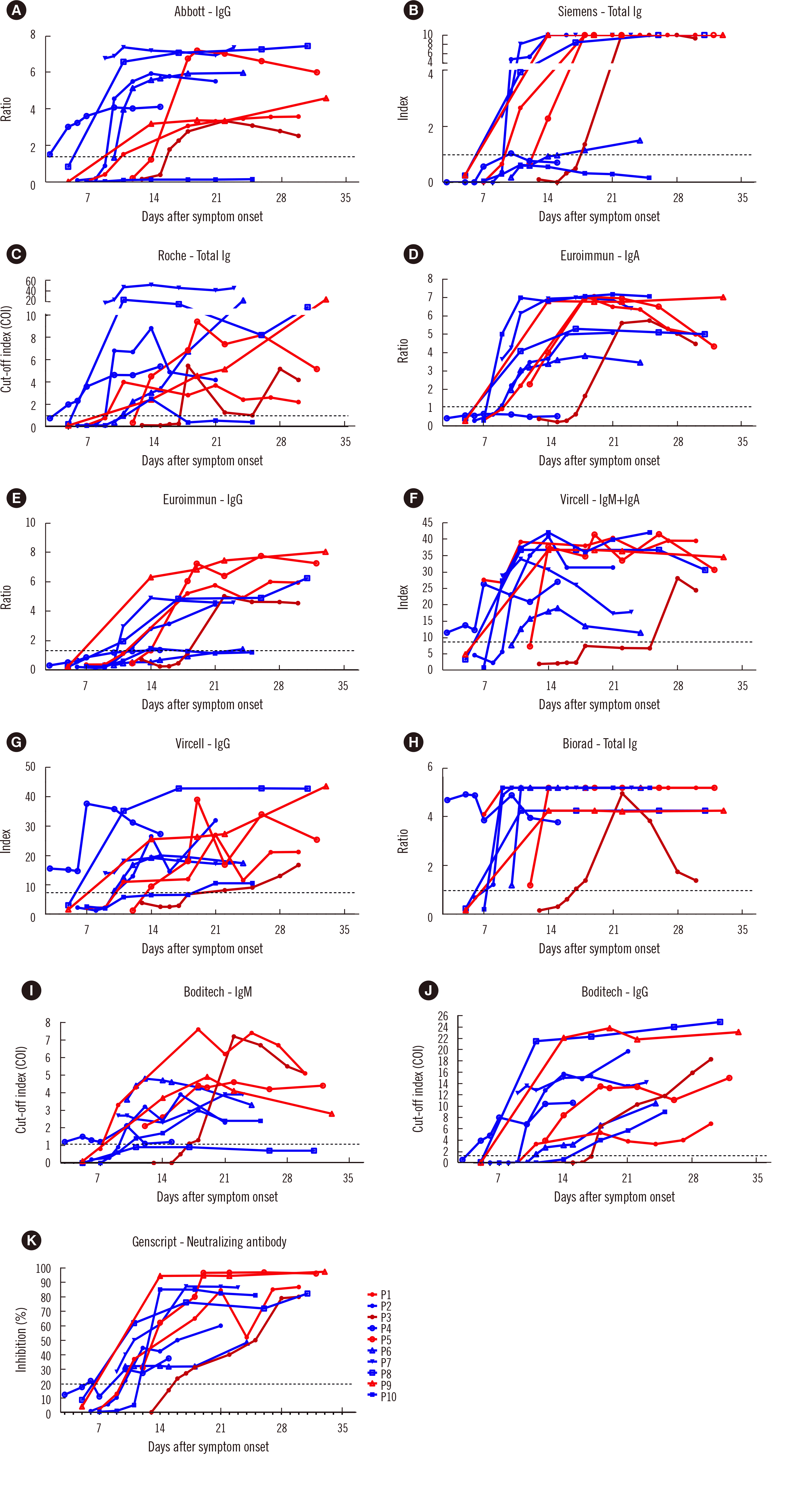
Fig. 1
Seroconversion detected using eight antibody assays with corresponding index values. Serial serum samples from 10 patients hospitalized for confirmed SARS-CoV-2 infection were tested. The index values against days from symptom onset are plotted. The dotted horizontal line represents the assay cut-off for positivity. Patients with a severe (patients 1, 5, 9) and critical (patient 3) disease course are indicated in red and dark red, respectively. Patients with a mild disease course (patients 2, 4, 6, 7, 8, 10) are indicated in blue. (A) Abbott–IgG, (B) Siemens - Total Ig, (C) Roche–Total Ig, (D) Euroimmun–IgA, (E) Euroimmun–IgG, (F) Vircell–IgM+IgA, (G) Vircell–IgG, (H) Biorad–Total Ig. (I) Boditech–IgM, (J) Boditech–IgG, (K) Genscript –Neutralizing antibody.
![]()
Correlation between SARS-CoV-2 binding antibody values and neutralizing antibody results
Fig. 2 shows the correlations between values of seven SARS-CoV-2 binding antibody assays (three CLIAs, three EIAs, and one LFIA) and the neutralizing antibody results (%) of the sVNT. Index values from binding antibody assays showed moderate to strong correlations with neutralizing antibody results (r=0.517–0.874). The neutralizing antibody results by sVNT correlated strongly with the results of binding antibody assays targeting S protein (Euroimmun, r=0.874; Siemens, r=0.839). Stronger correlations were found for patients with severe disease than for patients with mild disease.
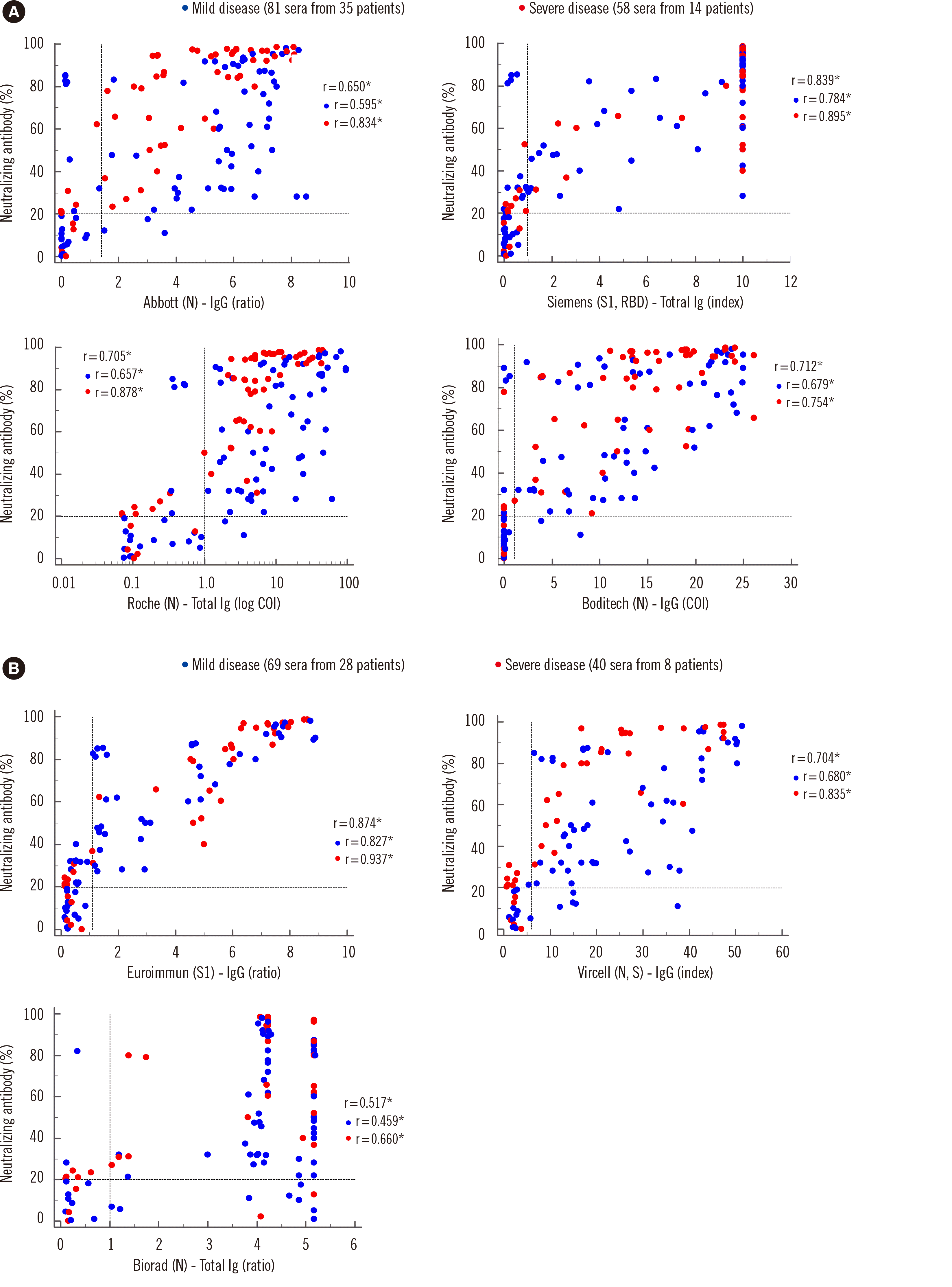
Fig. 2
Correlations between antibody results by SARS-CoV-2 binding assays (three CLIAs, three EIAs, and one LFIA) and the neutralizing antibody results by sVNT. (A) Correlation between index values of SARS-CoV-2 IgG (Abbott; ratio, Siemens; index, Roche; log COI, Boditech; COI) and neutralizing antibody results (%) measured in 139 samples plotted according to disease severity. (B) Correlation between index values of SARS-CoV-2 IgG (Euroimmun; ratio, Vircell; index) or total antibody binding assays (BioRad; ratio) and neutralizing antibody results (%) measured in 109 samples plotted according to disease severity. The dotted line represents the assay cutoff for positivity. *P<0.001.
Abbreviations: COI, cut-off index; r, Pearson correlation coefficient.
![]()
Agreement rates among 12 IgG or total antibody assays
Agreement rates among the 12 IgG or total antibody assays were evaluated using 139 serum samples (except for the three EIAs for which only 109 serum samples were analyzed) (Table 3). Agreement rates among assays were 80.0% or greater except for those of BioRad vs. Siemens (74.3%, kappa=0.393) and BioRad vs. Euroimmun (77.1%, kappa=0.433). There was particularly high agreement in the Roche vs. Abbott comparison (95.7%, kappa=0.883) and in the Sugentech vs. Rapigen comparison (96.4%, kappa=0.905). Comparing each binding assay and the sVNT, the agreement rates ranged from 83.5% to 91.4%, with the Boditech, PCL, and Roche assays showing higher agreement rates (91.4%, 89.9%, and 89.2%, respectively).
Table 3
Agreement rates between 12 SARS-CoV-2 antibody (total or IgG) assays using 139 samples from 49 COVID-19 patients
| CLIA | EIA* | LFIA | sVNT | ||||||||||
|---|---|---|---|---|---|---|---|---|---|---|---|---|---|
|
|
|
|
|
||||||||||
| Abbott | Siemens | Roche | Euroimmun | Vircell | Biorad | Boditech | SD biosensor | PCL | Sugentech | Rapigen | GenScript | ||
| CLIA | Abbott | 87.8%† | 95.7% | 80.7% | 89.0% | 83.5% | 90.6% | 93.5% | 87.8% | 92.8% | 93.5% | 87.8% | |
| 0.702‡ | 0.883 | 0.563 | 0.714 | 0.544 | 0.739 | 0.827 | 0.679 | 0.809 | 0.827 | 0.628 | |||
| Siemens | 90.6% | 89.9% | 83.5% | 74.3% | 85.6% | 87.1% | 89.9% | 86.3% | 85.6% | 84.2% | |||
| 0.772 | 0.784 | 0.621 | 0.393 | 0.642 | 0.686 | 0.759 | 0.671 | 0.651 | 0.586 | ||||
| Roche | 86.2% | 92.7% | 85.3% | 92.1% | 92.1% | 92.1% | 92.8% | 93.5% | 89.2% | ||||
| 0.688 | 0.809 | 0.594 | 0.779 | 0.788 | 0.792 | 0.809 | 0.827 | 0.672 | |||||
| EIA* | Euroimmun | 86.2% | 77.1% | 86.2% | 79.8% | 91.7% | 81.7% | 80.7% | 83.5% | ||||
| 0.673 | 0.433 | 0.677 | 0.545 | 0.815 | 0.587 | 0.563 | 0.600 | ||||||
| Vircell | 87.2% | 92.7% | 86.2% | 90.8% | 88.1% | 89.0% | 88.1% | ||||||
| 0.589 | 0.792 | 0.647 | 0.768 | 0.694 | 0.714 | 0.636 | |||||||
| Biorad | 87.2% | 80.7% | 81.7% | 80.7% | 83.5% | 84.4% | |||||||
| 0.605 | 0.476 | 0.509 | 0.476 | 0.544 | 0.470 | ||||||||
| LFIA | Boditech | 89.9% | 91.4% | 92.1% | 89.9% | 91.4% | |||||||
| 0.722 | 0.767 | 0.784 | 0.722 | 0.726 | |||||||||
| SD biosensor | 87.1% | 95.0% | 92.8% | 85.6% | |||||||||
| 0.663 | 0.868 | 0.809 | 0.569 | ||||||||||
| PCL | 90.6% | 89.9% | 89.9% | ||||||||||
| 0.759 | 0.738 | 0.707 | |||||||||||
| Sugentech | 96.4% | 86.3% | |||||||||||
| 0.905 | 0.596 | ||||||||||||
| Rapigen | 85.6% | ||||||||||||
| 0.569 | |||||||||||||
![]()
DISCUSSION
Expanding the testing capacity with accurate, validated, and reliable assays is critical in the response to the ongoing COVID-19 pandemic and vaccine trials [12–14]. We compared 12 commercially available assays with various platforms and method principles. More than 100 LFIAs are now commercially available. Although these LFIAs are convenient for testing and are useful in small or emergency laboratories with limited resources, their clinical performance remains limited with various sensitivities and specificities [15]. We evaluated five LFIAs authorized by the Korean Food and Drug Administration for export using serum samples instead of fingerstick whole blood.
For samples obtained more than 14 days after symptom onset, the overall PPA ranged from 84.0% to 98.5%. The three automated CLIAs showed similar sensitivities. Our PPA results were generally higher than those reported previously [16] and were comparable for all 12 assays. This might be related to the studied population, as we used serum samples only from symptomatic patients. Approximately 40% of the samples were from patients with severe disease courses, highlighting the importance of determining whether the performance of an antibody assay is affected by disease stage or severity. Overall, IgM or IgA isotype assays detected seroconversion earlier than IgG assays. This result is consistent with previous data showing early detection of acute patients using IgM-based assays [17, 18]. Patient 3 was an 83-year-old man who was suspected of having delayed seroconversion due to an age-related decrease in immunity. Patient 10, who had a mild disease course, showed early seroconversion with IgM assays (9–11 days), although this patient showed late seroconversion with IgG assays (14–25 days). Interestingly, the three automated CLIAs did not detect antibodies until day 25 after symptom onset in patient 10. This might be because CLIAs have defined thresholds to improve the negative likelihood ratio [19]. We determined the index values of eight assays and their correlation with disease severity. Assays targeting the S protein or RBD showed a tendency of early detection of seroconversion with higher index values, which supports previous reports demonstrating earlier seroconversion and higher titers in patients with severe disease using EIAs targeting the S protein or RBD [20–22]. However, the Roche and Abbott assays targeting the N protein antibody showed a different tendency, demonstrating later seroconversion (>14 days) and lower antibody titers in patients with severe disease (Fig. 1). This finding is similar to a previous report demonstrating delayed detection of nucleocapsid antibody in severely ill patients [20]. Although the number of patients was small, these findings suggested that the antibody response might be different according to the targeting antibodies and assay method. Since ACE2 expression varies in different ethnic populations, further studies are needed to understand the factors contributing to SARS-CoV-2 antibody responses, including genetic variability, age-related variation, and comorbidities [15].
The antibody-mediated humoral immune response is critical to prevent viral infections. The most useful information is the correlation between antibody values and a metric of protective immunity such as the neutralizing capacity [23]. The current gold standard is the conventional VNT, which shows a good correlation with the neutralizing antibody titer [7, 24]. We observed good agreement rates (>83.0%) between the 11 IgG or total binding antibody assays and the sVNT and found moderate to strong correlations (r=0.517–0.874) between the index values of binding assays and neutralizing antibody results. Neutralizing antibodies are primarily against the S1 domain, S2 domain, and RBD of the SARS-CoV-2 S protein [25, 26], suggesting that antibody assays targeting these regions might be better at predicting neutralizing capacities. As expected, the Euroimmun (targeting S1) and Siemens (targeting S1 and the RBD) assays tended to show better correlations with the sVNT (r=0.874 and r=0.839, respectively) compared with the Roche and Abbott assays (r=
0.705 and r=0.650, respectively). These results are in contrast to previous reports showing similar performances across antibody assays targeting the N and S proteins [25]. We also found that patients with severe infection showed better correlations between antibody levels and sVNT results than those with mild infection. This result confirmed previous reports showing a wide range of SARS-CoV-2 neutralizing antibody titers depending on disease severity [22, 27, 28]. Due to the limitation of using the sVNT instead of the conventional VNT, further investigation is needed to verify the immune response dynamics. Antibody detection differences can be associated with patient and assay characteristics [4–6].
This study has several limitations. First, because of the emergency isolation of patients with a positive molecular testing result and mild COVID-19 in Korea, serum samples of asymptotic patients were not included, and only a small number of patient samples were tested. We were also not able to evaluate the waning antibody response in each assay because of the lack of follow-up samples of discharged or transferred patients.
In summary, different SARS-CoV-2 antibody assays showed reliable performance, demonstrating a PPA of 84.0% or greater for samples tested more than 14 days after symptom onset. All assays detected seroconversion within less than two weeks for most patients without immune complications. However, their positivity rates and seroconversion of SARS-CoV-2 antibodies varied depending on the assay kits, disease severity, and antigen target. Commercial antibody assays should be further evaluated using serial samples over time. This study contributes to gaining a better understanding of the antibody response using currently available assays in symptomatic COVID-19 patients.
 XML Download