Journal List > Ann Hepatobiliary Pancreat Surg > v.25(2) > 1147111

Hwang, Jung, Lee, and Kim: Indication and surgical techniques of bypass choledochojejunostomy for intractable choledocholithiasis

Abstract

Despite development in endoscopic treatment and minimally invasive surgery for choledocholithiasis, there remains a small number of patients who require bypass Roux-en-Y choledochojejunostomy (RYCJ) because of the intractable occurrence of common bile duct (CBD) stones. We herein present the detailed procedures of open RYCJ customized for intractable choledocholithiasis. The first method is a side-to-end choledochojejunostomy with intraluminal closure of the distal CBD. This method was applied to a 79-year-old female patient who underwent endoscopic retrograde cholangiopancreatography (ERCP) more than 10 times in the past 14 years (Case No. 1). The distal CBD was explored through choledochotomy and then the distal CBD lumen was occluded with internal running sutures. A large-sized choledochojejunostomy was performed. The patient recovered uneventfully and has been doing well for the past 2 years. The second method is an end-to-end choledochojejunostomy with segmental CBD resection. It was applied to a 75-year-old male patient who underwent ERCP 9 times in the past 10 years (Case No. 2). The CBD was resected segmentally and a large-sized choledochojejunostomy was performed. The patient also recovered uneventfully and has been doing well for the past 2 years. In conclusion, the primary indication of bypass RYCJ is intractable choledocholithiasis which requires numerous sessions of endoscopic stone removal over a long period. Open RYCJ is the preferred procedure to date. If the papilla is patulous, the distal CBD should be occluded or resected to prevent reflux ascending cholangitis. We recommend to resect the intrapancreatic distal CBD if it is markedly dilated like choledochal cyst.

INTRODUCTION

Because of the recent development in endoscopic retrograde cholangiopancreatography (ERCP) with endoscopic therapeutic intervention, the common bile duct (CBD) stone or choledocholithiasis has not been regarded as an eligible indication of surgical treatment any more.1-9 However, a small number of patients has experienced the repeated occurrence of CBD stones, thus numerous sessions of endoscopic stone removal were required in such cases.9 Such an intractable occurrence of CBD stones may be associated with bottle-neck stasis of bile within the dilated CBD and reflux ascending cholangitis due to the patulous widening of the papilla. Bypass Roux-en-Y choledochojejunostomy (RYCJ) can be a definitive treatment for intractable choledocholithiasis as it can prevent stone-producing bile stasis. RYCJ had been performed in a non-negligible number of patients with unresectable periampullary cancer and benign distal CBD stricture in the past; however, currently, it is regarded as a surgical procedure that is performed only in rare incidences. The surgical procedure of RYCJ for intractable choledocholithiasis is rather different from that for distal CBD stricture.10,11 There exists an increasing number of case series regarding laparoscopic or robotic RYCJ in the literature, but such minimally invasive surgeries are not regarded as the preferred procedures primarily due to high medical cost and high risk of surgical complications.12-16 Thus, we herein present the detailed procedures of open RYCJ customized for intractable choledocholithiasis.

SIDE-TO-END CHOLEDOCHOJEJUNOSTOMY WITH INTRALUMINAL CLOSURE OF THE DISTAL CBD

If the CBD is not very largely dilated, segmental resection of the CBD is not essential to perform RYCJ. Considering that RYCJ is usually performed for very old-aged patients who cannot tolerate repeated endoscopic stone removal any more, the aim of the surgery is focused on effective biliary bypass drainage with minimal risk of operative complications.
Just after cholecystectomy, the CBD is incised longitudinally to open the lumen. The distal CBD is explored with stone forceps, by which the opening status of the papilla can be examined. If the papilla is patulous permitting free passage of the curved stone forceps, the distal CBD should be closed to prevent reflux ascending cholangitis. The anterior wall of the CBD should be excised to make a large opening that would be suitable for anastomosis.
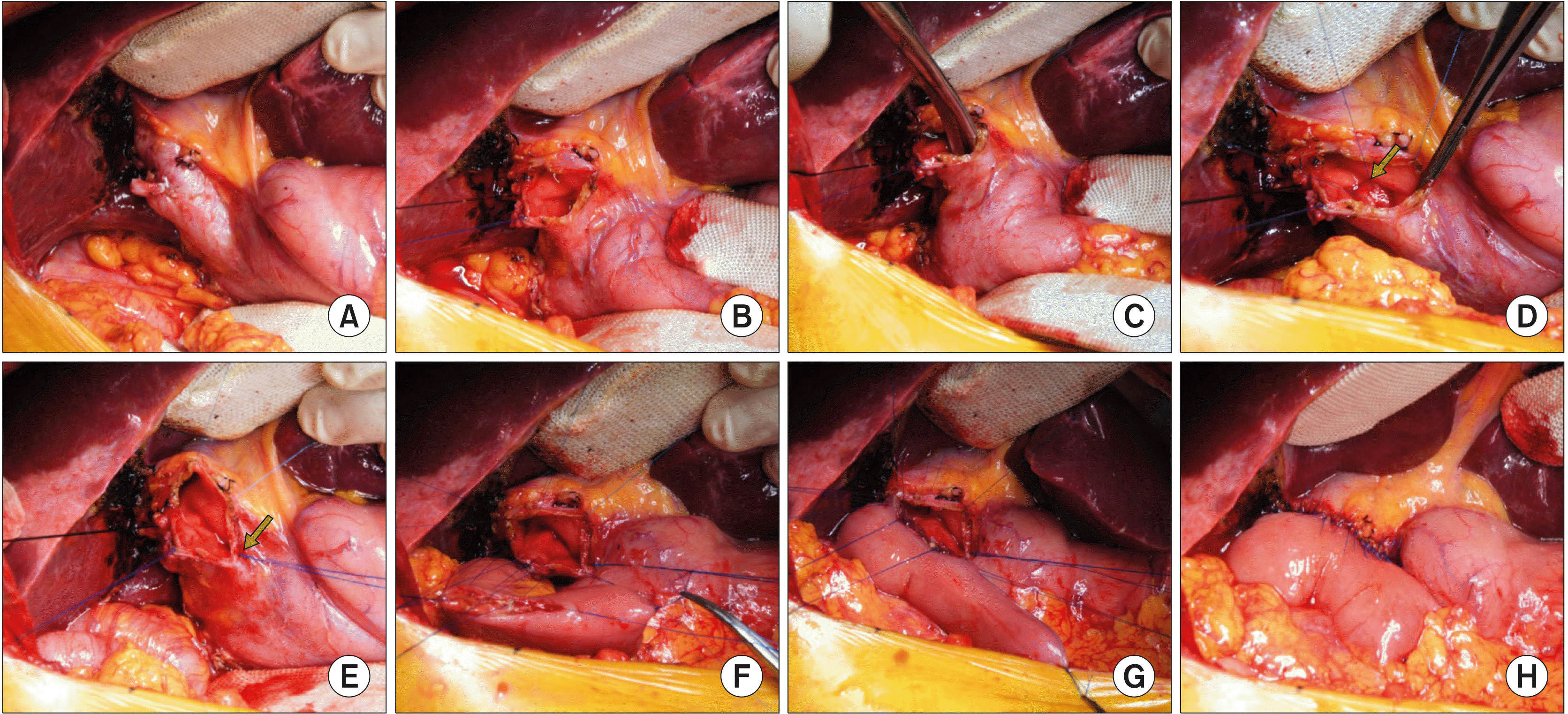
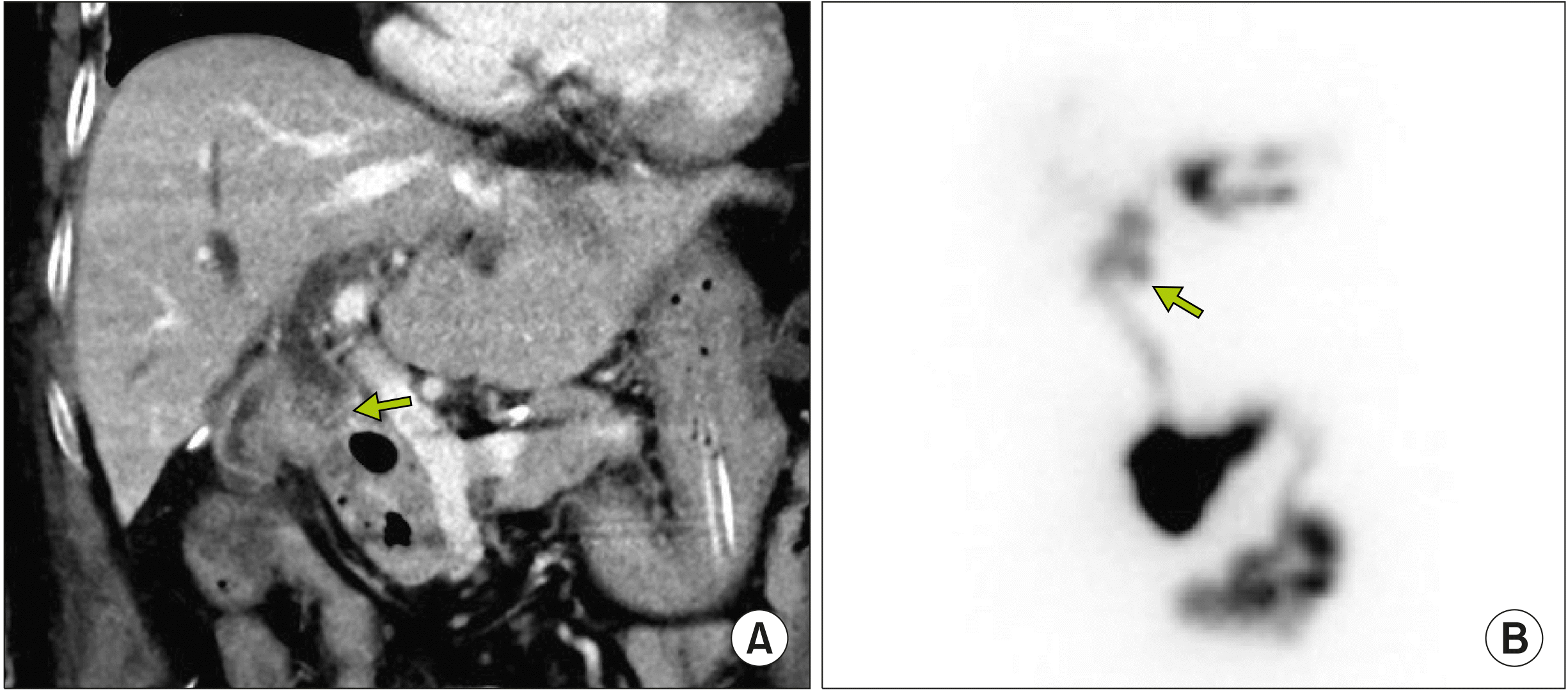
The detailed surgical procedures applied to a 79-year-old female patient who underwent ERCP more than 10 times in the past 14 years (Case No. 1) (Fig. 1) are as follows: after cholecystectomy, the mid-portion of the CBD was incised longitudinally and the distal CBD was explored with a curved stone forceps. As the sphincter of Oddi was patulous, the lumen of the distal CBD at the upper board of the pancreas was securely closed with 5-0 Prolene running sutures. Subsequently, a part of the anterior wall of the CBD was elliptically excised to make a large opening that enabled a large choledochojejunostomy. A Roux-en-Y jejunal limb was anastomosed to the CBD opening in a side-to-side fashion (Fig. 2). The patient recovered uneventfully (Fig. 3) and has been doing well without any biliary problems for the past 2 years. On follow-up imaging studies, a 1.5 cm-sized remnant portion of the distal CBD was visible within the pancreas head.

END-TO-END CHOLEDOCHOJEJUNOSTOMY WITH SEGMENTAL RESECTION OF THE CBD

If the CBD is markedly dilated, the abovementioned luminal closure can leave a large blind-end space within the intrapancreatic bile duct as like a choledochal cyst, which can be a source of stone formation or pancreatitis. Thus, it is reasonable to remove the dilated CBD portion which can remain as a small blind-end lumen at the intrapancreatic bile duct.
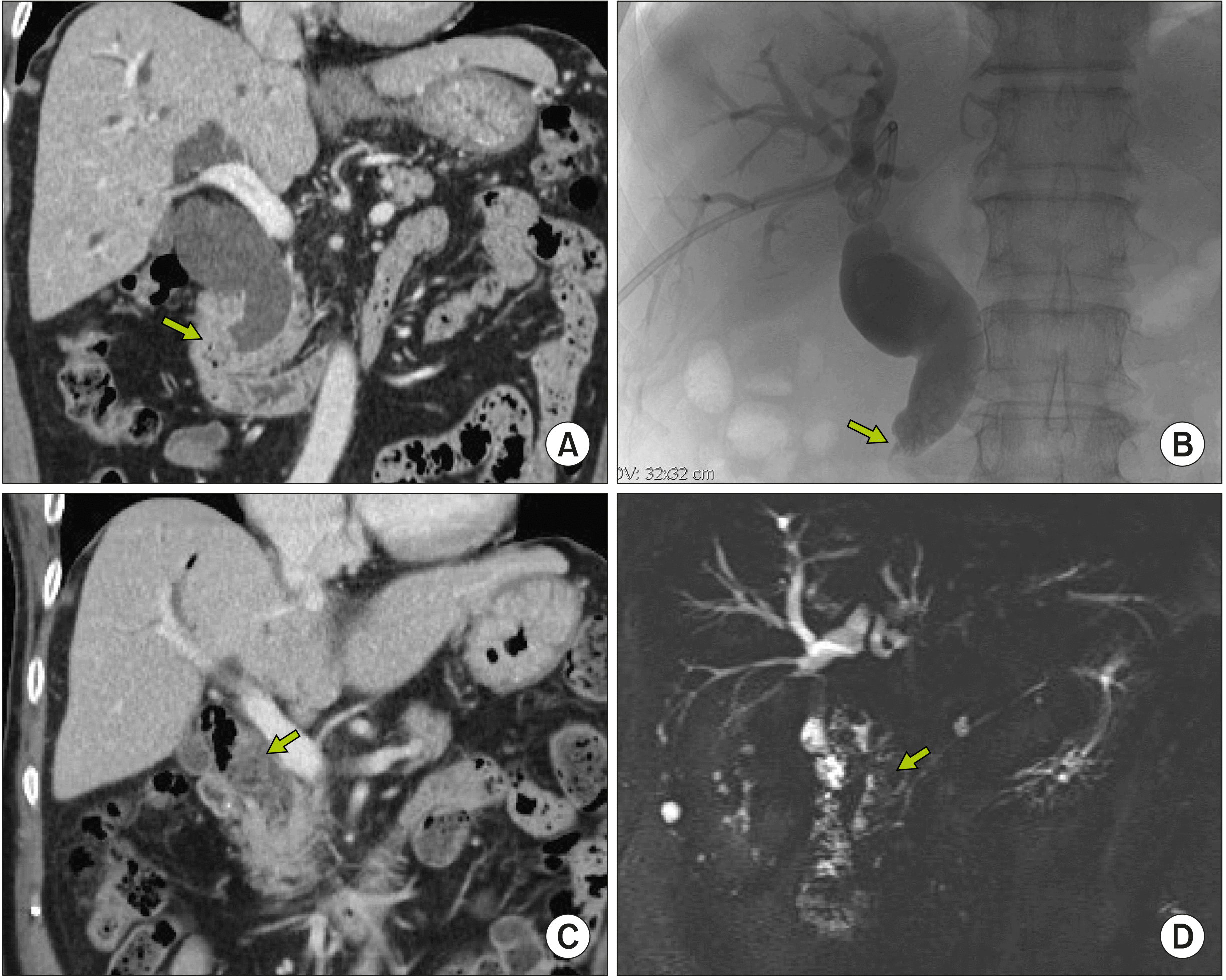
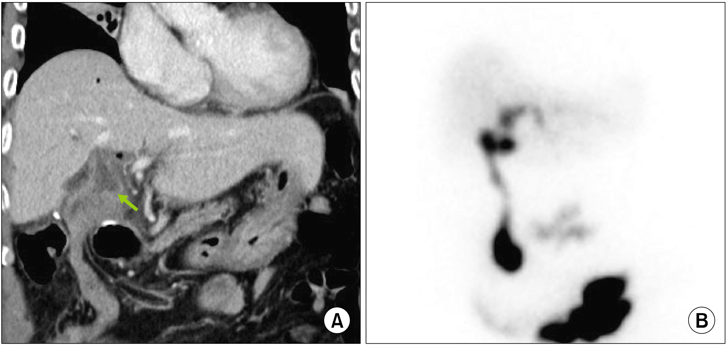
The detailed surgical procedures applied to a 75-year-old male patient who underwent ERCP 9 times in the past 10 years (Case No. 2) (Fig. 4) are as follows: after cholecystectomy, the CBD was incised longitudinally and the distal CBD was explored with a curved stone forceps. Multiple CBD stones and sludges mixed with food materials were removed from the distal CBD. As the distal CBD was markedly dilated and the papilla was patulous, the distal CBD was deeply dissected into the pancreas parenchyma until the narrow portion of the intrapancreatic bile duct, and then transected and securely repaired with 5-0 Prolene sutures. The proximal CBD was also transected at the level of the right hepatic artery to make a large proximal bile duct opening. A Roux-en-Y jejunal limb was anastomosed to the CBD opening in an end-to-side fashion (Fig. 5). The patient recovered uneventfully (Fig. 6) and has been doing well with any biliary problems for the past 2 years. On follow-up imaging studies, a small portion of the remnant distal CBD was identified within the pancreas head.

DISCUSSION

In the era of prevailing minimally invasive surgeries with laparoscopic or robotic approaches, open RYCJ is not a matter of concern.12-16 Moreover, recent development in ERCP with endoscopic therapeutic intervention has led choledocholithiasis to be placed beyond the territory of surgical indication.1-9 On the contrary, we should not forget that there remains a small number of patients who still require open RYCJ because of the intractable occurrence of CBD stones. Since such a surgical procedure has been rarely performed, many young surgeons majoring in hepatobiliary surgery do not have enough experience on the indications and surgical techniques for open RYCJ in the cases of patients with intractable choledocholithiasis.
The principles for surgical techniques toward achieving complication-free RYCJ include avoidance of unnecessary dissection of the CBD, making a large-sized biliary-enteric anastomosis according to the fluid dynamics, and prevention of reflux ascending cholangitis. To avoid unnecessary dissection of the CBD, the luminal occlusion technique was used instead of segmental bile duct resection in Case No. 1. As the wall of the dilated CBD is usually thickened over 2 mm, the elliptical side-wall excision should be performed to make a large opening since a simple longitudinal incision can be eventually narrowed due to the slit closing effect.
Unlike the abovementioned two points, some background knowledge is necessary to understand why reflux ascending cholangitis should be prevented. If RYCJ is performed without interruption of distal CBD lumen, food materials would pass in a retrograde manner through the patulous papilla, which can be a source of frequent ascending cholangitis and CBD stone formation. Based on our experience with 8 cases of conventional side-to-end RYCJ without distal CBD interruption, episodes of reflux ascending cholangitis occurred repeatedly in at least 2 patients. A prospective study to determine the incidence of duodenobiliary reflux and acute cholangitis after placement of self-expanding metal stent across the main duodenal papilla revealed that reflux of duodenal contents is a universal phenomenon.17 A patulous papilla following multiple sessions of ERCP is vulnerable to reflux similar to the papilla state with a self-expanding metal stent. On the contrary, distal CBD interruption is not necessary for patients with distal CBD stenosis from benign or malignant diseases.
In the cases of usual patients with intractable choledocholithiasis, segmental bile duct resection is theoretically not necessary. However, if the CBD is markedly enlarged, leaving a large lumen within the distal CBD can make a blind-ended pool inducing infection or stone formation by food material regurgitation through the patulous ampulla. Thus, it is reasonable to resect the excessively dilated portion of the distal CBD, as demonstrated in Case No. 2.
We had used a refined technique of clustered biliary-enteric anastomosis when the bile duct opening was greater than 2 cm in diameter. Routine Roux-en-Y jejunal limb was prepared and pulled up through the retrocolic tunnel. A longitudinal incision was made at the antemesenteric border of the jejunal limb and the posterior wall anastomosis was performed with 2 or 3 segmented continuous running sutures with 5-0 Prolene. Anterior wall anastomosis was performed through multiple interrupted sutures.18 With the application of such customized techniques for intractable choledocholithiasis, until date, we did not experience any noticeable surgical complications yet 15 patients with age of 63-82 years.
In conclusion, we contemplate that the primary indication of bypass RYCJ is intractable choledocholithiasis which requires numerous sessions of ERCP over a long period. So far, open RYCJ has been the preferred procedure as it is much more reliable than the minimally invasive procedures regarding patient safety. If the papilla is patulous, distal CBD should be occluded or resected to prevent reflux ascending cholangitis. We recommend to resect the intrapancreatic distal CBD if it is markedly dilated like a choledochal cyst because food material can regurgitate through the patulous ampulla.

Notes

CONFLICT OF INTEREST

None of the authors has any conflict of interest.

AUTHOR CONTRIBUTIONS

Conceptualization: SH. Data curation: SH, DHJ. Formal analysis: SH. Methodology: SH, SKL, MHK. Project administration: SH. Visualization: SH. Writing - original draft: SH. Writing - review & editing: All.

REFERENCES

1. Costi R, Gnocchi A, Di Mario F, Sarli L. 2014; Diagnosis and management of choledocholithiasis in the golden age of imaging, endoscopy and laparoscopy. World J Gastroenterol. 20:13382–13401. DOI: 10.3748/wjg.v20.i37.13382. PMID: 25309071. PMCID: PMC4188892.
crossref
2. Nathanson LK, O'Rourke NA, Martin IJ, Fielding GA, Cowen AE, Roberts RK, et al. 2005; Postoperative ERCP versus laparoscopic choledochotomy for clearance of selected bile duct calculi: a randomized trial. Ann Surg. 242:188–192. DOI: 10.1097/01.sla.0000171035.57236.d7. PMID: 16041208. PMCID: PMC1357723.
3. Urbach DR, Khajanchee YS, Jobe BA, Standage BA, Hansen PD, Swanstrom LL. 2001; Cost-effective management of common bile duct stones: a decision analysis of the use of endoscopic retrograde cholangiopancreatography (ERCP), intraoperative cholangiography, and laparoscopic bile duct exploration. Surg Endosc. 15:4–13. DOI: 10.1007/s004640000322. PMID: 11178753.
4. Pring CM, Skelding-Millar L, Goodall RJ. 2005; Expectant treatment or cholecystectomy after endoscopic retrograde cholangiopancreatography for choledocholithiasis in patients over 80 years old? Surg Endosc. 19:357–360. DOI: 10.1007/s00464-004-9089-1. PMID: 15645324.
crossref
5. Maple JT, Ikenberry SO, Anderson MA, Appalaneni V, Decker GA, Early D, et al. 2011; The role of endoscopy in the management of choledocholithiasis. Gastrointest Endosc. 74:731–744. DOI: 10.1016/j.gie.2011.04.012. PMID: 21951472.
crossref
6. Shim CS. 2010; How should biliary stones be managed? Gut Liver. 4:161–172. DOI: 10.5009/gnl.2010.4.2.161. PMID: 20559517. PMCID: PMC2886934.
crossref
7. Sugiyama M, Atomi Y. 2000; Endoscopic sphincterotomy for bile duct stones in patients 90 years of age and older. Gastrointest Endosc. 52:187–191. DOI: 10.1067/mge.2000.107285. PMID: 10922089.
crossref
8. Moon JH, Cha SW, Ryu CB, Kim YS, Hong SJ, Cheon YK, et al. 2004; Endoscopic treatment of retained bile-duct stones by using a balloon catheter for electrohydraulic lithotripsy without cholangioscopy. Gastrointest Endosc. 60:562–566. DOI: 10.1016/S0016-5107(04)02012-7.
crossref
9. Sugiyama M, Suzuki Y, Abe N, Masaki T, Mori T, Atomi Y. 2004; Endoscopic retreatment of recurrent choledocholithiasis after sphincterotomy. Gut. 53:1856–1859. DOI: 10.1136/gut.2004.041020. PMID: 15542528. PMCID: PMC1774317.
crossref
10. Kadaba RS, Bowers KA, Khorsandi S, Hutchins RR, Abraham AT, Sarker SJ, et al. 2017; Complications of biliary-enteric anastomoses. Ann R Coll Surg Engl. 99:210–215. DOI: 10.1308/rcsann.2016.0293. PMID: 27659373. PMCID: PMC5450270.
crossref
11. Vogt DP, Hermann RE. 1981; Choledochoduodenostomy, choledochojejunostomy or sphincteroplasty for biliary and pancreatic disease. Ann Surg. 193:161–168. DOI: 10.1097/00000658-198102000-00006. PMID: 7469551. PMCID: PMC1345035.
crossref
12. Hori T, Aisu Y, Yamamoto M, Yasukawa D, Iida T, Yagi S, et al. 2019; Laparoscopic approach for choledochojejunostomy. Hepatobiliary Pancreat Dis Int. 18:285–288. DOI: 10.1016/j.hbpd.2019.04.004. PMID: 31023579.
crossref
13. Hori T. 2019; Comprehensive and innovative techniques for laparoscopic choledocholithotomy: a surgical guide to successfully accomplish this advanced manipulation. World J Gastroenterol. 25:1531–1549. DOI: 10.3748/wjg.v25.i13.1531. PMID: 30983814. PMCID: PMC6452235.
crossref
14. Han HS, Yi NJ. 2004; Laparoscopic Roux-en-Y choledochojejunostomy for benign biliary disease. Surg Laparosc Endosc Percutan Tech. 14:80–84. DOI: 10.1097/00129689-200404000-00006. PMID: 15287605.
crossref
15. Ruurda JP, van Dongen KW, Dries J, Borel Rinkes IH, Broeders IA. 2003; Robot-assisted laparoscopic choledochojejunostomy. Surg Endosc. 17:1937–1942. DOI: 10.1007/s00464-003-9008-x. PMID: 14569457.
crossref
16. Benzie AL, Sucandy I, Spence J, Ross S, Rosemurgy A. 2019; Robotic choledochoduodenostomy for benign distal common bile duct stricture: how we do it. J Robot Surg. 13:713–716. DOI: 10.1007/s11701-019-00957-8. PMID: 30989618.
crossref
17. Misra SP, Dwivedi M. 2009; Reflux of duodenal contents and cholangitis in patients undergoing self-expanding metal stent placement. Gastrointest Endosc. 70:317–321. DOI: 10.1016/j.gie.2008.12.054. PMID: 19539920.
crossref
18. Hwang S, Ha TY, Song GW, Jung DH. 2016; Cluster hepaticojejunostomy with radial spreading anchoring traction technique for secure reconstruction of widely opened hilar bile ducts. Korean J Hepatobiliary Pancreat Surg. 20:66–70. DOI: 10.14701/kjhbps.2016.20.2.66. PMID: 27212993. PMCID: PMC4874047.
crossref

Fig. 1
The preoperative finding of Case No. 1. Computed tomo-graphy (CT) and endoscopic retrograde cholangiopancreato-graphy (ERCP) taken 7 years before surgery (A, B) show marked dilatation of the com-mon bile duct with the patulous sphincter. CT scan and ERCP taken 1 month before surgery (C, D) show further dilatation of the common bile duct filled with air and sludges, indicating reflux ascending cholangitis. The spine was externally fixed 11 years before due to spine fracture.
ahbps-25-2-259-f1.tif
Fig. 2
Intraoperative photograph showing the procedure of side-to-end choledochojejunostomy with intraluminal closure of the distal common bile duct (CBD) in Case No. 1. (A) The dilated CBD is visible after cholecystectomy. (B) The mid-portion of the CBD is incised longitudinally, showing markedly thickened CBD wall indicating repeated episodes of cholangitis. (C) The distal CBD is explored with curved stone forceps to examine the opening status of the sphincter. (D, E) The lumen of the distal CBD at the upper board of the pancreas is securely closed with 5-0 Prolene running sutures twice. The suture knots are made at the outer surface of the CBD (arrow). A part of the anterior wall of the CBD is excised to widen the opening. (F-H) A Roux-en-Y jejunal limb is anastomosed to the CBD opening in a side-to-side fashion.
ahbps-25-2-259-f2.tif
Fig. 3
Postoperative findings at two weeks after surgery in Case No. 1. (A) Computed tomography scan shows uneventful drainage of the bile from to proximal common bile duct (CBD) to the jejunal limb. The distal CBD is occluded (arrow). (B) Hepato-biliary scintigraphy shows a normal biliary excretion rate of 92.5% at 90 minutes. The distal CBD is occluded (arrow).
ahbps-25-2-259-f3.tif
Fig. 4
The preoperative finding of Case No. 2. Computed tomo-graphy (CT) and direct cholan-giography taken 10 years before surgery (A, B) show marked dilatation of the common bile duct with an obstructing mass at the sphincter (arrows), which was later proven to be a benign stricture. CT scan and magnetic resonance cholangiography taken 1 month before surgery (C, D) show that the common bile duct is filled with stones and sludges (arrows).
ahbps-25-2-259-f4.tif
Fig. 5
Intraoperative photograph showing the procedure of end-to-end choledochojejunostomy with segmental resection of the common bile duct (CBD) in Case No. 2. (A) The dilated CBD is visible after cholecystectomy. (B) The mid-portion of the CBD is incised longitudinally, showing multiple CBD stones and sludges. (C) The distal CBD is dissected and transected at the narrow portion of the intrapancreatic bile duct, and then securely closed with 5-0 Prolene sutures. (D) The proximal CBD was also transected at the level of the right hepatic artery to make a large proximal bile duct opening. (E, F) A Roux-en-Y jejunal limb is anastomosed to the CBD opening in an end-to-side fashion.
ahbps-25-2-259-f5.tif
Fig. 6
Postoperative findings at 10 days after surgery in Case No. 2. (A) Computed tomogra-phy shows uneventful drainage of the bile from to proximal common bile duct to the jejunal limb. (B) Hepatobiliary scinti-graphy shows a biliary excre-tion rate of 89.2% at 90 min-utes.
ahbps-25-2-259-f6.tif
TOOLS
Similar articles