

 PDF
PDF Citation
 Print
Print


Introduction
The incidence of thyroid cancer has increased in recent decades.1)Cervical lymph node (LN) metastases in differentiated thyroid carcinoma (DTC) are common. The reported incidence of metastatic LNs is 20-46% at the initial diagnosis of DTC and local recurrences is reported 5-20% of cases during surveillance after the initial treatment.2)Although DTC is considered as a cancer with a low mortality rate,3)long-term follow-up studies of DTC patients with LN metastasis showed poor prognosis associated with low survival and high recurrence rates.4,5) Ultrasonography (US) and US-guided fine needle aspiration cytology (FNAC) have been the gold standard modality for the diagnosis of LN metastases before surgery or during cancer surveillance.6)However, the results of FNAC can be inadequate in small LNs and are limited by false negative findings in cystic metastatic LNs.7)
The direct measurement of thyroglobulin (Tg) in FNA washout fluid (washout Tg) has been proposed to improve the diagnostic outcomes of FNAC.6,8,9) Tg is a 660kD glycoprotein which is produced only by the thyroid follicular cells thus, its detection in nonthyroidal tissue could be evidence of persistent or recurrent disease after complete surgery and/or radioiodine therapy.10)Current clinical practice guidelines for thyroid cancer already incorporate washout Tg as an essential additive to FNAC in suspicious LNs.11-13) However, washout Tg levels could be detected at high concentration before surgery due to the presence of a normal thyroid, leading to false positive results. In the case of lobectomy, contralateral lobe might affect the false positivity rate. Serum Tg antibodies (Ab) could also interfere with washout Tg.14,15) The accuracy of washout Tg and FNAC of LNs has been evaluated in selected patients with total thyroidectomy or near total thyroidectomy. Few studies have assessed discordant results in a wide range of clinical conditions from real-world practice.
This study aimed to 1) evaluate the diagnostic performance of FNAC combined with washout Tg by applying different cut-off Tg values to preoperative and postoperative LNs and 2) assess the clinical outcome of discrepant results between FNAC combined with washout Tg and final histopathologic diagnosis.
Go to : 
Materials and Methods
Subject
From January 2015 to December 2017, 566 patients with 774 LNs suspicious for metastatic diseases underwent US-guided FNAC and washout Tg measurements at initial diagnosis of thyroid cancer prior to thyroidectomy or during cancer surveillance after thyroidectomy and/or radioiodine ablation at a tertiary referral hospital (Fig. 1). The exclusion criteria were as follows: 1) LNs from primary lesions other than DTC such as anaplastic thyroid carcinoma, medullary thyroid carcinoma, follicular adenoma, and parathyroid adenoma (26 LNs in 19 patients); 2) metastatic LN from malignancy other than thyroid cancer (one LN in one patient; adenocarcinoma from lung metastasis); 3) LNs without serum Tg and TgAb at the time of FNAC and washout Tg measurement (nine LNs in eight patients); 4) LNs without follow-up at our institution after the first FNAC and washout Tg measurement (29 LNs in 24 patients); and 5) LNs without any decision after FNAC and washout Tg measurement, no LNs dissection or no imaging follow-up such as thyroid US, neck computed tomography (CT), magnetic resonance imaging (MRI), and positron emission tomography (PET)-CT (two LNs in two patients) during the study periods. Finally, the remaining 707 LNs in 512 patients were evaluated in this study. This study complied with the ethical standards of the Declaration of Helsinki and was approved by the Catholic University of Korea, Catholic Medical Center, Seoul St. Mary’s Hospital Institutional Review Board (No. KC17TESI0328). The requirement for informed consent explaining the study purpose and procedures was waived due to the retrospective design of this study.

US-Guided FNAC of Suspicious LNs and Washout Tg Measurement
US-guided FNAC of suspicious LNs was performed by one of several experienced radiologists. Thyroid and neck US was performed with an HDI 3000 scanner (Advanced Technology Laboratories, Bothell, WA, USA) and an HDI 5000 diagnostic sonography system (Philips Medical Systems, Bothell, WA, USA) with a CL10-5 MHz compact linear array transducer. The criteria and technique for FNAC of suspicious LNs were based on the consensus statement of the Korean Society of Thyroid Radiology.16)Suspicious LNs with cystic change, calcification, hyperechogenicity, abnormal vascularity, and loss of central hilar echogenicity on thyroid and neck US received FNAC. After aspiration, the samples were immediately smeared on slides, fixed in 95% ethanol, and processed by both hematoxylin and eosin and Papanicolaou stains.17)As described in a previous study,15)the same needle and syringe were rinsed with 2 mL of normal saline, and the washout fluid was submitted for washout Tg measurement.
Biochemical Analysis: Tg and TgAb
Serum Tg and washout Tg levels were measured using a monoclonal antibody immunoradiometric assay (Cisbio Bioassays, Codolet, France). The analytical sensitivity, which indicates the lowest detectable concentration with a probability of 95%, was 0.2 ng/mL. The functional sensitivity at which the efficiency variation was equal to 20% was 0.7 ng/mL. Serum TgAb levels were measured using a competitive radioimmunoassay kit (ZenTech, Angleur, Belgium) with a functional sensitivity of <15 IU/mL. The washout Tg was normalized with dividing by serum Tg (washout Tg/serum Tg ratio).
FNAC Results and Final LN Diagnosis
The interpretation of FNAC was performed by experienced pathologists who specialized in thyroid cytology. The FNAC results were classified into two groups; 1) metastatic LNs with the presence of highly suspicious atypical cells, or with cytological feature of DTC and 2) benign LNs which were documented as negative for malignancy, reactive hyperplasia, or other benign lymphadenopathies and non-diagnostic including inadequate results due to the absence of cells and the presence of blood only.18)
Generally, the final LN diagnosis was confirmed by postoperative histopathologic results. Metastatic LNs were defined as those with postoperative metastasis of histology in the same compartment that was previously noted in FNAC.19)If patients did not undergo LNs dissection, metastatic LNs were defined based on metastatic FNAC results combined with high washout Tg level and lesions that were revealed by imaging studies such as thyroid US, neck CT, PET/CT, or MRI. Otherwise, repeat FNAC results showing metastatic cancer cells were included as final metastatic LNs. Benign LNs were confirmed by benign histology after postoperative review. If the target LNs were not dissected and follow-up imaging showed decreased size or no interval change in size during a follow-up of at least two years, the final LNs were considered to be benign.20)If repeat FNAC results revealed benign LNs, the final LNs were also defined as benign LNs.
Statistical Analysis
All statistical analyses were conducted using SPSS software, version 14 (Chicago, IL, USA). Continuous variables with normal distribution were expressed as means and standard deviation. Variables with non-normal distributions were presented as medians (interquartile range). Comparisons of the basic clinical characteristics between benign and metastatic LNs were performed by independent t-tests for continuous variables and by χ2 or Fisher exact tests for categorical variables in preoperative LNs and postoperative LNs, respectively. A receiver operating characteristic (ROC) curve analysis was used to confirm the cut-off levels of washout Tg in our data. The diagnostic performances of FNAC and washout Tg were evaluated with respect to sensitivity, specificity, positive predictive value (PPV), negative predictive value (NPV), and diagnostic accuracy. McNemar’s test was used for statistical comparison of their diagnostic performances. All p values <0.05 were considered significant, and two-sided tests were used.
Go to :
Results
Clinical Characteristic of Patients and LNs in Metastatic and Benign LNs according to Final LNs Diagnosis
Female gender was significantly more prevalent in both metastatic and benign LNs (p=0.020). There was no significant difference in age between patients with metastatic LNs and those with benign LNs (mean age 47.6 vs. 47.3 years, p=0.771).
According to the final LN diagnosis, 214 of 707 (30.3%) LN were metastatic; the remaining 493 (69.7%) were confirmed as benign LNs. Among 214 metastatic LNs, 91 (42.5%) were evaluated before thyroidectomy with or without LNs dissection and 123 (57.5%) were postoperative LNs. 204 of 493 (41.4%) benign LNs were preoperative and 289 (58.6%) were postoperative. There were significant differences between metastatic and benign LNs in US findings (p=0.000). Metastatic LNs showed more suspicious feature than benign LN in US findings. Washout Tg was higher in metastatic LNs with significance (523.4±385.5 vs. 31.4±140.9 ng/mL, p=0.000). Normalized washout Tg with serum Tg (washout Tg/serum Tg ratio) was higher in metastatic LN than in benign LN. However, there was no significance (p=0.084). The difference of serum Tg and serum TgAb levels were not significant. Discordant rates between FNAC and washout Tg were higher in metastatic LNs than in benign LNs (19.6% vs. 13.0%, p=0.000) (Table 1).
Table 1
Clinical characteristics of 707 lymph nodes (LNs) and 512 patients according to final LNs diagnosis
| Characteristics | Metastatic LNs | Benign LNs | p value |
|---|---|---|---|
| Patients (n=512) | |||
| Gender male/female (%) | 55/83 (39.9/60.1) | 107/267 (28.6/71.4) | 0.020 |
| Age (years) | 47.6±16.7 | 47.3±14.3 | 0.771 |
| LNs (total=707), (n, %) | 214/707 (30.3) | 493/707 (69.7) | |
| Time of FNAC and washout Tg | 0.777 | ||
| Preoperative LNs (n/total, %) | 91/214 (42.5) | 204/493 (41.4) | |
| Postoperative LNs (n/total, %) | 123/214 (57.5) | 289/493 (58.6) | |
| US finding* | 0.000 | ||
| Suspicious (n/total, %) | 122/214 (57.0) | 80/493 (16.2) | |
| Intermediate (n/total, %) | 73/214 (34.1) | 299/493 (60.7) | |
| Benign (n/total, %) | 19/214 (8.9) | 114/493 (23.1) | |
| Serum Tg (ng/mL) | 27.1±76.6 (0.0-868.2) | 22.5±91.2 (0.0-805.7) | 0.516 |
| Serum TgAb (IU/mL) | 73.5±249.5 (0.1-1896.7) | 68.1±164.6 (0.1-9480.1) | 0.873 |
| Washout Tg (ng/mL) | 523.4±385.5 (0.0-1251.7) | 31.4±140.9 (0.0-1074.0) | 0.000 |
| Washout Tg/serum Tg ratio | 933.3±3185.9 (0.0-29802.3) | 377.3±5247.9 (0.0-11277.3) | 0.084 |
| Discordant results between FNAC and washout Tg (n, %) | 42 (19.6) | 64 (13.0) | 0.000 |
![]()
Table 2 summarizes the clinical characteristics of LNs according to surgery status (preoperative vs. postoperative). Serum Tg and serum TgAb levels were higher in preoperative LNs than those in postoperative LNs (p=0.002 and p=0.008). The rate of discordant results between FNAC and washout Tg was higher in metastatic LNs than that in benign LNs in both preoperative and postoperative LNs. Overall, the discordant results between FNAC and washout Tg did not differ in preoperative and postoperative LNs (15.3 vs. 14.8%, p=0.321).
Table 2
Clinical characteristics of 707 lymph nodes (LNs) according surgery status and final outcome
| Characteristics | Preoperative LNs (n=295) | Postoperative LNs (n=412) | p value | ||
|---|---|---|---|---|---|
|
|
|
||||
| Metastatic | Benign | Metastatic | Benign | ||
| LNs (n, %) | 91 (30.8) | 204 (69.2) | 123 (29.9) | 289 (70.1) | |
| US finding* | 0.572 | ||||
| Suspicious (n, %) | 59 (64.8) | 31 (15.1) | 63 (51.2) | 49 (17.0) | |
| Intermediate (n, %) | 25 (27.5) | 124 (60.8) | 48 (39.0) | 175 (60.6) | |
| Benign (n, %) | 7 (7.7) | 49 (24.1) | 12 (9.8) | 65 (22.4) | |
| Serum Tg (ng/mL) |
35.8±66.5 (0.1-461.7) |
37.0±100.1 (0.0-753.5) |
20.6±83.0 (0.0-868.2) |
12.1±83.0 (0.0-805.7) |
0.002 |
| Serum TgAb (IU/mL) |
91.8±170.4 (3.4-750.6) |
111.4±674.8 (0.1-6480.7) |
59.6±295.7 (0.1-1896.7) |
37.4±200.5 (0.1-2826.6) |
0.008 |
| Washout Tg (ng/mL) |
574.7±366.1 (0.0-1251.7) |
26.4±122.6 (0.0-1002.4) |
485.5±396.4 (0.0-1248.5) |
35.0±152.6 (0.0-1074.0) |
0.088 |
| Washout Tg/serum Tg ratio |
138.8±1366.5 (0.0-14307.3) |
194.6±568.9 (0.0-4309.6) |
1479.8±4095.5 (0.0-29802.3) |
545.6±6757.6 (0.0-11277.3) |
0.001 |
| Discordant results between FNAC and washout Tg (n, %) | 16 (17.6) | 29 (9.8) | 26 (21.1) | 35 (12.1) | 0.321 |
![]()
Cut-off Values for Washout Tg in All LNs, Preoperative LNs, and Postoperative LNs
In the present study, the diagnostic performance of washout Tg was determined using an optimal cut-off value of 2.9 ng/mL in all LNs (area under the receiver operating characteristic curve [AUC] 0.919, sensitivity 89.3%, and specificity 88.0%, 95% confidence interval [CI] 0.892-0.945) and the washout Tg/serum Tg ratio was 3.0 (AUC 0.873, sensitivity 82.2%, and specificity 83.8%, 95% CI 0.842-0.904). The cut-off value of preoperative washout Tg was 9.6 ng/mL (AUC 0.943, sensitivity 89.0%, and specificity 91.7%, 95% CI 0.911-0.975) and that of postoperative washout Tg was 2.1 ng/mL (AUC 0.902, sensitivity 80%, and specificity 87.8%, 95% CI 0.863-0.942). The washout Tg/serum Tg ratio for preoperative and postoperative status was 7.0 (AUC 0.919 sensitivity 85.7%, and specificity 89.7%, 95% CI 0.886-0.953), and 2.9 (AUC 0.845 sensitivity 82.1%, and specificity 83.7%, 95% CI 0.796-0.893). When applying the cut-off value for washout Tg (2.9 ng/mL) regardless of surgery status, the sensitivity, specificity, PPV, NPV, and accuracy of FNAC combined washout Tg were lower than those for the cut-off values according to surgery status (9.6 ng/mL in preoperative LNs and 2.1 ng/mL in postoperative LNs, respectively) (Table 3). The discordant results between FNAC combined washout Tg and final diagnosis were reduced from 45 (15.3%) to 34 (11.5%) LNs in 295 preoperative LNs after applying the cut-off for washout Tg of 9.6 ng/mL. In postoperative LNs, the discordant rate decreased by 1.5%, from 61 of 412 LNs (14.8%) to 55 of 412 LNs (13.3%) after applying the cut-off value of 2.1 ng/mL.
Table 3
Cut-off values of FNAC with washout Tg measurement and diagnostic performance according to surgery status
![]()
Final Diagnosis of All LNs and of the Results of FNAC with Washout Tg
We analyzed discordant results in FNAC combined with washout Tg and final diagnosis and decision making. Fig. 1 demonstrated the final diagnosis of all LNs with different cut-off values of washout Tg according surgery status. In preoperative LNs (cut-off of washout Tg; 9.6 ng/ml), FNAC with washout Tg showed concordant in 78 out of 88 (88.6%) metastatic LNs (FNAC+washout Tg+; metastatic cytology and high titer of washout Tg). All these FNAC+washout Tg+ 78 LNs were dissected with thyroi-dectomy. 74 of 78 dissected LNs were metastatic LNs based on result of postoperative histopathology. Four of 78 FNAC+washout Tg+ LNs were revealed as benign LNs in histopathologic results of same compartment LNs. These four benign LN showed cytology of atypia of undetermined significance (AUS) and metastatic PTC prior to thyroidectomy. 10 LNs with discordant results (FNAC+washout Tg−) were dissected with thyroidectomy. Three of 10 dissected LNs were proven as benign LNs from histopathology.
FNAC with washout Tg showed consistent results in 183 of 207 (88.4%) benign LNs by FNAC results (FNAC−washout Tg−; benign cytology and low titer of washout Tg). Discordant result (FNAC−washout Tg+) were found in 24 of 207 LNs prior to thyroidectomy. Twenty-one of these 24 LN were dissected, of which seven were proven as metastatic LNs. Four of these seven metastatic LNs presented cystic change on thyroid US and results of FNAC were cystic fluid only in two LNs and blood only in two LNs. The remaining three LNs were reported as benign but suspiciously metastatic in FNAC samples. 23 of 183 LNs with FNAC−washout Tg−were resected with thyroidectomy. Final diagnoses in 3 LNs with FNAC−washout Tg− were metastatic LNs. Among these 3 LNs two LNs had nondiagnostic cytology due to insufficient cellularity in FNAC with macrocalcification and one LN showed only blood in FNAC.
Final Diagnosis of Postoperative LNs and of the Results of FNAC with Washout Tg (Fig. 1)
Among postoperative LNs, FNAC with washout Tg showed concordant results (FNAC+washout Tg+) in 100 of 119 (84.0%) metastatic LNs. Three of the 100 LNs were proven to be benign LNs in same compartment neck. Among the 19 (of 119) with metastatic LNs by FNAC and low washout Tg levels (FNAC+ washout Tg−), 15 were metastatic and four were benign in the final diagnosis. Among the 15 LNs that were metastatic in the final diagnosis, two LNs in one patients (washout Tg of 0.05 and 0.15 ng/dL) were poorly DTC with high TgAb titer (1896.7 IU/mL) and this patient underwent radiotherapy. One LN (washout Tg 0.02 ng/dL, TgAb 7.73 IU/mL) was finally diagnosed with anaplastic thyroid cancer (ATC). FNAC with washout Tg showed concordant results (FNAC−washout Tg−) in 257 of 293 (87.7%) benign LNs. Two postoperative LNs in one patient showed metastatic histology. The FNAC results in these two LNs presented as diffuse sclerosis combined with high TgAb (157.21 IU/mL). Finally, these LNs were proven to be metastatic PTC by histopathology. Among 36 LNs with FNAC−washout Tg+, nine were finally proven to be metastatic LNs. Two of nine metastatic LNs in final diagnosis showed insufficient cellularity of cytology. Two LNs consisted of colloid in FNAC sample. Three LNs of nine metastatic LN showed negative cytology with only cystic fluid and diagnosed with metastatic PTC by repeat FNAC. Two LNs showed benignity with degenerated inflammatory cells in FNAC.
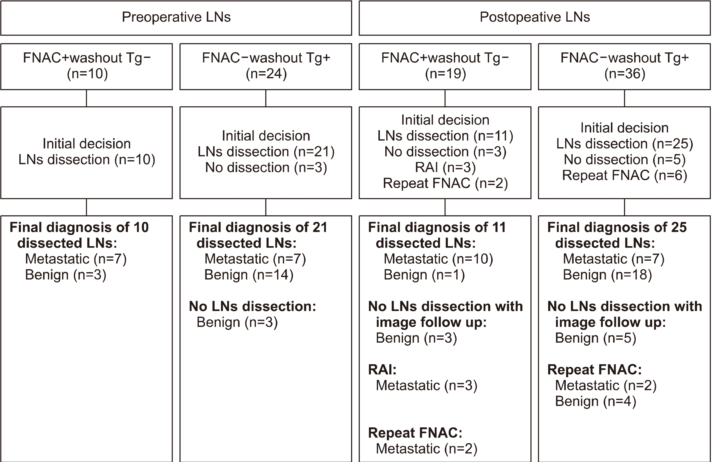
Decision Making for Discordant LNs between FNAC and Washout Tg (Fig. 2)
Among preoperative LNs, the majority of FNAC+ or washout Tg+ LNs was dissected with thyroidectomy. All 10 of FNAC+washout Tg− and 21 of 24 FNAC-washout Tg+ LN were dissected (Fig. 2, right panels). Among 19 postoperative FNAC+washout Tg− LNs, three LNs were not managed with any other diagnostic or therapeutic methods such as dissection, radioiodine ablation (RAI), or repeat FNAC; Two cystic metastases in FNAC were diminished after FNAC and not detected in follow-up imaging studies. One LN showed atypia in FNAC but was not detected in neck CT and follow-up thyroid US during study period. Three of 19 postoperative FNAC+washout Tg− LNs received RAI. 25 of 36 FNAC−washout Tg+ postoperative LNs were dissected (Fig. 2, rightmost panels). Five FNAC−washout Tg+ were not dissected and decreased in size in follow-up imaging during the study period. Six of 36 LNs had repeated FNAC; four LNs were benign and two were metastatic.
Overall, 8.9% (63 of 707) of LN findings were not in agreement between FNAC with washout Tg and the final LN diagnosis or were inconclusive results from FNAC with washout Tg. In postoperative LNs, discrepancies were less frequent compared to those in preoperative LNs. Inconclusive results were observed in 9.2% (27/295) of preoperative LNs and 8.7% (36/412) of postoperative LNs. 12.8% preoperative LNs (17/132 dissected LNs) and 15.0% of postoperative LNs (19/127 dissected LNs) were performed unnecessary dissection due to false positive of washout Tg and false positivity in FNAC. To confirm diagnosis, there are needed repeat invasive FNAC, image study including neck CT, and PET-CT.
Go to :
Discussion
Washout Tg measurement in FNA has been suggested as a supplementary diagnostic tool to FNAC since it was first performed by Pacini et al.8)in 1992. Although the diagnostic performance has been improved with combined diagnostic modalities,21)diagnostic cut-off values of washout Tg have not been established, and there are discrepancies between FNAC and washout Tg levels due to a wide range of washout Tg cut-off values and cytological insufficiency. To our knowledge, no study has evaluated the discordant rates of FNAC and washout Tg levels in both preoperative and postoperative settings simultaneously and in real-world practice.
In this study, discordant or inconclusive results occurred in any LNs for FNAC with atypia or cystic metastasis. four of 78 LNs with FNAC+washout Tg+ had benignity in final LN diagnosis. These four benign LN in histopathology presented AUS and metastatic PTC in FNAC before thyroidectomy. Three LNs from 10 preoperative FNAC+washoutTg− LNs were benign result of histopathology. There were no metastatic LN among all dissected LN in same compartment and at the biopsy location. It is worth mentioning that tissue from near intact thyroid lesion during FNAC before surgery would be obtained. Cystic transformation could be another challenge to diagnosis for metastatic LNs and was reported in primary DTC and in metastatic LNs.22)In preoperative 24 LNs with FNAC−washout Tg+ and postoperative 36 LNs with FNAC−washout Tg+, some of these LNs were metastatic on final diagnosis and they presented cystic change on thyroid US. Cystic changes in metastatic LNs are a diagnostic challenge and may show false negative results because of their low cellularity.23)Hatabu et al.24)reported that results of FNAC were metastatic in only 33% LNs with cystic change. Therefore, close examination for the detection of cystic LNs from DTC is needed. Majority of discordant cases between FNAC and final LN diagnosis came from inadequate or low cytology due to calcification or cystic portion.
In the present study, the optimal cut-off value for washout Tg in all LNs was 2.9 ng/dL. The cut-off value for washout Tg (9.6 ng/mL) for malignant diagnosis in the preoperative setting was higher than that for the postoperative setting (2.1 ng/mL). This result was consistent with previous studies.25,26) Several studies have suggested a wide range of preoperative washout Tg levels, 0.2 to 39.3 ng/dL.18,27) To improve the diagnostic performance of both FNAC and washout Tg, several centers use the washout Tg/serum Tg ratio to adjust the influence of serum Tg in preoperative LNs.7,12,28) However, the diagnostic performance was not significantly higher than that for FNAC with washout Tg and serum Tg level did not differ significantly between metastatic and benign LNs, regardless of surgery. Therefore, we used FNAC with washout Tg for the evaluation of discrepant results. A previous study in our center on preoperative LNs with or without TgAb reported washout Tg cut-off values ranging from 2.5 ng to 8.5 ng/mL according to TgAb posi-tivity.15)This difference may be due to the different study period and lack of classification according to TgAb titer in this study. In LNs awaiting thyroidectomy, a higher washout Tg cut-off value (9.6 ng/mL) showed high performance in sensitivity, PPV, NPV, and accuracy. Our washout Tg threshold for metastatic LNs was higher than those of previous studies (0.6–1.1 ng/mL).28,29) However, these previous studies were not based on a large number of LNs but based on only small case series. Moreover, higher cut-off values to 2.1 ng/mL and 9.6 ng/mL showed good diagnostic performance without decreasing sensitivity.
Our study has several strengths. First, the large-scale patient samples included real-world data. Secondly, all patients included in this study had a complete biochemical data for serum Tg and TgAb levels. Thirdly, we used different cut-off values for washout Tg according to surgery status. Therefore, the diagnostic performance was better than that in other studies.
This study also has several limitations. The retrospective study design might have led to selection bias in the interpretation of the results. Second, we included LNs without dissection, the final outcomes of which were decided only by follow-up imaging.
Washout Tg measurement in FNA washout is useful with, good diagnostic performance and no need for additional invasive procedures. However, false positive or false negative findings are not infrequent with FNAC in real-world clinical conditions and there is a need for accurate diagnostic tools in real-world practice.
In real-world practice, discordant results between FNAC with washout Tg and final LN outcomes may occur in up to 10% of cases, which can lead to unnecessary neck dissection or additive procedures. To improve the diagnostic accuracy of FNAC with washout Tg, washout Tg cut-off values should be applied depending on preoperative or postoperative LN status. Further research to identify new biomarkers in washout fluid is necessary to resolve discordant findings.
Go to :
 XML Download