This article has been
cited by other articles in ScienceCentral.
Abstract
The phosphorous balance is clinically important in increasing the long-term outcomes and preventing complications of end-stage renal disease. Sevelamer is a phosphate binder used widely to regulate hyperphosphatemia. On the other hand, gastrointestinal side effects increase with increasing sevelamer intake. A 29-year-old male with end-stage renal disease of IgA nephropathy on maintenance hemodialysis was admitted for diffuse alveolar bleeding and pneumonia. He presented with a low-grade fever and watery diarrhea tinged with blood. Initially, a Clostridioides difficile-associated diarrhea treatment was started with positive findings of Clostridioides difficile toxin and culture. Despite this, there was no improvement in the symptoms even with the appropriate antibiotic treatment. Computed tomography of the abdomen and pelvis revealed an occlusive mass in the rectum and secondary obstructive changes in the sigmoid colon. The initial suspicion was a malignancy or fungal infection. Sigmoidoscopy with a biopsy identified the mass as a lump of mucous material with the entire lumen covered with exudate. The subsequent histopathology examination revealed a colonic mucosal injury and characteristic “fish scale”-like sevelamer crystals in the exudate. The diagnosis of a sevelamer-induced rectal ulcer was made. We report this case of a sevelamer-associated rectal ulcer of the sigmoid.
Go to :

Keywords: Clostridioides difficile infection, Kidney failure, chronic, Sevelamer, Gastrointestinal hemorrhage
서 론
고인산혈증은 말기 신질환 환자에서 심혈관계 질환 발병 및 mortality의 독립적인 위험인자로 알려져 있으며 말기 신질환 환자에서 혈중 인농도의 조절은 매우 중요하다.
1,2 현재 국내 투석 환자의 고인산혈증 치료를 위한 인산염 결합제 중 비칼슘기반 인산염 결합제인 sevelamer (renvela)의 사용이 점차 증가하고 있다. 저자들은 만성 신장 질환으로 투석하는 환자에서 대장 종괴로 오인하게 한 Sevelamer-associated rectal ulcer를 경험하여 치료하였기에 보고하는 바이다.
Go to :

증 례
29세 남자 환자가 미만성 폐포 출혈과 폐렴으로 서울아산병원 호흡기내과에 입원하였다. 항생제 치료 중, 입원 4일째부터 점액 양상의 혈성 설사가 발생하여
Clostridioides difficile toxin과 배양 검사를 시행하였고, 양성 소견으로
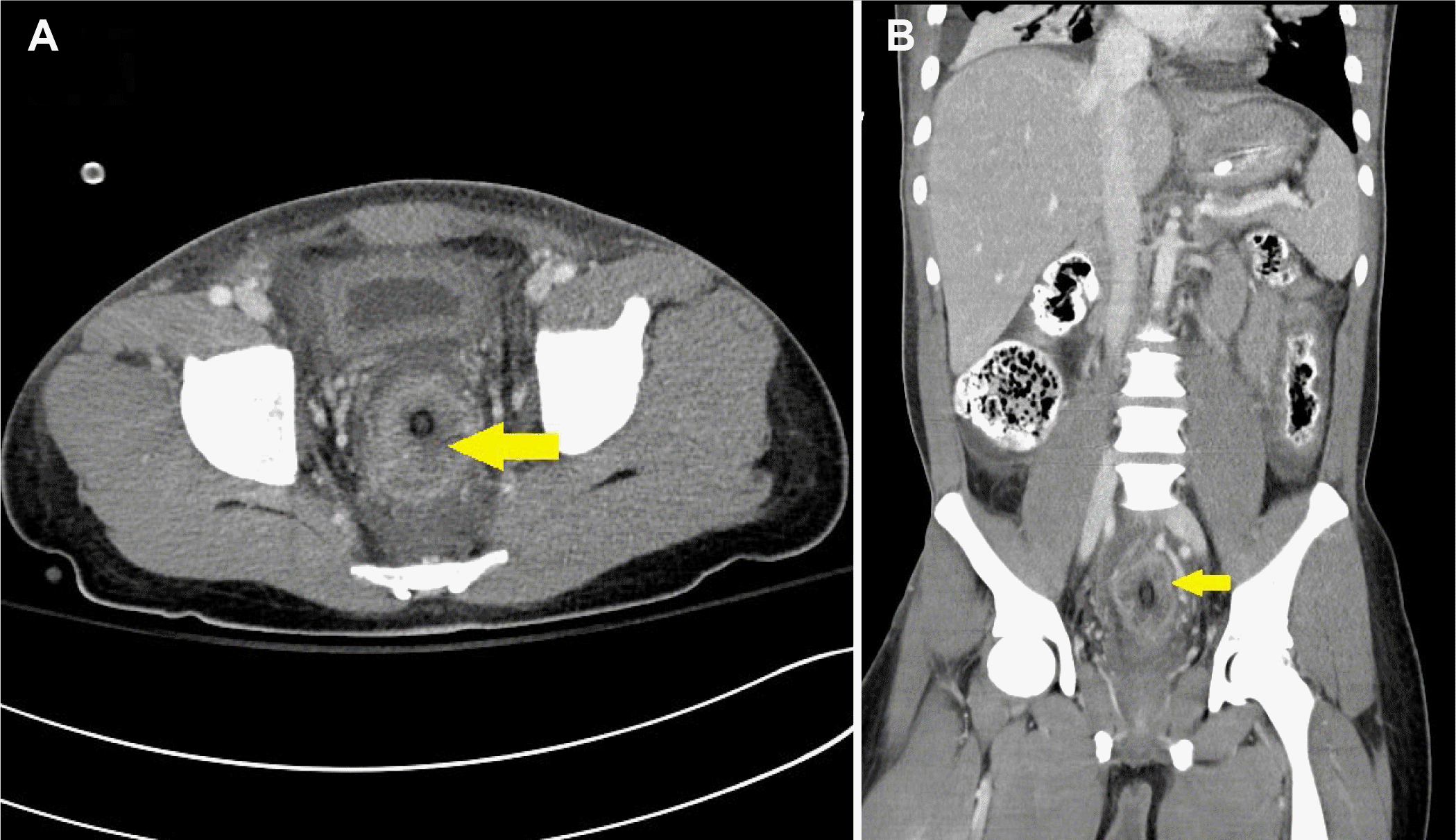
Clostridioides difficile 관련 설사로 진단하고, 이에 대해 경구 vancomycin 투약을 시작하였다. 약제 치료 후 5일이 지나도 임상 경과에 호전이 없어 경구 vancomycin 용량을 증량하고, 주사제로 metronidazole도 투약을 시작한 뒤, 소화기내과에 향후 치료 계획 수립과 관련하여 상의 되었다. 환자는 다운증후군을 진단받았으며, 내원 1년 전 IgA 관련 만성 신질환으로 투석을 유지하는 중이었고, 6개월 전부터 비칼슘기반 인산염 결합제인 sevelamer (renvela)를 복용하였다. 신체 진찰에서는 복부 팽만이 관찰되었으며, 촉지되는 종괴나 압통은 없었다. 일반혈액 검사에서 백혈구 23,700/uL, 혈색소 8.2 g/dL였고, Na 134 mmol/L, K 5.3 mmol/L, P5.2 mg/dL, CRP 8.69 mg/dL였다. 이에 복부 전산화단층촬영을 시행하였으며, 직장에 약 23 mm 크기의 종괴가 확인되었다(
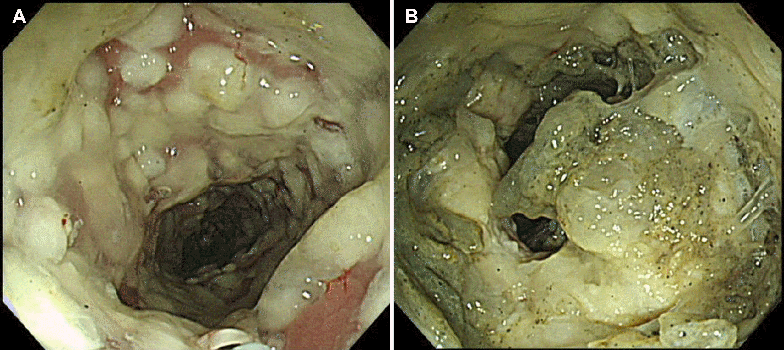
Fig. 1). 이에 구불결장 내시경 검사를 시행하였으며, 구불결장 전체에 위막성 변화가 관찰되고(
Fig. 2A), 항문연 5-9 cm에 걸쳐 위막으로 덮여 있는 종괴가 확인되어 조직 검사를 시행하였다(
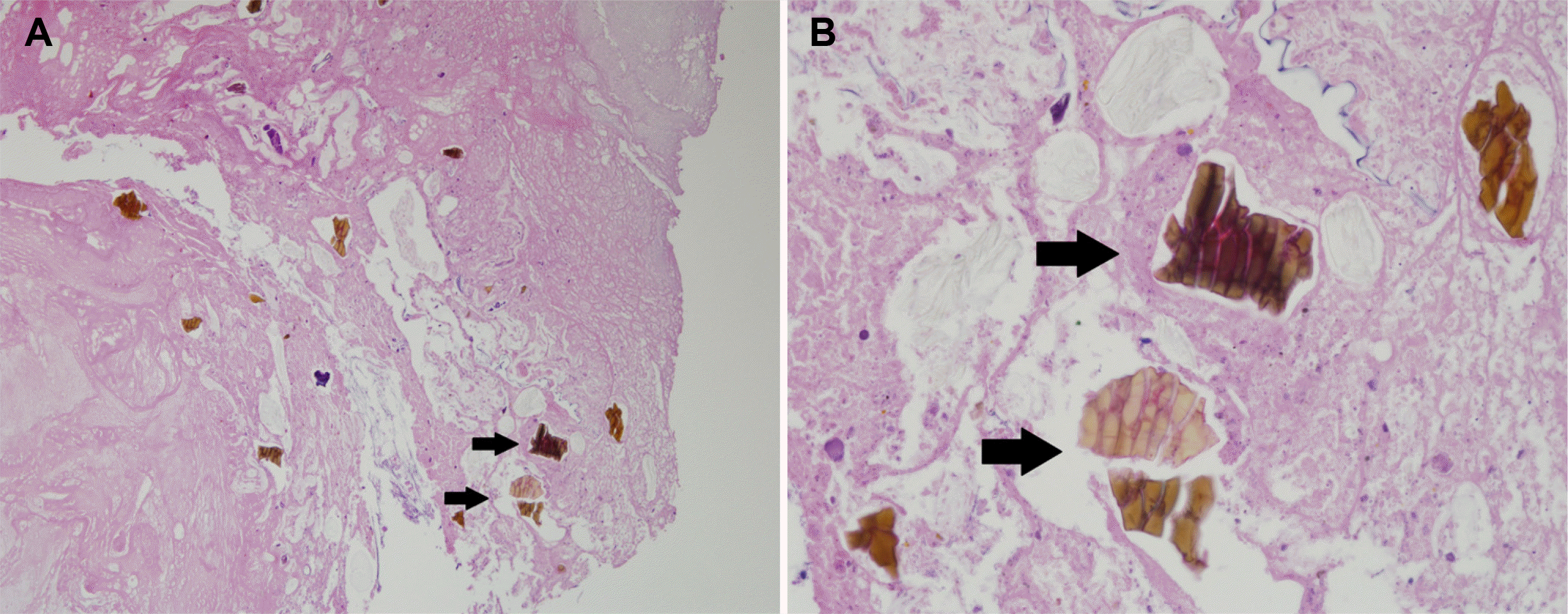
Fig. 2B). 대장 조직 검사 결과, "fish-scale" 형태가 특징적인 sevelamer crystal이 대장 점막손상에 동반된 것이 확인되어 sevelamer-associated rectal ulcer로 진단할 수 있었다(
Fig. 3).
 | Fig. 1Abdominal pelvis computed tomography (CT). The rectal lumen is narrow, and the rectal wall is thickened. CT showed a defined, irregular, low attenuating mass with heterogeneous attenuation in the rectum (arrow of A and B). (A) Axial view. (B) Coronal view. 
|
 | Fig. 2Sigmoidoscopy. (A) Raised, yellowish-white pseudomembranous plaques on the sigmoid. (B) A mass covered with whitish exudate on the rectum. 
|
 | Fig. 3Histology examination. (A) The sections show necrotic debris and sevelamer crystals (black arrows) (H&E, ×100) (B) H&E stain with a magnified view of the sevelamer crystals. The fish-scale appearance is characteristic (black arrows) (H&E, ×400). 
|
Go to :

고 찰
Sevelamer-associated rectal ulcer는 sevelamer의 복용이 확대되고 있는 상황에서 약제로 인한 장관 손상의 발생을 보여준 보기 드문 진단명이다. 비칼슘기반 인산염 결합제인sevelamer는 처음에는 담즙산과 결합하여 담즙 배출을 증가시키고 LDL-cholesterol을 낮추는 역할로 사용되었으나,
3 투석 환자나 신부전 환자에서 고칼슘혈증의 위험을 높이지 않으면서 장관에서 인산과 흡착하여 혈중 인산 농도를 낮추는 효과를 나타내어 2008년에 Food and Drug Administration에서 투석 환자의 고인산혈증을 치료하는 치료제로 승인되었다.
4 이후 Kidney Disease Improving Global Outcomes guideline에서도 고칼슘혈증이 동반된 환자에서 고인산혈증치료제로 권유하고 있어 널리 쓰이고 있다.
5
고분자 음이온 교환 수지인 sevelamer의 작용 기전은 위의 산성 환경에 의해 해리되어 장내 인산염과 결합하여 체내 흡수를 감소시키고 혈청 인산염의 농도를 낮춘다.
6 인산염과 결합된 고분자는 인산염 결정 결석으로 배설된다. 보고된 소화기계 부작용으로 구토, 오심, 설사, 소화불량, 복통, 변비 등이 있으며, 최근 sevelamer 관련 숙변성 궤양이나 점막 손상으로 인한 위장관 염증, 혈변 등의 증례가 보고되고 있어
7,8 장폐색이 있는 환자에서는 금기이다.
9
Sevelamer 결정이 위장관에서 점막의 염증이나 궤양, 허혈성 변화 및 궤사와 관련이 있다고 언급하였으며,
9 실제 sevelamer 결정으로 인한 위장관 궤사가 발생한 조직에서 호중구 괴사와 염증을 일으키는 호중구 세포 밖 덫(neutrophil extracellular traps)이라는 물질이 발현하는 것이 증명되었고, 이로 인한 장 점막의 염증은 방어벽 기능을 악화시키고,약제가 고농도로 사용될 때 장 점막에 세포독성으로 작용하여 점막의 세포 사멸을 유도할 수 있어 sevelamer로 인한 위장관 점막의 염증, 궤사 및 천공 등을 설명 가능하게 하였다.
10그러나 sevelamer로 유발된 위장관 부작용을 분석한 논문에 따르면 약제 농도와의 상관관계는 분명하지 않다고 하였다.
11
이 논문에 따르면 16명의 사례를 분석한 결과, 가장 흔한 위장관 관련 임상증상은 위장관 출혈(44%)이었고, 이어서 복통 혹은 복부 불편감 호소(19%)였으며, 무증상 환자(18%)도 있었다. Sevelamer 결정이 확인된 생검 조직의 위치로는 상행결장과 하행결장이 가장 많았으며(50%), 소장(37%), 식도(25%), 위(18.8%) 순서로 발생한 것으로 되어 있어 위장관 내 모든 위치에서 발생이 가능하다. 생검 조직 소견으로는 궤양(n=9), 궤사(n=6) 그리고 급성 염증(n=5) 및 허혈성 손상(n=3)과 염증성 폴립(n=3)이 뒤를 이었다. 본 증례의 환자는 구불결장 내시경에서 흰색의 삼출물이 덮여 있는 종괴가 확인되었고, 조직 검사에서 괴사성 조각 및 sevelamer 결정이 확인되어 진단이 되었다. 내시경 소견에서 볼 때, 위막성 대장염과 동반되어 육안적으로 대장 점막의 손상 여부를 확인하지 못하였고, 병리 조직에서도 점막의 손상 여부가 불분명하였으나, 처음에는 점액 양상의 혈성 설사가 나타난 것으로 미루어 볼 때, 종괴 형성과 더불어 점막의 손상이 동반되었을 것으로 생각해 볼 수 있었다.
Sevelamer 결정이 유도한 종괴를 형성한 증례 3예
12-14 모두 만성 신장 질환으로 고인산혈증 치료를 위해 sevelamer를 복용하였으며 그중 한 예
14는 대장 폐색으로 수술적 치료 후 허혈성 손상을 받은 대장 점막에서 sevelamer 결정이 확인되어 진단되었다. 이와 같이 만성 신질환 환자에서 위장관의 운동 기능 저하와 점막의 분비 및 흡수 기능의 장애로 약제결정화 위험성이 더 증가할 수 있어,
15 기왕력 및 약제 복용력의 확인이 중요하겠다.
Sevelamer crystal의 병리적인 특징은 H&E 염색에서 짙은 노란색부터 갈색을 나타내며, PAS 염색에서는 보라색을 특징으로 하는, 널찍하고 불규칙한 간격의 물고기 비늘 또는 나무껍질 모양을 나타낸다.
9 본 증례에서도 H&E 염색에서 전형적인 물고기 비늘 모양과 약제 복용력으로 sevelamer-associated rectal ulcer를 진단할 수 있었다.
치료에 대해서는 명확하게 밝혀진 바가 없지만, 원인 약제 중단 후 추적 관찰한 내시경에서 점막 손상이 회복되었다는 보고가 있어
12 원인 약제 중단 후 추적 관찰하는 것이 중요하겠다.
만성 신부전 환자에서 고인산혈증에 대해 sevelamer의 약제 복용이 증가하고 있으며, 임상증상이 다양하고, 영상이나 내시경 소견 등이 비특이적이기 때문에 사전에 약제 유발 점막 손상 가능성을 인식하는 것이 중요하다. 또한 특징적인 병리 소견과 환자의 병력 등을 고려한다면 적절하게 진단하고 치료하는 데 도움이 될 수 있을 것이다. 저자들은 직장에서 sevelamer로 유도된 대장 종괴 1예를 경험하여 이를 보고하는 바이다.
Go to :


 PDF
PDF Citation
 Print
Print


 XML Download