

 PDF
PDF Citation
 Print
Print


Iodinated contrast-induced encephalopathy (CIE) is a rare complication of cerebral angiography. Iodinated contrast media disrupts the blood brain barrier (BBB) temporarily, causing an encephalopathy that is usually self-limiting.1 Imaging is important in confirming the diagnosis of CIE and excluding thromboembolic and hemorrhagic complications of cerebral angiography. Typical computed tomography (CT) findings include abnormal cortical contrast enhancement and edema, subarachnoid contrast enhancement, and striatal contrast enhancement. 2 The absence of diffusion restriction or hyperintensity without a change in the apparent diffusion coefficient (ADC) on diffusion-weighted imaging (DWI) of magnetic resonance imaging (MRI) is an important finding.3–5 This relatively excludes the possibility of acute ischemia secondary to shower of emboli during the angiographic procedure.3 Here, we report a rare case of CIE mimicking acute cerebral infarction (ACI) following cerebral angiography, showing diffusion restriction on DWI with a low ADC value in post-angiography MRI.
CASE
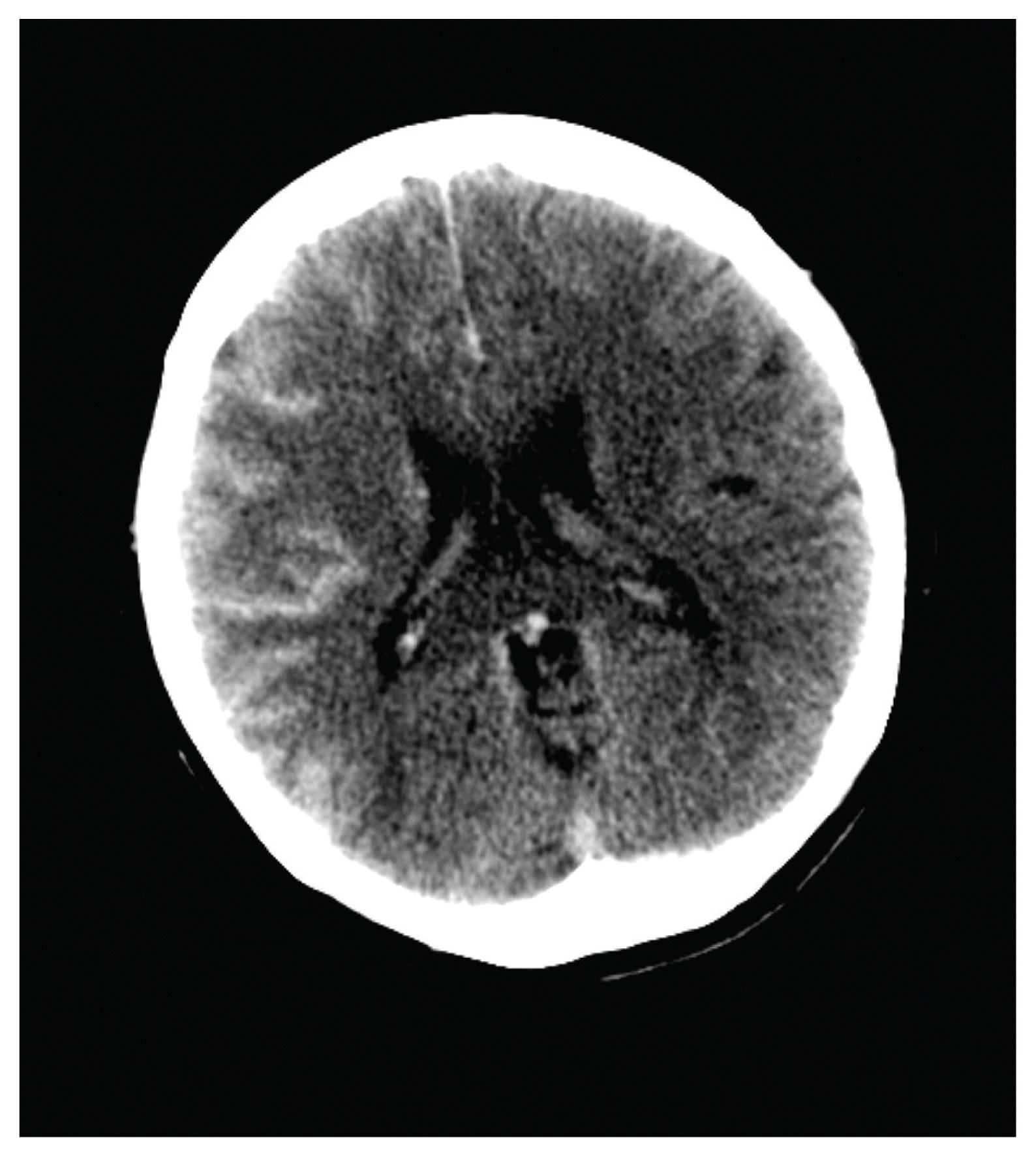
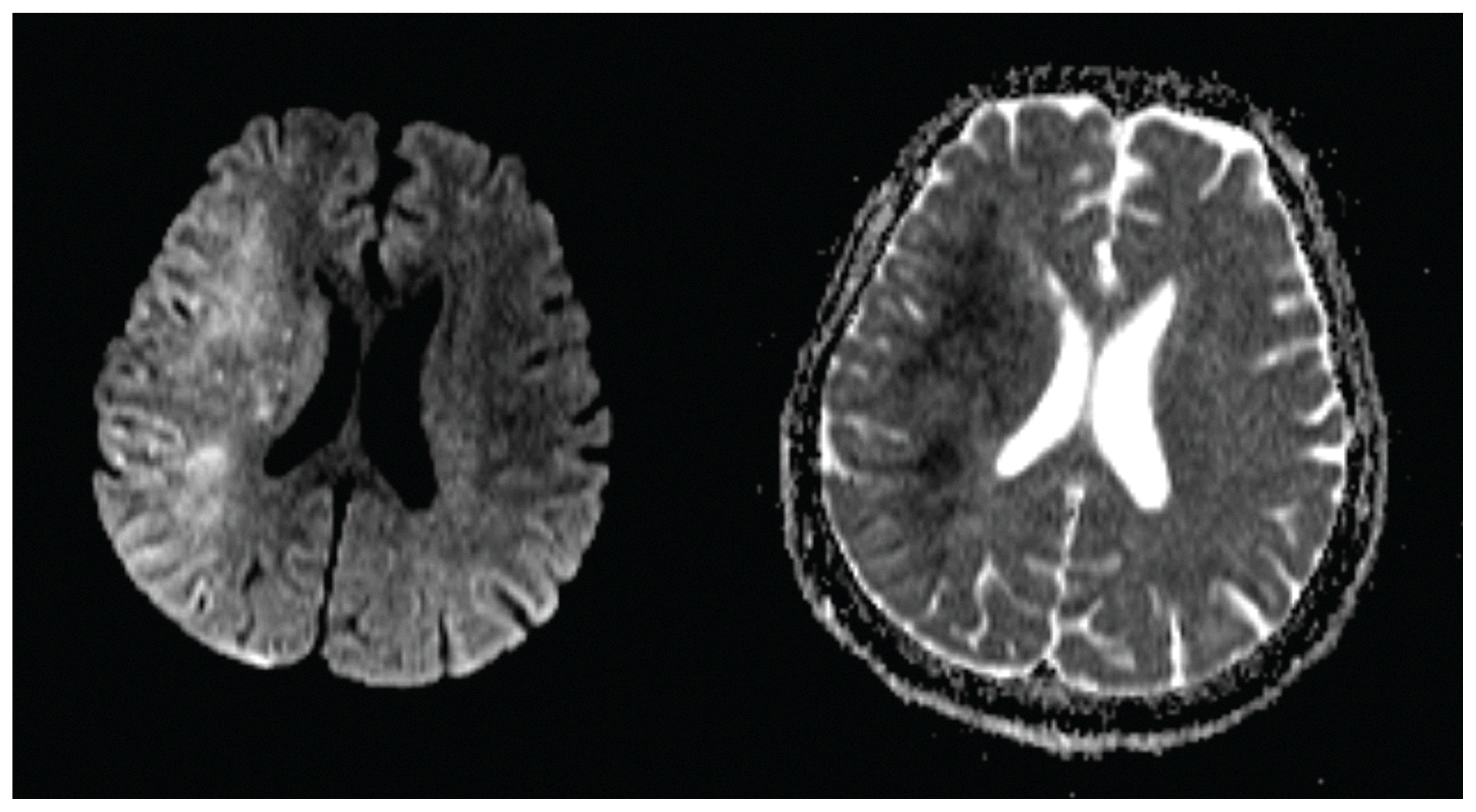
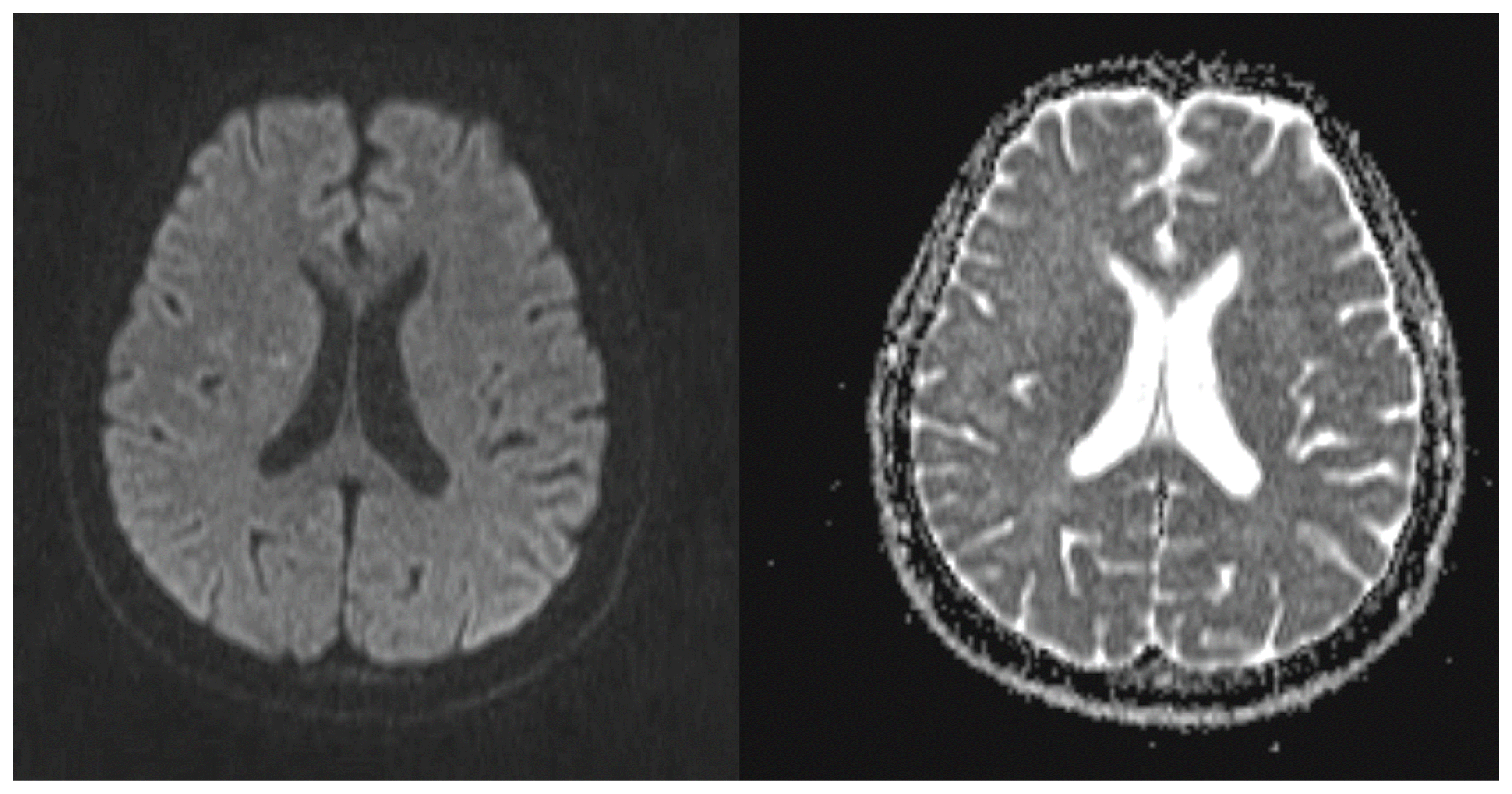
A 44-year-old woman who underwent uneventful coiling for intracranial aneurysm 1 month previously was readmitted to our hospital for additional coiling of a right paraclinoid internal carotid artery (ICA) aneurysm. Risk factors for aneurysm formation included a 17-year history of systemic lupus erythematous and hypertension. During the right ICA angiography just before coiling, the patient developed rapidly worsening mentality and left hemiparesis on the table. She had received a total of 40 mL of iopromide (Ultravist 300, Bayer Healthcare Pharmaceuticals, INc., Wayne, NJ, USA), which is a low-osmolar non-ionic contrast agent. After confirming no abnormal findings on right ICA angiography, we stopped the procedure without coiling the aneurysm. Non-contrast brain CT, performed immediately after stopping the procedure, showed marked enhancement throughout the right cerebral cortex and right basal ganglia with concordant diffuse swelling of the right cerebral hemisphere, indicative of CIE (Fig. 1). To exclude the possibility of acute ischemic complications, MRI was immediately performed. It showed extensive cortical hyperintensity within the right hemisphere on DWI and corresponding hypointense signal alterations on the ADC map, nearly matching with the findings of CT (Fig. 2). Overall, a poor clinical outcome was predicted. However, with dexamethasone injections and supportive care, the patient gradually recovered without additional neurologic signs. Follow-up MRI performed 1 week after the angiography showed a normal findings (Fig. 3). She was discharged from the hospital with a modified Rankin scale score of 0.
DISCUSSION
Although with exceptions, CIE is usually a transient and reversible phenomenon. Neurological recovery occurs over at a median duration of 2.5 days.1 The definitive diagnosis of CIE is important as it may have a similar presentation to ischemic or hemorrhagic complications of cerebral angiography. Imaging is essential in confirming the diagnosis and excluding thromboembolic and hemorrhagic complications of cerebral angiography. If brain CT and MRI performed immediately after clinical presentations shows typical findings of CIE, a rapid clinical and radiological recovery is predicted. However, if MRI findings suggest ACI, as in this case, a poor clinical outcome is expected by most clinicians.
Hyperintensity on DWI and hypointense signal alterations on the ADC map correspond to a decreased movement of highly mobile extracellular water molecules resulting from their shift into the intracellular compartment. This diffusion restriction reflects the cellular edema, which is considered to cause an irreversible lesion and cellular death. Cellular edema and swelling associated with ischemia have been defined as “cytotoxic”. When cellular edema and swelling occur without ischemia, the case may be defined as “neurotoxic”. 6 Neurotoxicity results from contrast media penetrating BBB with resultant cerebral edema and altered neuronal excitability. Most cases related to the breakdown of the BBB are reported as a transient and reversible. Therefore, contrast-induced BBB breakdown may only cause vasogenic edema in most cases and rarely lead to cytotoxic edema. In previous MRI findings of patients with CIE, the vasogenic edema was considered the most probable underlying cause of the reversible lesion.1
Although reversal of diffusion restriction is uncommon, it has been described in cases of status epilepticus and the postictal state, venous infarction, and thrombosis, hemiplegic migraine, transient global amnesia, transient ischemic attacks, and early reperfusion of ischemic brain parenchyma.7 Theories have been proposed to explain how the reversal occurs; however, the definitive mechanism is not completely understood yet. In this case, extravasation of a large amount of contrast media due to BBB breakdown might have resulted in cytotoxic effects on the affected neurons. Although contrast-induced neurotoxicity is more likely to cause vasogenic edema because of BBB breakdown, it may cause cytotoxic edema by an idiosyncratic reaction to contrast media or the individual’s predisposition for brain tissue edema. In our case, the initial MRI suggested extensive ACI; however, the diffusion restriction resolved completely with supportive care, and related neurologic deficits disappeared without permanent neurologic deficits. Based on the current knowledge, this complication appears to be an idiosyncratic reaction to contrast media. This fact makes avoidance of contrast encephalopathy difficult. Further investigations are required to understand why some patients experience contrast gyral enhancement whereas others do not, why only a minority of patient is symptomatic, and why some patients have persistent deficits or die whereas others do not.
We reported a rare case of CIE with diffusion restriction on MRI following cerebral angiography that mimicked MRI findings of ACI. Our unique case highlights that occasionally, diffusion restriction on DWI due to CIE may show reversal, and good clinical outcome can be achieved.
 XML Download