

 PDF
PDF Citation
 Print
Print


At autopsy, the incidence of primary cardiac tumors is 0.1 – 0.3%.1 Approximately three quarters are benign tumors, most of which are myxomas or fibroelastomas.2 Blood cysts are presumptively congenital and are relatively common in infants, but extremely rare in adults.3,4 Intracardiac blood cysts have a thin-walled membrane and a homogeneously echolucent center on echocardiography.2
We experienced an adult patient with a right atrium (RA) cyst with heterogeneous echogenicity. This cyst initially resembled vegetation when the patient presented with sepsis.
CASE
A 79-year-old woman presented to another hospital with complaints of right leg pain for one week. Abdominal computed tomography (CT) and spinal magnetic resonance imaging (MRI) was performed at that hospital, which showed an abscess in the right iliacus muscle. She was referred to our hospital because of a mass in the RA detected by echocardiography. At the time of admission, an electrocardiogram showed normal sinus rhythm with a right bundle branch block, and all cardiac markers were within normal range. Inflammatory markers were elevated (white blood cell, 22,750 /μL, C-reactive protein 19.14 mg/dL), and Staphylococcus aureus was identified in blood cultures.
Transthoracic echocardiography (TTE) at our hospital revealed a hypermobile mass (1.7 × 0.6 cm) on the interatrial septum in the RA, similar to the previous examination which was performed at another hospital. We thought that it was a vegetation (yellow arrow, Fig. 1A, 1B). However, transesophageal echocardiography (TEE) showed another aspect to the mass, a hypoechoic mass. It was 1.9 × 1.7 cm and looked like a myxoma or cyst (yellow arrow, Fig. 1C). The hyperechoic mass, which appeared on TTE, was attached to the surface of that mass. A patent foramen ovale was confirmed by color Doppler (Fig. 1D). The shunt flowed toward the RA mass, where there was hyperechoic mass. We thought that it was a newly developed vegetation on the previously existing RA mass. A cardiac CT image appeared to show a myxoma, which was the RA mass without enhancement (Fig. 2). The differential diagnosis of this mass included a blood cyst, myxoma, and vegetation. We surgically removed the mass, which was pathologically confirmed as a myxoid with cystic changes (Fig. 3). There was no evidence of vegetation on the surface of the cyst. The inflammatory marker was improved gradually as the abscess in the right iliacus muscle was improved.
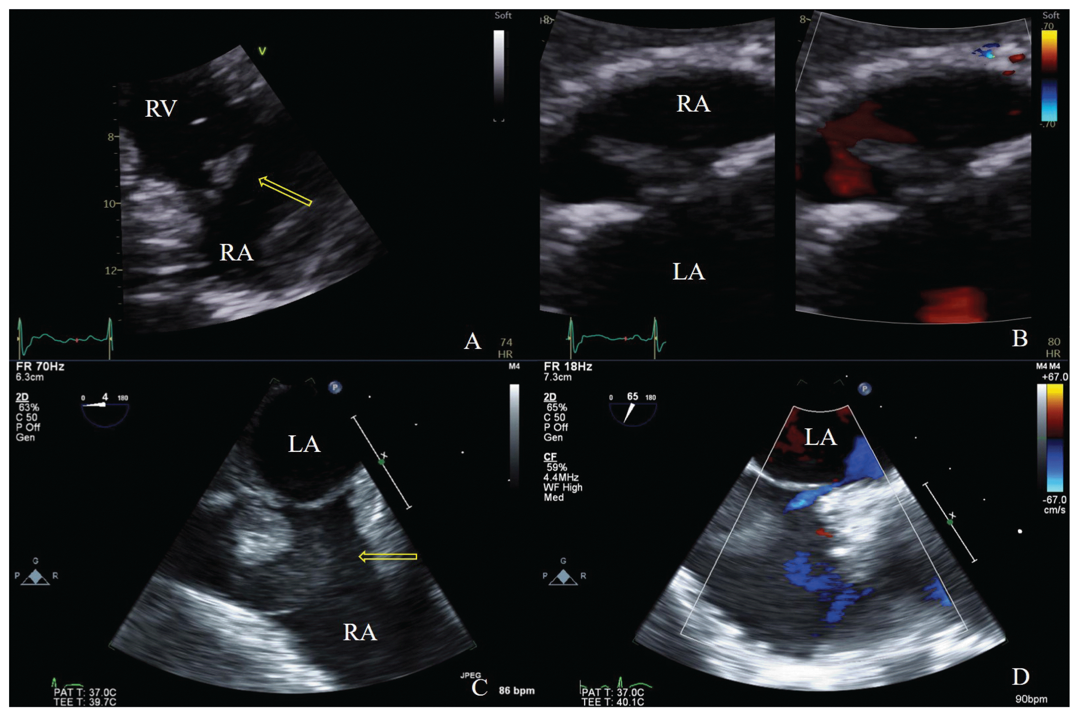 | Fig. 1(A) TTE in the parasternal RV inflow view showing a shaggy-appearing mass. (B) TTE in the subxiphoid view, showing a mass with color Doppler. (C) TEE shows a large hypoechoic mass and shaggy-appearing mass. (D) A patent foramen ovale is seen on TEE.
TTE, transthoracic echocardiography; RV, right ventricular; TEE, transthoracic echocardiography
|
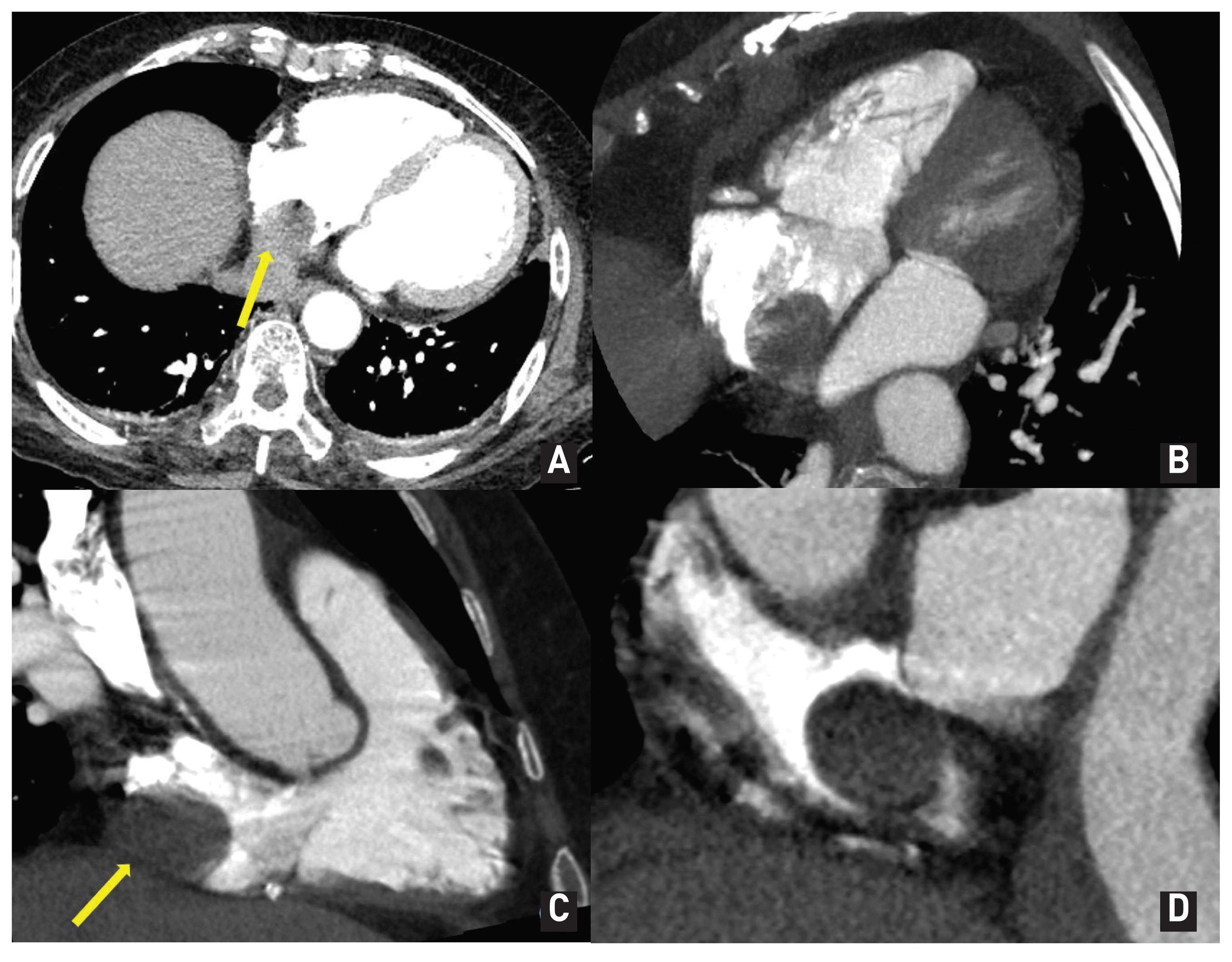 | Fig. 2Heart CT image showing a 2.5 cm mass arising from the interatrial septum of the RA.
CT, computed tomography; RA, right atrium
|
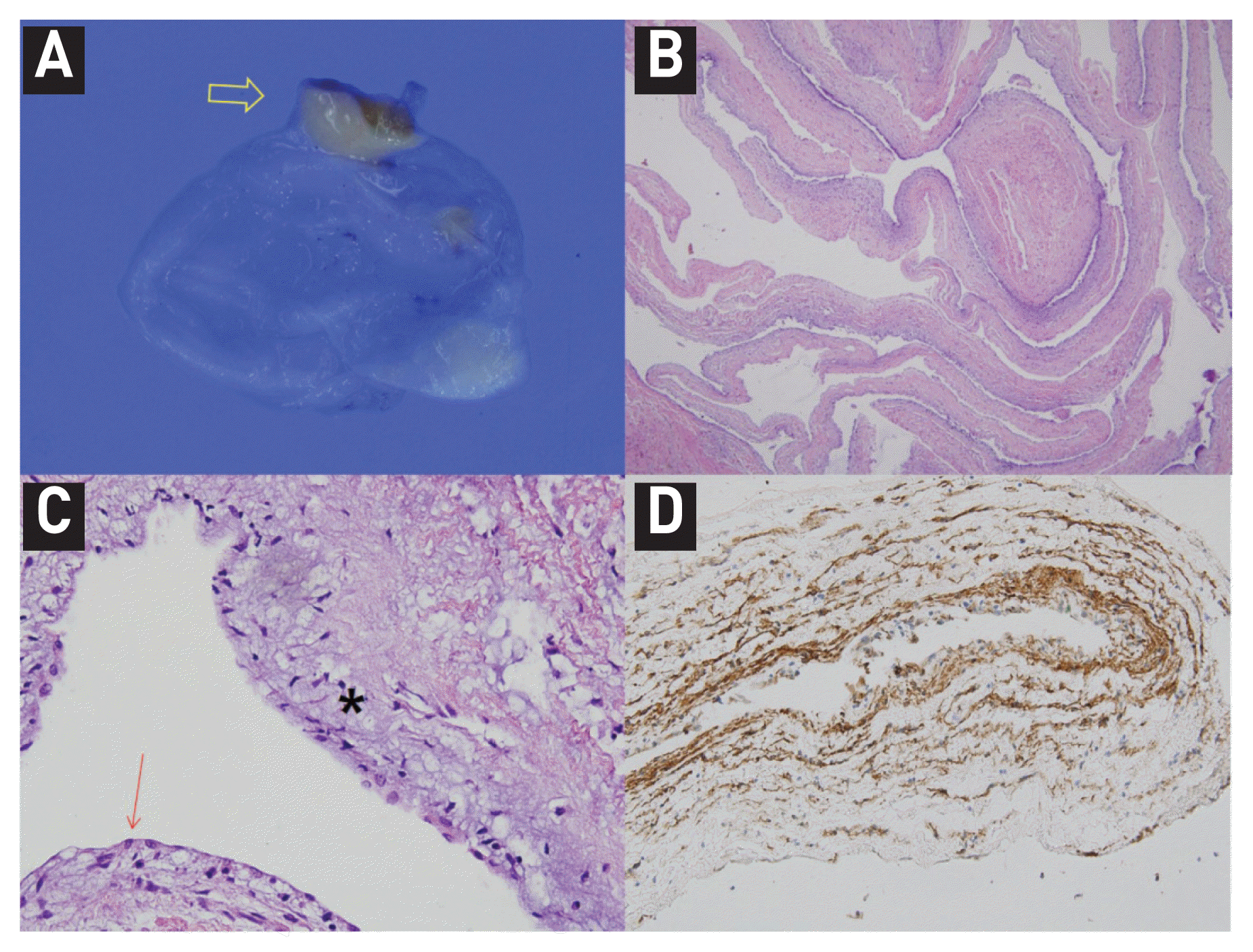 | Fig. 3(A) Gross findings of the resected RA mass. The cystic mass shows a translucent cystic wall without a solid nodule. The pedicle of the cystic mass is identified (arrow). (B) Microscopic examination of the resected specimen. A. At low magnification, the cystic wall shows myxoid changes in the surface aspect and is supported by a fibromuscular wall (H-E stained, x40). (C) At a high-power view (x400), the cystic wall appears composed of the flat surface epithelium and subepithelial myxoid stroma (flat surface epithelium; red arrow, myxoid stroma; star). (D) The surface epithelium and stellate or spindle-shaped stromal cells appear focally positive for CD34 which was often positive in myxoma10.
RA, right atrium; H-E, hematoxylin and eosin
|
Go to : 
DISCUSSION
Primary benign cardiac tumors are rare, and approximately 50% are myxomas. Other benign cardiac tumors, including cysts, have an incidence of 0.0017% in the general population. Intracardiac cysts are observed less frequently than pericardial cysts.2,5 Blood cysts in the heart were first reported in 1844 by Elsasser. 6 These cysts are congenital in origin and rare in adults. Cardiac blood cysts are typically found on the closure of the valvular endocardium, but can grow in all heart cavities.3,7 A patient with a blood cyst, high fever, and chills was previously reported by Dumantepe et al.8 In that report, the patient’s fever was attributed to a pulmonary embolism and there was no bacteremia.
We encountered a difficult case where a blood cyst mimicked vegetation in a patient with Staphylococcus aureus bacteremia. There are no recommendations for the treatment of asymptomatic blood cysts. Some authors have monitored the cyst size and found that it did not increase in size over time.9 However, in this case, the patient had fever and bacteria were identified in repeated blood cultures; therefore, surgical treatment was necessary.
Go to :
 XML Download