This article has been
cited by other articles in ScienceCentral.
Abstract
Idiopathic intracranial hypertension (IIH) is a syndrome defined by elevated intracranial pressure without any abnormal findings. In the present study, we report a rare case of IIH in a patient after ventriculoperitoneal shunt (VPS) due to infant hydrocephalus. A 13-year-old girl with a history of VPS due to infant hydrocephalus was admitted to emergency room with the complaint of severe headache and visual disturbance. Brain computed tomography showed normal findings. However, based on the measurement by lumbar puncture, her cerebrospinal fluid (CSF) pressure was observed to be very high. The shunt function test revealed a VPS malfunction. Thus, we conducted VPS revision in this patient. All symptoms improved immediately after the revision. Thus, it is proposed that IIH should be considered for patients with visual disturbance and severe headache after VPS due to infant hydrocephalus without ventriculomegaly.
Go to :

Keywords: Idiopathic intracranial hypertension, Infant hydrocephalus, Ventriculoperitoneal shunt
Idiopathic intracranial hypertension (IIH) is a syndrome defined by elevated intracranial pressure without any clinical, laboratory, or radiographic evidence of responsible infection, vascular abnormality, space-occupying lesion, or hydrocephalus.
1 Treatment options for IIH include physical modalities (weight loss), acetazolamide, furosemide, and lumbar puncture. Ventriculoperitoneal shunt (VPS) is considered as safe and very effective in patients with uncontrolled IIH.
2 Infant hydrocephalus shows ventricle size enlargement due to elevated resistance to cerebrospinal fluid (CSF) resorption (approximately three times the normal)
3 that can mainly occur due to hemorrhage and infection. VPS can be performed as a treatment.
4,
5 However, cases of IIH without progression to hydrocephalus are limited even if VPS is performed due to infant hydrocephalus. Herein, we report a rare case of IIH in a patient after VPS malfunction due to infant hydrocephalus.
CASE
A 13-year-old girl was admitted to our emergency department with the complaint of severe headache, seizure, and visual disturbance for the past month. There were no other neurological abnormalities, such as facial palsy. She had a history of VPS due to infant hydrocephalus at 6 months after birth. One year before the emergency department visit, the patient underwent VPS (Strata) revision due to distal catheter shortening in another hospital.
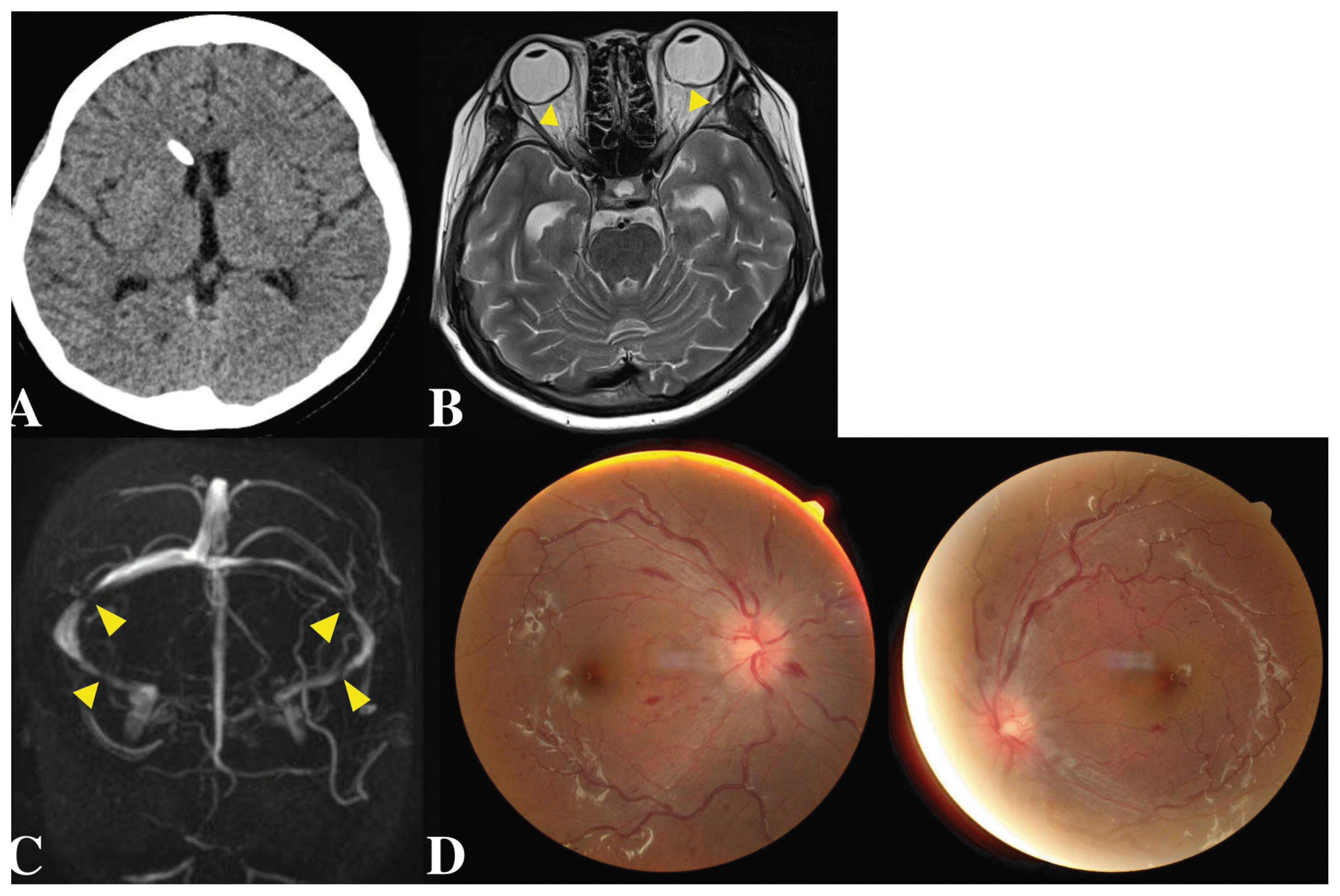
Brain-computer tomography (CT) imaging finding was not consistent with hydrocephalus (
Fig. 1A). Brain magnetic resonance (MR) imaging showed flattening of the posterior sclera and intraocular protrusion of both the optic nerve heads, depicting papilledema (
Fig. 1B). MR venography showed multifocal severe stenosis of bilateral transverse and sigmoid sinuses (
Fig. 1C).
 | Fig. 1(A) Preoperative brain computer tomography (B) Brain magnetic resonance T2 image showing intraocular protrusion of both the optic nerve heads (yellow arrows) (C) Brain magnetic resonance venography showing multifocal stenosis of bilateral transverse and sigmoid sinuses (yellow arrows) (D) Papilledema with marked circumferential disc elevation, and obstruction of vessels on the disc with multiple retinal hemorrhages in both the eyes were observed in fundus photograph. 
|
Her visual acuity was 20/125 in the right eye and 20/200 in the left eye. Her light reflex was normal. Her bilateral optic discs were elevated bilaterally, with retinal veins dilated and tortuous. Multiple retinal hemorrhages were observed (
Fig. 1D). Her body mass index was normal (20.9 kg/m
2).
We taped a ventricular catheter reservoir to rule out shunt infection. Her CSF study was normal (protein concentration: 51.3 g/l; glucose: 105 g/l; and 3 white cells/mm3). The shunt pressure was changed from 1.0 to 0.5 (3 cm H2O). However, her symptoms showed no improvement.
Lumbar drainage was performed to decrease intracranial pressure (ICP). The opening pressure was 39.5 cm H2O.
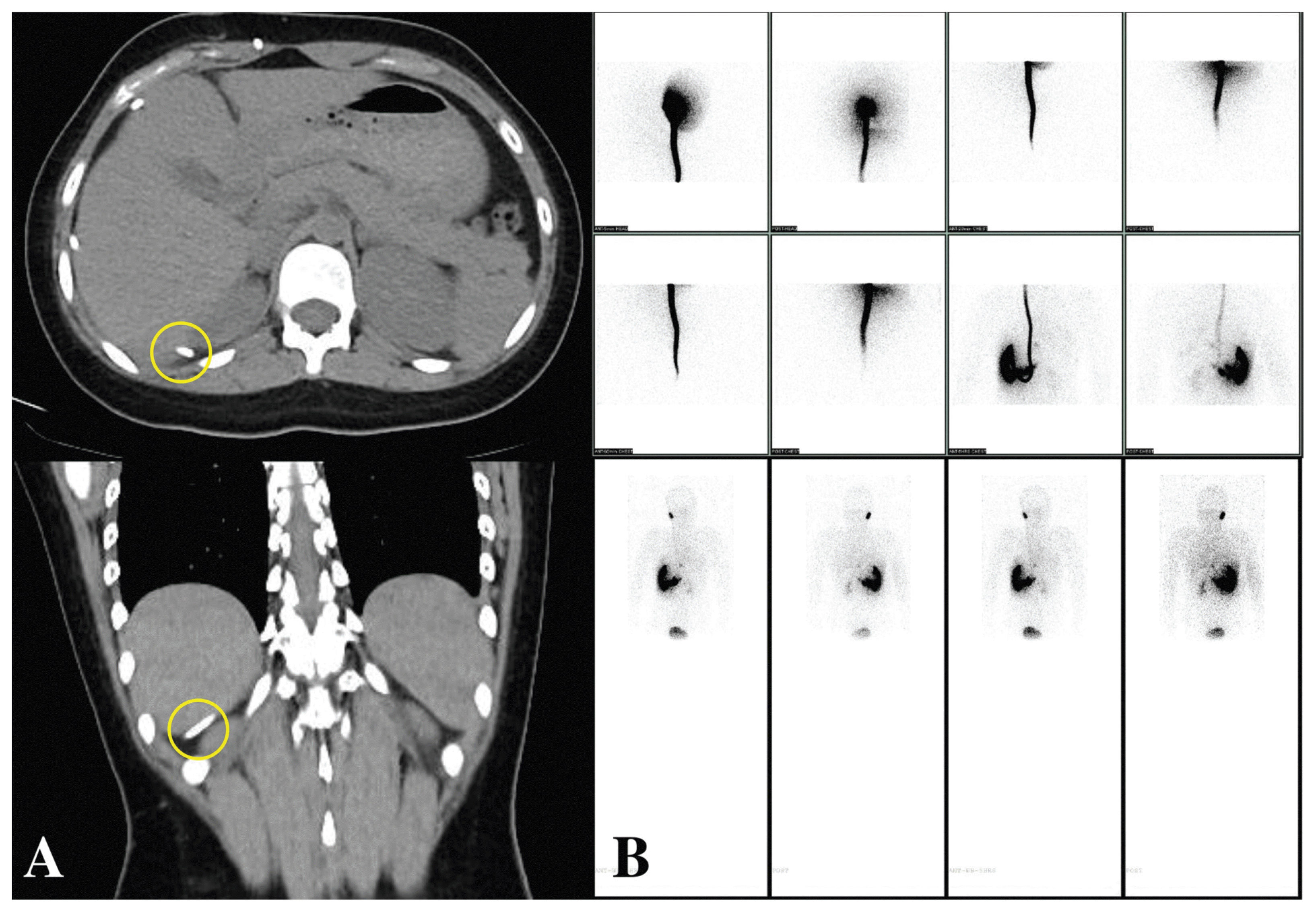
After CSF drainage, improvement in headache and visual disturbance was observed. An abdomen X-ray was performed to check the location of the distal catheter. It was observed to be shifted to a little upper side and located around the liver. Abdomen CT imaging surprisingly revealed that the tip of the distal catheter had found its way through the perihepatic space to the retroperitoneum (
Fig. 2A). VPS function test (Tc-99m DTPA) suspected malfunction of both distal and proximal VPS catheters (
Fig. 2B). We suspected that these IIH symptoms were caused by the malfunction of VPS. Thus, we decided to perform a VPS revision.
 | Fig. 2(A) Abdomen computer tomography axial and coronal images showing catheter tip (yellow circles) stuck in perihepatic space (B) Tc-99m diethylene-triamine-pentaacetate (DTPA) image showing the malfunction of both proximal and distal catheters. 
|
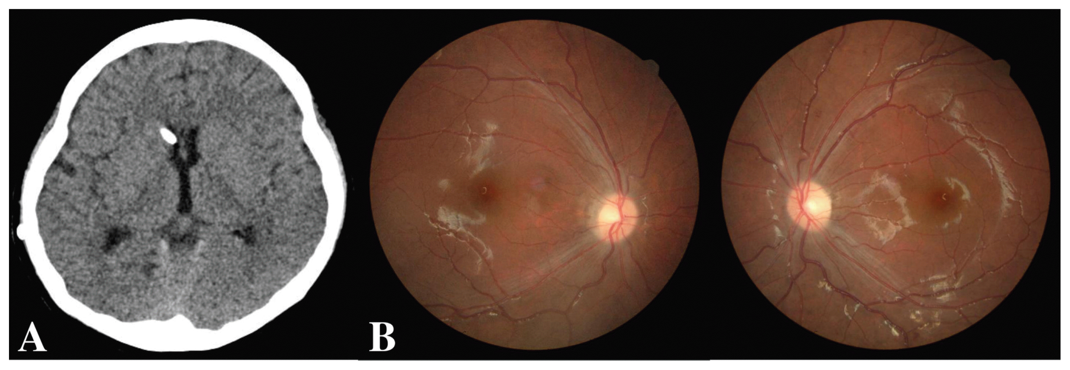
During surgery, the proximal catheter was observed to function well. Thus, it was maintained and we changed the distal catheter since valve (ProGAV). After reaffirming its function, the distal catheter was inserted into the peritoneal cavity. Her headache improved immediately on a postoperative day one. The overall ventricular size was unchanged (
Fig. 3A). Her visual acuity was also improved to 20/32 in the right eye and 20/80 in the left eye. She was discharged on post-operative day 10. Her papilledema and retinal vascular abnormalities also subsided. Her optic disc was pale on postoperative one month (
Fig. 3B). She is currently being followed up in the outpatient department without any symptoms.
 | Fig. 3(A) There is no significant change in postoperative brain computer tomography (B) Optic disc swelling subsided and retinal hemorrhages were disappeared postoperatively. 
|
Go to :

DISCUSSION
IIH is a syndrome related to elevated intracranial pressure of unknown cause. It is sometimes a cerebral emergency. It occurs in all age groups, especially in children and young obese women. Patients with IIH may show several symptoms including visual disturbance without underlying expansive intracranial lesion.
2 A definite diagnosis of IIH requires these symptoms and signs listed in the modified Dandy criteria (
Table 1).
1 The present case met the above criteria. The patient underwent VPS. However, she showed severe headache and visual disturbance without any size change in the ventricle.
Table 1
Modified Dandy criteria of idiopathic intracranial hypertension
|
Variable |
-
- Symptoms of increased intracranial pressure (ICP)
Headache, nausea, vomiting, transient visual obscuration, or papilledema
-
- No localizing findings in neurological examination
Except for false localizing signs such as abducens or facial palsies
- Normal CT/ MRI finding without evidence of dural sinus thrombosis - ICP of > 250mm H2O with normal cerebrospinal fluid cytological and chemical findings - No other cause of increased ICP
|

There are various causes of IIH, including endocrine diseases, infections, anemia, head trauma, and drugs. The choice of treatment must be preceded by the correction of these underlying causes. Treatment options of IIH include physical modalities (weight loss), medical treatment for lowering intracranial pressure, and surgical treatment such as CSF shunting. Acetazolamide and furosemide (a carbonic anhydrase inhibitor drug) are mainly used as the first-line medical treatment to reduce the rate of CSF production.
2 Topiramate (an antiepileptic drug), zonisamide (another drug with secondary carbonic anhydrase activity), and methylprednisolone (an anti-inflammatory drug) can also be used. Surgical treatment is considered when medical management fails. Surgical methods include optic nerve sheath fenestration, venous stenting, and CSF shunting. The purpose of all these procedures is to prevent progression of vision loss by reducing ICP. CSF shunting is considered safe and very effective in patients with uncontrolled IIH.
2 Because of the small size of the ventricle, numerous difficulties arise in targeting it for the placement of the proximal catheter. Lumbo-peritoneal shunt (LPS) is preferred over VPS. Although LPS is effective in improving the symptoms, it tends to have a high risk of re-operation due to shunt malfunction. Chumas et al.
6 reported that 9 out of 10 patients with LPS showed improvement of symptoms. However, 7 patients needed revision due to shunt migration, construction, and catheter fracture. Currently, due to the development of cranial electromagnetic navigation system, the number of cases undergoing VPS is increasing as VPS has many advantages for treating IIH, including its safety and accuracy.
7 In addition, patients without ventriculomegaly may develop a slit-ventricle syndrome after VPS due to over drainage of CSF.
8 To prevent this, we changed the valve to proGAV, a gravity-assisted differential pressure valve, during the revision.
9
It is well established that shunt obstructions constitute the majority of shunt system failures, especially for those with distal catheter obstruction.
10,
11 In the present case, we found that the tip of the distal catheter was trapped in the perihepatic space and blocked at CT. Even if the location of the distal catheter on the abdomen X-ray is assumed to be appropriate, abdomen CT examination is necessary in such a case.
We evaluated the function of the shunt with Tc-99m DTPA shuntography that is known to be very useful in patients with suspected catheter malfunction. Tc-99m DTPA shuntography could guide surgical revision with specificity and a positive predictive value of 100% in surgical findings.
12 In the present case, 99m DTPA shuntography showed obstruction of both the catheters. However, only the distal catheter was blocked during VPS, which could probably be due to a high CSF pressure of 35 cm H
2O or more, which prevented the radionuclide from entering the ventricle. Thus, it is necessary to check the function of both the catheters without changing the catheters during the revision.
Infant hydrocephalus can be caused by multiple etiologies such as congenital anomalies and genetic disorders. The most common cause is intra-ventricular hemorrhage.
4 In 20% of pediatric patients who underwent shunt surgery, the cause has been related to post-hemorrhagic hydrocephalus.
13 The mechanism of occurrence remains unclear for both infant hydrocephalus and IIH. An elevation in response to CSF response due to significant elevation on venous pressure might be the underlying mechanism.
3 This same mechanism might lead to IIH over time.
3 In the present case, the shunt remained functional and passed unknowingly. Later, IIH was diagnosed because the shunt was dysfunctional. Some studies have demonstrated that IIH in patients with infant hydrocephalus is due to ventriculitis.
14,
15 However, the present patient had no history of such infection. Thus, for patients who have undergone VPS due to infant hydrocephalus, IIH should be suspected even if CT shows no ventriculomegaly. Obese women should be especially considered as apprehensive.
In conclusion, IIH should be considered for patients with visual disturbance and severe headache after VPS due to infant hydrocephalus without ventriculomegaly. Proper identification of the cause and management for IIH are needed to prevent irreversible critical outcomes.
Go to :

ACKNOWLEDGEMENTS
This work was supported by a clinical research grant from Pusan National University Hospital in 2019.
Go to :

REFERENCES
1. Rangwala LM, Liu GT. Pediatric idiopathic intracranial hypertension. Surv Ophthalmol. 2007; 52:597–617.

2. Wall M. Idiopathic intracranial hypertension. Neurol Clin. 2010; 28:593–617.

3. Bateman GA, Smith RL, Siddique SH. Idiopathic hydrocephalus in children and idiopathic intracranial hypertension in adults: two manifestations of the same pathophysiological process? J Neurosurg. 2007; 107:439–44.

4. Tully HM, Dobyns WB. Infantile hydrocephalus: a review of epidemiology, classification and causes. Eur J Med Genet. 2014; 57:359–68.

5. Rekate HL. Hydrocephalus in infants: the unique biomechanics and why they matter. Childs Nerv Syst. 2020; 36:1713–28.

6. Chumas PD, Kulkarni AV, Drake JM, Hoffman HJ, Humphreys RP, Rutka JT. Lumboperitoneal shunting: a retrospective study in the pediatric population. Neurosurgery. 1993; 32:376–83.
7. Hermann EJ, Polemikos M, Heissler HE, Krauss JK. Shunt Surgery in Idiopathic Intracranial Hypertension Aided by Electromagnetic Navigation. Stereotact Funct Neurosurg. 2017; 95:26–33.

8. Epstein F, Lapras C, Wisoff JH. ‘Slit-ventricle syndrome’: etiology and treatment. Pediatr Neurosci. 1988; 14:5–10.

9. Freimann FB, Sprung C. Shunting with gravitational valves--can adjustments end the era of revisions for overdrainage-related events?: clinical article. J Neurosurg. 2012; 117:1197–204.
10. Sainte-Rose C, Piatt JH, Renier D, Pierre-Kahn A, Hirsch JF, Hoffman HJ, et al. Mechanical complications in shunts. Pediatr Neurosurg. 1991; 17:2–9.

11. Blount JP, Campbell JA, Haines SJ. Complications in ventricular cerebrospinal fluid shunting. Neurosurg Clin N Am. 1993; 4:633–56.

12. Tsai SY, Wang SY, Shiau YC, Yang LH, Wu YW. Clinical value of radionuclide shuntography by qualitative methods in hydrocephalic adult patients with suspected ventriculoperitoneal shunt malfunction. Medicine(Baltimore). 2017; 96:e6767.

13. Riva-Cambrin J, Kestle JR, Holubkov R, Butler J, Kulkarni AV, Drake J, et al. Risk factors for shunt malfunction in pediatric hydrocephalus: a multicenter prospective cohort study. J Neurosurg Pediatr. 2016; 17:382–90.

14. Engel M, Carmel PW, Chutorian AM. Increased intraventricular pressure without ventriculomegaly in children with shunts: “normal volume” hydrocephalus. Neurosurgery. 1979; 5:549–52.
15. Shapiro K, Fried A. Pressure-volume relationships in shunt-dependent childhood hydrocephalus. The zone of pressure instability in children with acute deterioration. J Neurosurg. 1986; 64:390–6.
Go to :


 PDF
PDF Citation
 Print
Print


 XML Download