

 PDF
PDF Citation
 Print
Print


INTRODUCTION
Vestibular schwannomas (VSs) are benign tumors arising from the vestibular component of the eighth cranial nerve.12 Surgical resection is the mainstay of VS treatment, especially for large tumors.34 Although gross total resection (GTR) of VS is associated with long-term tumor control,56 it is not always achievable, and considerable risk of facial nerve (FN) dysfunction remains a concern.789 In this situation, the concept of intended subtotal resection (STR) followed by adjuvant stereotactic radiosurgery such as gamma knife radiosurgery (GKRS) has been introduced as a good treatment option for FN preservation with favorable tumor control.101112131415 The strategy of intended STR in VS surgery is based on the concept that aggressive resection to achieve GTR poses a higher risk of damage to the FN.816 A previous review article reported 85.7–100% good FN outcomes in VS patients who underwent adjuvant GKRS following intended STR.12 However, the definition of STR is not standardized and varies with each study.34817 As residual tumor volume (TV) has been associated with tumor recurrence after adjuvant GKRS,131819 the optimal extent of STR not only for functional FN preservation but also for long-term tumor control has been discussed in many studies.6192021 Still, no standard evidence-based definition of STR has been established.22 If there is a standard cut-off value for residual TV that would predict favorable outcomes in both tumor control and FN preservation, it will provide useful information regarding the optimal extent of intended STR and help physicians predict the behaviors of residual tumors after adjuvant GKRS.
Therefore, this study retrospectively analyzed data from patients who underwent adjuvant GKRS for residual VS after microsurgery. Volumetry of the residual tumor was conducted, and its serial changes were investigated to identify the optimal cut-off volume associated with long-term tumor control and favorable clinical outcomes.
METHODS
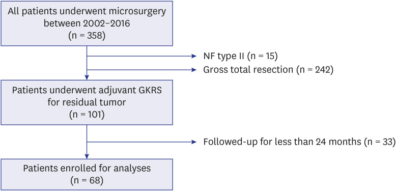
Between January 2002 and December 2016, 358 patients with VS underwent microsurgery at a single institute (shown in Fig. 1). This retrospective study assessed the patients who underwent adjuvant GKRS for residual VS after microsurgery. Fifteen patients with neurofibromatosis type 2 were excluded due to the aggressive nature of the disease. Except for the 242 patients with complete resection, 101 patients underwent adjuvant GKRS for residual tumors. All patients were intraoperatively and histopathologically confirmed to have VS. Patients who had been followed up for ≥ 24 months after GKRS were included, and 68 patients were finally enrolled.
Fig. 1
Flow chart showing the numbers of included and excluded patients.
A total of 68 patients were enrolled for the analyses.
NF = neurofibromatosis, GKRS = gamma knife radiosurgery.
Concerning tumor extent, intraoperative neuromonitoring (IOM) systems including motor-evoked potentials, somatosensory-evoked potentials, and brain stem auditory-evoked potentials were used, as well as other related cranial nerve electromyographies (EMGs). We generally pursued maximal safe resection of tumors, which is especially focused on the anatomical and functional preservation of the FN. Identification of the FN and tumor resection were performed under free-running EMG tracing and direct FN electrical stimulation. The decision of incomplete resection was made intraoperatively.2324 When surgeons felt that further tumor resection would jeopardize FN function, incomplete resection was determined. Two different surgical approaches (retrosigmoid and translabyrinthine approach) were used as described previously.25 The majority of patients underwent surgery via the retrosigmoid (RS) approach. Except for cases involving a high jugular bulb and/or superficial location of the posterior semicircular canal, unroofing of the internal auditory canal (IAC) was performed to facilitate maximal resection.26272829 The translabyrinthine (TL) approach was preferred when a substantial cerebellar retraction was expected, such as when tumors were deeply invaginated in the cerebellar peduncle or brain stem.
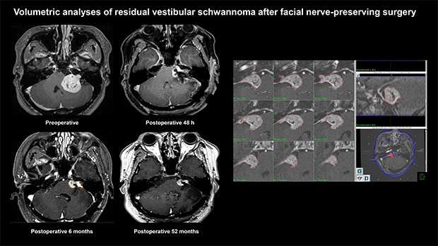
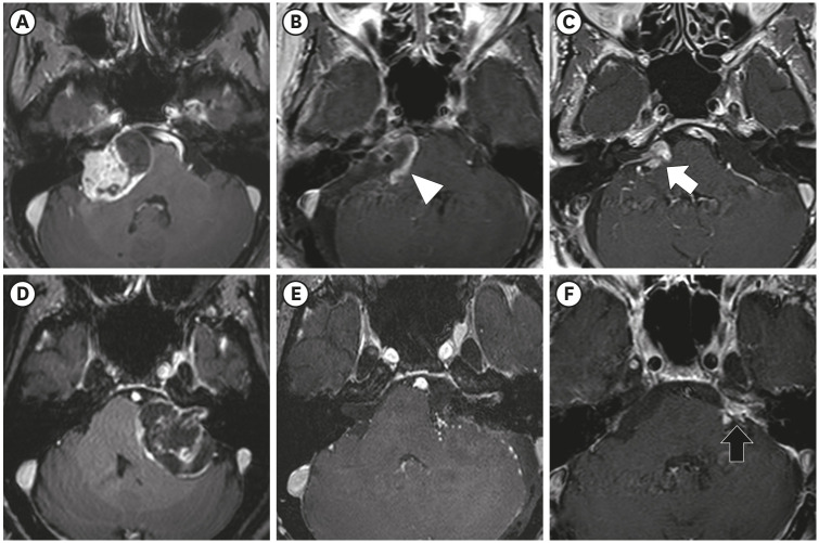
After microsurgery, patients generally underwent immediate postoperative IAC magnetic resonance imaging (MRI) within 48 hours for the evaluation of their baseline postoperative status. Considering the postoperative changes in residual VS, follow-up MRI was performed 3–6 months after surgery.142023 Subsequently, upfront GKRS was scheduled if there was definite evidence of a residual tumor (Fig. 2).30 If it was difficult to determine or clarify any residual tumor on serial postoperative MRI, a follow-up MRI was conducted 1 year later.
Fig. 2
Examples of postoperative changes in the residual tumor on serial MRIs. Preoperative (A), immediate postoperative (B), and 6-month postoperative (C) T-1 weighted contrast-enhanced MRI of right VS. The thin tumor capsule covering the facial nerve was left during the surgery (white arrowhead). Postoperative image taken at 6 months showing progressive closure of the tumorectomy cavity, and the residual tumor was changed to a shape more suitable for gamma knife radiosurgery (white arrow). Preoperative (D), immediate postoperative (E), and 6-month postoperative (F) MRI of left VS. The residual tumor (black arrow) left around the porus acusticus was observed on 6-month postoperative images, but it was not visualized on immediate postoperative imaging.
MRI = magnetic resonance imaging, VS = vestibular schwannoma.
Stereotactic radiosurgery was performed using the Leksell Gamma Knife type B, type C, Perfection, and Icon (Elekta AB, Stockholm, Sweden). Treatment planning was conducted by expert neurosurgeons with specialized experience in radiosurgery using the Leksell Gammaplan® planning software. Patients were followed up in the outpatient clinic at 6 months, 1 year, and 2 years after GKRS using serial IAC MRI scans. Afterwards, follow-up was continued at 2- or 3-year intervals. Tumor progression was defined as a 20% or greater increase in TV as compared to that in the previous serial MRIs during the follow-up period.1931 Transient swelling within 6 months after GKRS due to radiation-induced tumor necrosis was considered as pseudoprogression.3233 Additional treatment for tumor progression was determined based on multiple factors: the size of the tumor, previous surgical findings, the patient's symptoms, and other medical conditions. Generally, repeat GKRS was conducted for small-to-medium-sized recurrent tumors. For large tumors, surgical treatment was recommended.
TV was measured using the Leksell Gammaplan® planning software version 11.1.1 (Leksell Gammma Plan; Elekta AB) on a series of contrast-enhanced T1-weighted IAC MRI scans. T2-weighted images were also used to clarify the tumor margin and surrounding tissues. Residual TV was measured at the time of the first adjuvant GKRS using MRI with a GKRS protocol of 1-mm slice thickness with no gap.
FN function was categorized according to the House-Brackmann (H-B) grading system.34 If there were any discrepancies in grading, the worst grade was considered. H-B grades I and II were considered to indicate good FN function.619 Hearing function was categorized using the Gardner-Robertson (G-R) scale.35 According to the audiometry result, G-R grades I and II were considered as serviceable and those higher than II as non-serviceable. Immediate postoperative function was defined as the status measured during the 7–14 days of hospital stay after microsurgery. Functional preservation was defined as the maintenance of good and serviceable status in each FN and cochlear nerve function at the last follow-up.
Statistical analyses
Data are presented as medians or means (with ranges) for continuous variables and as frequencies and percentages for categorical variables. Univariate statistical analyses (Cox and logistic regression analyses) were performed to assess categorical and continuous variables. Variables with P < 0.20 in the univariate analysis were selected for multivariate models using multiple Cox regression analyses. The sensitivity and specificity of the cut-off values were analyzed using the receiver operating characteristic (ROC) curve. MaxStat package of R (MaxStat Software, Jever, Germany) was used to identify the optimal cut-off values of the variables. A Kaplan-Meier curve was plotted for progression-free survival (PFS) from the time of GKRS to the last follow-up. Mean and frequency comparisons were performed using the Student's t-test, χ2 test, Mann-Whitney U test, or Fisher's exact test, as appropriate. Statistical analyses were performed using the IBM SPSS Statistics version 25 software (IBM Corporation, Armonk, NY, USA). A P value < 0.05 was considered significant.
Ethics statement
This study was approved by the hospital Institutional Review Board (IRB No. 2019-08-023-001). All procedures performed in studies involving human participants were in accordance with the ethical standards of the institutional and/or national research committee (Samsung Medical Center Institutional Review Board) and with the 1964 Helsinki declaration and its later amendments or comparable ethical standards. For this type of study, formal consent is not required.
RESULTS
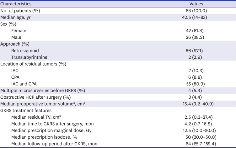
Characteristics of the 68 patients enrolled in this study are described in Table 1. One patient received fractionated GKRS (20 Gy in 4 fractions) because the large surface of the residual tumor touched the brain stem. Four patients had a previous history of multiple microsurgeries before the first adjuvant GKRS. One patient underwent 2 surgeries using the TL approach at another hospital. This patient was referred to our institute, and we operated on the patient using the RS approach. The others underwent 2 surgeries using the RS approach. No patient had a previous history of radiosurgery. Three (4%) patients underwent ventriculo-peritoneal shunt (VPS) surgery before adjuvant GKRS due to postoperative obstructive hydrocephalus (HCP). The median preoperative TV in the available 65 patients was 15.4 cm3 (range: 3.2–40.9). The median residual TV in all patients was 2.5 cm3 (range: 0.3–27.4). The median follow-up period after the first adjuvant GKRS was 64 months (range: 25.7–152.4).
Table 1
Characteristics of the study population
Tumor control
Tumor progression was observed in 8 (12%) patients, all of whom received additional treatments (Table 2). The median residual TV of these 8 patients was 7.0 cm3 (range: 0.5–27.4), and the median time to progression after the first GKRS was 15.8 months (range: 3.2–66.0). Although 3 patients (cases 5, 6, and 7) showed decreased TV after the first GKRS, additional treatment was determined as necessary because continuous regrowth of the residual tumors was detected on serial imaging.
Table 2
Summary of 8 patients who underwent additional treatments due to tumor progression
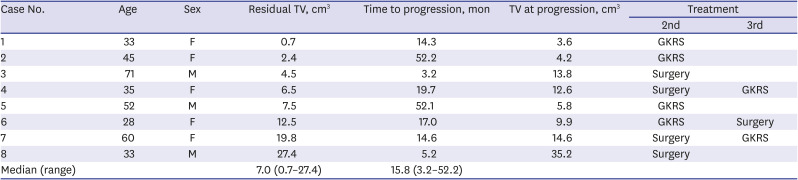
Four of the 8 patients underwent a second GKRS. In 1 patient (case 6), additional surgery was performed 12 months after the second GKRS owing to further tumor progression. No regrowth of the tumor was observed in the other 3 patients during the 74.6-month (range: 48.0–126.6) median follow-up period after the second GKRS. The remaining 4 patients underwent subsequent surgery as a second line treatment. In the second surgery, a more conservative approach was taken to preserve FN function. One patient (case 3) showed acute deterioration due to tumor bleeding 3.2 months after the first GKRS. The measured TV (13.8 cm3) at progression included the volume from a peritumoral hematoma. GTR of the residual tumor was achieved in only 1 patient (case 3). Additional adjuvant GKRS was performed in another 2 patients (cases 4 and 7). The other patient (case 8) received regular follow-up for the residual lesion after the second surgery.
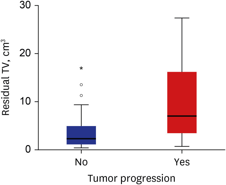
In the 60 patients without tumor progression, the median residual TV was 2.3 cm3 (range: 0.3–17.0). The distribution of residual TV according to the status of tumor progression is shown in Fig. 3. The median time interval between the surgery and first adjuvant GKRS in patients with tumor progression was 4.6 months (range: 1.8–7.2) and in patients without tumor progression was 4.2 months (range: 0.7–16.2) (P = 0.338).
Fig. 3
Box and whisker plots of residual TV distribution according to tumor progression The boxes indicate the 25th and 75th percentiles. The whiskers indicate the minimum and maximum values, dots indicate the outliers, asterisk indicates extreme values, and thick horizontal lines indicate the median value.
TV = tumor volume.
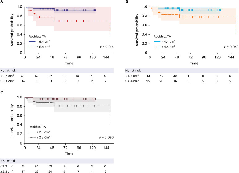
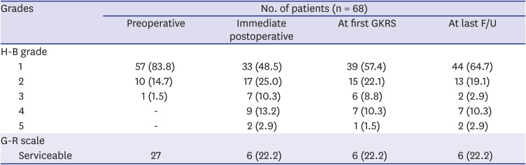
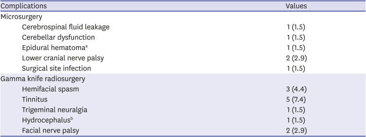
Residual TV was associated with tumor progression in both uni- (P = 0.002; hazard ratio [HR], 1.173; 95% confidence interval [CI], 1.062–1.296) and multivariate analyses (P = 0.003; HR, 1.229; 95% CI, 1.075–1.405) (Table 3). The ROC curve showed that the residual TV can be used as the cut-off variable to predict tumor progression, and several cut-off values of residual TV were proposed (P = 0.021; 95% CI, 0.546–0.960; area under the curve, 0.753) (Fig. 4). MaxStat package of R identified that 6.4 cm3 was the optimal cut-off of residual TV showing the greatest difference in PFS. The Kaplan-Meier plot showed the PFS in the 2 groups stratified according to several cut-off values of the residual TV (Fig. 5). The 5-year PFS rates in the group with residual TVs of < 6.4 cm3 (54 patients) and that with residual TVs of ≥ 6.4 cm3 (14 patients) were 93.3% and 69.3%, respectively (P = 0.014). Patients with a lower residual TV showed better 5-year PFS (93.6% in patients with a residual TV of < 4.4 cm3 and 96.8% in patients with a residual TV of < 2.3 cm3).
Table 3
Factors affecting tumor progression based on uni- and multivariate analyses
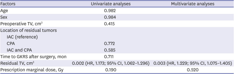
Fig. 4
Sensitivity and specificity analysis. Analyses were performed using the cut-off value of residual TV to predict tumor progression.
TV = tumor volume, CI = confidence interval.
*P = 0.021; 95% CI, 0.546–0.960; area under the curve, 0.753.
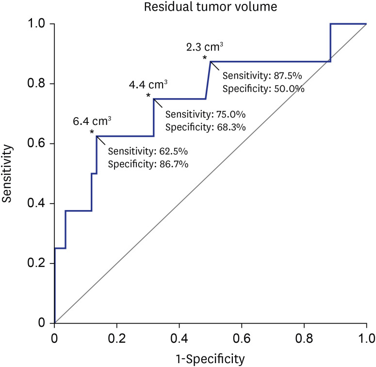
Fig. 5
Kaplan-Meier plot of PFS according to residual TV cut-offs. (A) The 5-year PFS rates in the group with residual TVs < 6.4 cm3 (54 patients) and that with residual TVs ≥ 6.4 cm3 (14 patients) were 93.3% and 69.3%, respectively (P = 0.014). (B) The 5-year PFS rates in the group with residual TVs < 4.4 cm3 (43 patients) and that with residual TVs ≥ 4.4 cm3 (25 patients) were 93.8% and 78.6%, respectively (P = 0.049). (C) The 5-year PFS rates in the group with residual TVs < 2.3 cm3 (31 patients) and that with residual TVs ≥ 2.3 cm3 (37 patients) were 96.8% and 81.7%, respectively (P = 0.096).
PFS = progression-free survival, TV = tumor volume.
Functional outcomes
Sixty-seven (99%) patients had a preoperative good FN function (Table 4). A good FN function was observed in 50 (74%) patients in the immediate postoperative period and 54 (81%) patients at the time of the first GKRS. Two patients experienced worsening of the FN function after GKRS. One patient (case 3) showed worsened FN function (H-B grade III → IV) due to the tumor bleeding after the first GKRS. Another patient (case 4) showed worsened FN function (H-B grade III → V) after the second GKRS after the additional surgery. At the last follow-up, 57 (84%) patients maintained a good FN function.
Table 4
Functional outcomes
In the 8 patients with tumor progression, 6 patients maintained good FN function. The functional preservation rates of the FN in the group with a residual TV of < 6.4 cm3 and ≥ 6.4 cm3 were 85% and 79%, respectively (P = 0.684). Residual TV was not associated with functional preservation of the FN during the immediate postoperative period (P = 0.695; odds ratio [OR], 1.024; 95% CI, 0.908–1.156) or at the last follow-up (P = 0.755; OR, 0.980; 95% CI, 0.866–1.110).
Serviceable hearing was observed in 27 patients before surgery. Postoperative hearing preservation was achieved only in 6 (22%) patients. These findings did not change over the follow-up period. None of the patients maintained serviceable hearing in the tumor progression group.
Complications of microsurgery
After initial surgery, 1 (1%) patient underwent wound revision due to cerebrospinal fluid leakage (Table 5). One (1%) patient with diffuse cerebellar hematoma had to receive long-term rehabilitation. One (1%) patient underwent another craniotomy due to an epidural hematoma associated with a perioperative external ventricular drain. After emergent hematoma evacuation, the patient recovered without permanent neurologic sequelae. Two (3%) patients underwent swallowing rehabilitation due to transient dysphagia caused by lower cranial nerve palsy. A surgical site infection was observed in 1 (1%) patient after a second surgery.
Table 5
Complications
Complications of GKRS
After the first adjuvant GKRS, 3 (4%) patients complained of hemifacial spasm and 5 (7%) patients had transient tinnitus (Table 5). One (1%) patient underwent a second GKRS due to newly developed trigeminal neuralgia. One (1%) patient received a VPS due to HCP caused by radiation-induced necrosis of a residual tumor 8 months after the second GKRS. However, no further treatment was required as the tumor regressed gradually. Two (3%) patients showed aggravated FN palsy, as described above.
DISCUSSION
Functional preservation of the FN is one of the main goals of VS surgery. Many studies have revealed that intended STR of VS had superiority in terms of good FN outcomes as compared to GTR.41136 However, the concerns related to regrowth of residual tumors remain.537 Several previous studies demonstrated that postoperative residual TV was associated with tumor regrowth.192438 Adjuvant GKRS following intended STR of VS has been discussed as an effective treatment option in terms of FN preservation and long-term tumor control.91011141517 In our previous study of VS, the tumor control rate in the GTR group was 91.9%, whereas the rates in the NTR and STR groups were 84.3% and 75.2%, respectively. However, 92.1% tumor control was achieved in the NTR and STR groups when NTR and STR were followed by adjuvant radiotherapy or radiosurgery.39 A recent meta-analysis also revealed favorable tumor control and functional outcomes from this “nerve-centered” treatment strategy.15 However, the volume of residual tumor after STR was also associated with tumor recurrence after adjuvant GKRS.131819 The risk of tumor progression increases with the size of the residual tumor.14 In the present study, residual TV was the only factor associated with tumor progression after GKRS. When surgeons plan to do intended STR of VS for functional preservation of the FN, the optimal extent of the residual tumor associated with long-term tumor control using adjuvant GKRS needs to be discussed.481223
This study aimed to identify the optimal cut-off volume of residual VS that can predict a favorable outcome in terms of both tumor control and FN preservation. A recent multicenter study demonstrated that a good FN outcome immediately post-operation was associated with a larger residual TV.18 Radwan et al.21 revealed that a postoperative residual TV of > 3 cm3 was associated with good FN outcomes after adjuvant GKRS. However, some authors assert that FN preservation is not correlated with the extent of resection if the dissection is performed with a priority given to FN preservation.440 In the present study, residual TV was not associated with either early or late good FN outcomes. However, this result can be affected by the technical bias related to individual surgeons and the surgical strategy. There are 2 ways of performing intended STR: planned STR and unplanned STR.22 In planned STR, the decision to pursue STR is made preoperatively. The plane between the tumor capsule and cranial nerves is not dissected.14 Our treatment strategy for VS was unplanned STR, as described above. We tried to maximize the extent of tumor resection to minimize the residual tumor load. If we had tried to dissect the tumor more conservatively, different outcomes may have been obtained. Resection guided by intraoperative FN monitoring may play a significant role in FN preservation during VS surgery.82241 Regarding tumor control, better outcomes were obtained in patients with smaller residual TVs (Fig. 5). If we use a smaller TV as a cut-off value for STR, more favorable long-term outcomes would be achieved. Moreover, the amount of residual tumor as measured by the surgeon's subjective judgment during microsurgery can be different from postoperative MRI-based volumetric measurements. Second salvage operations have been frequently required for large recurrent tumors after radiosurgery.4243 Functional preservation of the FN in revision surgery of recurrent VS is more challenging.40444546 Therefore, optimal volume reduction is an important factor in VS surgery, even if the intended STR was planned. Based on our result, favorable outcomes of FN preservation can be achieved without leaving significant amounts of residual tumor.
Both revision surgery and repeat GKRS can be used for patients with recurrent tumors after adjuvant GKRS. Repeat GKRS can be a reasonable treatment option for small-to-medium-sized recurrent tumors after adjuvant GKRS.47 A recent review article demonstrated that radiosurgery showed better FN outcomes than a repeat surgical resection for recurrent VS after primary surgery.48 Iorio-Morin et al.49 reported a 10-year tumor control rate of 92.2% in 76 patients who underwent repeat GKRS for recurrent VS. In the present study, favorable long-term tumor control was achieved in patients with small- to medium-sized recurrent VS using repeat GKRS. Further investigation through larger population studies will be needed to establish the role of repeat GKRS in the prevention of recurrent VS.
There are several limitations of this study. First, this was a retrospective study. Second, the small patient population limited the statistical power. The area under the ROC curve was only 0.753. While it supports the results of our study that larger residual TV are associated with a higher risk of tumor progression, it also shows that residual TV is a relatively weak predictor of the treatment failure. The sensitivity and specificity of the suggested cut-off volume (6.4 cm3) were also insufficient to make a clinical decision. Third, sufficiently longer follow-up periods will be required, as tumor progression can occur at various times and have diverse clinical courses.101219 Finally, other factors that can affect the FN outcomes such as aberrant pathways of the FN, consistency of the tumor, surgical techniques of individual surgeons, and the loss of anatomical continuity of the FN during surgery were not evaluated in this study.50
In this study, residual TV was associated with tumor progression in VS after adjuvant GKRS following STR. As preservation of FN function was not correlated with the extent of resection, optimal volume reduction is imperative to achieve long-term tumor control. Our findings will help surgeons predict the prognosis of residual VS after FN-preserving surgery.
 XML Download