

 PDF
PDF Citation
 Print
Print


INTRODUCTION
Neuromyelitis optica spectrum disorder (NMOSD), an inflammatory disease of the central nervous system (CNS), is associated with neuropathic pain, which arises as a direct consequence of inflammatory lesions in the somatosensory nervous system.1,2 Neuropathic pain can be usually resistant to therapeutic approaches, but there is no standardized objective quantitative biomarker for neuropathic pain in individuals with NMOSD. Identifying such biomarkers may be helpful for monitoring neuropathic pain, which can affect the quality of life of these patients in clinical practice.3
Cytoskeletal proteins of astrocytes (like glial fibrillary acidic protein [GFAP]) could be candidates for biomarkers of neuropathic pain since the pathophysiology of NMOSD is primarily astrocytopathy.4 Because NMOSD is an inflammatory condition of the CNS, cytokines involved in the inflammatory process could also be candidates for biomarkers of neuropathic pain.5 In this preliminary study, we aimed to evaluate potential serological biomarkers of neuropathic pain in individuals with NMOSD.
Go to : 
MATERIALS AND METHODS
A total of 38 sera samples from 38 individuals with NMOSD who visited the National Cancer Center (NCC) were analyzed. The Korean version of the painDETECT questionnaire was investigated as previously described at remission status (after at least 3 months of relapses).3,6 Participants with available stored serum samples, collected at remission periods were randomly enrolled; 26 were simultaneously obtained with questionnaire and 12 were obtained after administering questionnaire but within 3 month-interval of sampling and administering questionnaire.3,6 All samples were stored at -80°C prior to analysis. All participants satisfied the 2015 diagnostic criteria for NMOSD and were positive for AQP4 antibodies tested by in-house live cell-based assays.7,8 Pain-DETECT scores ranged from 0 to 38, and pain is classified 3 categories; 19-38: strongly suggestive of neuropathic pain (definite neuropathic pain), 13-18: suggestive of pain with neuropathic components, and < 13: suggestive of non-neuropathic pain.6 Among the 38 participants with NMOSD, 22 had experienced pain with neuropathic components assessed by painDETECT (score ≥ 13), but 16 had non-neuropathic pain (score < 13). Additionally, 17 participants had experienced definite neuropathic pain (score ≥ 19), and these participants were also compared to 16 participants with non-neuropathic pain. This study was approved by the Institutional Review Board of the NCC.
Several candidate biomarkers of neuropathic pain in NMOSD participants, including GFAP and cytokines (tumor necrosis factor-alpha [TNF-α], interleukin [IL]-6, IL-10, IL-17A) were investigated. GFAP levels were assessed using a commercial single-molecule array assay (Simoa, Quanterix Corp., Billerica, MA, USA). Cytokines were measured by a multiplex bead-based immunoassay (cytometric bead array) according to the manufacturer’s protocol (BD Bioscience, San Jose, CA, USA). GFAP and cytokine levels were independently examined by a blinded investigator. The Student’s t-test and Mann-Whitney test were used to compare the levels of serum biomarkers.
Go to :
RESULTS
Demographics of participants
Table 1 shows the demographics of participants with NMOSD. The female-to-male ratio and age at sampling of 22 participants with (score ≥ 13) and 16 participants without (score < 13) neuropathic pain components (non-neuropathic pain) were 20:2 and 16:0, and 46.5 ± 6.0 and 42.8 ± 10.5 years, respectively. The disease duration of NMOSD participants with and without neuropathic pain components were 10.3 ± 5.3 and 11.0 ± 6.4 years, respectively. The median current Expanded Disability Status Scale values for participants with and without neuropathic pain components were 3.5 and 3.0, respectively. Additionally, the demographics of 17 participants who had definite neuropathic pain (score ≥ 19) were also compared with those of 16 participants who had non-neuropathic pain. There were no statistically significant differences in demographics between the two groups.
Table 1.
Demographics
![]()
Levels of serum biomarkers in NMOSD participants with neuropathic pain components (or definite neuropathic pain) and non-neuropathic pain
Fig. 1 and Table 2 show a comparison of the levels of individual serum biomarkers between NMOSD participants with and without neuropathic pain components (non-neuropathic pain). The absolute mean values of serum GFAP (148.3 ± 1.5 vs. 120.1 ± 9.4 pg/mL, p = 0.082), TNF-α (10.8 ± 5.3 vs. 2.4 ± 0.3 pg/mL), IL-6 (11.1 ± 2.3 vs. 7.5 ± 1.7 pg/mL), and IL-10 (5.3 ± 1.2 vs. 3.5 ± 0.4 pg/mL) levels were higher in NMOSD participants with neuropathic pain components than in those without neuropathic pain components but the difference did not reach statistical significance.
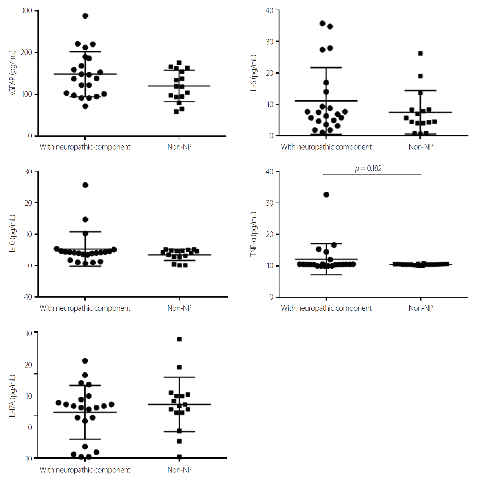 | Fig. 1.Comparison of levels of serum biomarkers between neuromyelitis optica spectrum disorder participants with neuropathic pain component and non-neuropathic pain (NP). GFAP, glial fibrillary acidic protein; IL, interleukin; TNF-α, tumor necrosis factor-alpha.
|
Table 2.
Individual levels of serum biomarkers
![]()
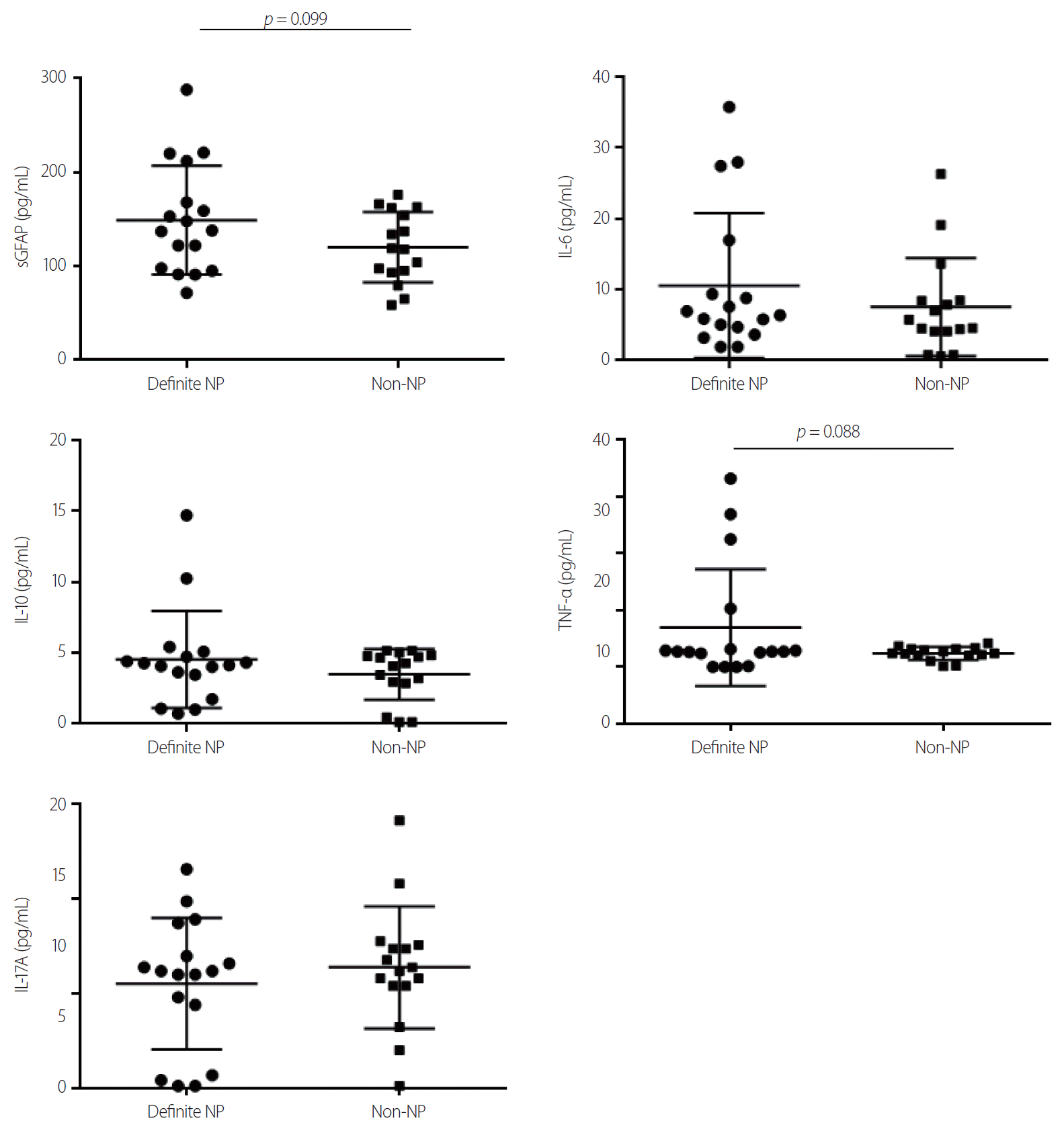
Fig. 2 and Table 2 show a comparison of the levels of individual serum biomarkers between NMOSD participants with definite neuropathic pain and non-neuropathic pain. The absolute mean values of serum GFAP (149.1 ± 14.1 vs. 120.1 ± 9.4 pg/mL, p = 0.099), TNF-α (6.9 ± 2.5 vs. 2.4 ± 0.3 pg/mL, p = 0.088), IL-6 (10.5 ± 2.5 vs. 7.5 ± 1.7 pg/mL), and IL-10 (4.5 ± 0.8 vs. 3.5 ± 0.4 pg/mL) levels were higher in NMOSD participants with definite neuropathic pain than in those with non-neuropathic pain but the results did not reach statistical significance.
Go to :
DISCUSSION
In comparison of NMOSD participants with neuropathic pain components (or definite neuropathic pain) and those without neuropathic pain, the non-significant higher tendency of serum GFAP, TNF-α, IL-6, and IL-10 levels were observed in the group with neuropathic pain components (or definite neuropathic pain). Future larger-scale investigations of serum GFAP, TNF-α, IL-6, and IL-10 levels as potential biomarkers for neuropathic pain in NMOSD are required to find surrogate serological indicators.
Neuropathic pain is an important symptom in NMOSD and can affect the quality of life.3 PainDETECT is a useful tool for detecting neuropathic pain with qualitative (categorical), but not fully objective or quantitative information in individuals with NMOSD.6,9 GFAP is a candidate for an objective and quantitative biomarker for neuropathic pain in individuals with NMOSD because aquaporin-4 (AQP4) is mainly located in astrocytes and anti-AQP4 antibody is a pathogenic component in NMOSD.10 Changes in GFAP, which is a cytoskeletal component of astrocytes, in body fluids such as serum could reflect the extent of astrocytopathy-associated inflammation which can cause neuropathic pain.4 This may explain the non-significant trend toward higher GFAP levels in NMOSD participants with neuropathic pain components (or definite neuropathic pain) than in those with non-neuropathic pain.
In the context of neuropathic pain, TNF-α is a broadly investigated inflammatory cytokine.11 In rodents, administration of exogenous TNF-α could cause allodynia, whereas a TNF-α antagonist could decrease pain.12,13 In individuals with lumbar radiculopathy, a positive correlation between low back pain disability index and TNF-α levels was observed.14,15 TNF-α inhibitors, such as etanercept and infliximab, are prescribed for painful disorders including rheumatoid arthritis, psoriatic arthritis, and ankylosing spondylitis, but the clinical efficacy of TNF-α inhibitors in neuropathic pain has shown mixed results.11,16 TNF-α inhibitors can cause CNS demyelination; 17 thus TNF-α is not suitable as a therapeutic target but rather would be a candidate for a simple objective quantitative biomarker in NMOSD individuals with neuropathic pain.
Association between neuropathic pain and IL-6, IL-17A, and IL-10 levels have been demonstrated in the previous studies.18-23 IL-6 knockout mice showed less mechanical allodynia after nerve injury compared to control, and administration of an anti-IL-6 antibody conferred similar pain alleviation in rodents.18,19 In a pilot study including 7 individuals with NMOSD, treatment with a monoclonal antibody for the IL-6 receptor (tocilizumab) reduced neuropathic pain.20 Administration of exogenous IL-17 induced neuropathic pain, while IL-17 knockout mice showed a reduced response to pain after nerve injury.21,22 IL-10 elevation in the ventrolateral orbital cortex of the rat with allodynia induced by nerve injury was observed.23 However, in this study, there was no significant difference in IL-6, IL-10, or IL-17A levels between NMOSD participants with and without neuropathic pain.
The main limitations of current study are the small sample size and the possibility of unintentional selection bias, including the preferential inclusion of participants with high disease activity because of the enrollment from a single referral center. Additionally, diverse control groups and modalities to detect pain beyond painDETECT could not be included. Further larger multicenter studies with various control groups and parameters for pain measurement should be designed to establish robust serological biomarkers for neuropathic pain in NMOSD.
Go to :
 XML Download