Abstract
Objectives
Histology, the study of tissue structure under a microscope, is one of the most essential yet least engaging topics for health professional students. Understanding tissue microanatomy is crucial for students to be able to recognize cellular structures and follow disease pathogenesis. Traditional histology teaching labs rely on light microscopes and a limited array of slides, which inhibits simultaneous observation by multiple learners, and prevents in-class discussions. We have developed an interactive web-based microscopy tool called “Histoscope” for oral histology in this context.
Methods
Good quality microscope slides were selected for digital scanning. The slides were scanned with multiple layers of z-stacking, a method of taking multiple images at different focal distances. The digital images were checked for quality and were archived on Histoscope. The slides were annotated, and self-assessment questions were prepared for the website. Interactive components were programmed on the website to mimic the experience of using a real light microscope.
Histology, the study of tissue structure under a microscope, has always been the principal approach to the structural analysis of living organisms at the cellular level. Understanding tissue microanatomy is essential to train medical, dental, and other health professional students to recognize cellular structures, comprehend the structure-function relationship of cells and tissues, and follow disease pathogenesis. However, this branch of science has been described as one of the least engaging topics by health professional students [1]. The traditional method of teaching histology relies on light microscopes, which does not allow simultaneous observation by multiple people, prevents interactive in-class discussions, and thus fails to motivate students [2].
At the School of Dentistry at the University of Alberta, students extensively study oral histology to learn about teeth and surrounding oral structures, their development, and developmental anomalies. Besides regular didactic lectures, students attend histology lab sessions in which they share a limited number of light microscopes and glass slides to view tooth and oral structures. The microscopic slides used for teaching are decades old, with some being extremely rare and irreplaceable.
A growing number of universities and medical schools worldwide are embracing digital slides as an alternative to traditional light microscopes [3,4]. The incorporation of virtual microscopy in histology and pathology courses has improved in-class teaching environments [3,5] and student performance [6]. The number of websites archiving digital histology slides is also growing internationally. For example, “The SecondLook” is a collection of histology slides developed by the University of Michigan [7]. “Oral Histology” created by the University of Kentucky College of Medicine contains a collection of annotated but low-resolution images of oral histology slides [8]. However, the existing web-based histology databases lack sufficient, high-quality resources from the oral cavity, which are critical for dentistry students. Most of the publicly available histology databases provide either annotated or non-annotated images without the ability to switch from one to the other, which does not allow learners to perform active memory recall after learning. Other drawbacks of the existing virtual microscopy websites are the quality of graphical resolution [9] and lack of z-axis information, which is essential for reconstructing the three-dimensional visualization of cellular structure [10]. Moreover, in most online sources, users cannot manipulate images to observe structural details. The few websites that provide the ability to zoom in and out permit users to expand a picture only at a predefined location. The inability to magnify any region of interest limits learners’ freedom to explore the histological section and restricts their curiosity, reducing the student-centeredness of their learning. Furthermore, the COVID-19 pandemic has moved most university teaching online, necessitating an alternative way to teach histology virtually yet in an interactive and engaging manner.
In this context, we have developed a web-based interactive microscopy tool called “Histoscope.” We have scanned our existing oral histological slides in high resolution and archived them on our website. The site allows users to interact with the digital slides and self-assess their knowledge of oral histology.
The School of Dentistry has a collection of oral histology glass slides to teach histology labs with light microscopes. We curated those slides for quality and rarity and selected the best slides for whole-slide scanning using an Aperio Digital Slide Scanner (Leica Biosystems, Wetzlar, Germany). We chose the slides to represent the oral mucosa, oral structures, tooth, tooth development, and facial structure development. The slides were scanned with 25 layers of z-stacking, an image-processing method of taking multiple images at different focal distances and combining them to make a composite image. This technique, also known as focus stacking, is useful for capturing in-focus images of objects under high magnification as the depth of field decreases with magnification [11].
The histology glass slides were scanned under 20× or 40× magnification depending on the specimen’s size and the structural details required for teaching oral histology. The slides scanned under 40× magnification has a resolution of 0.247 microns per pixel (MPP); the resolution was 0.497 MPP for the slides scanned under 20× magnification. The image bit depth was 8 bits for all the digital slides.
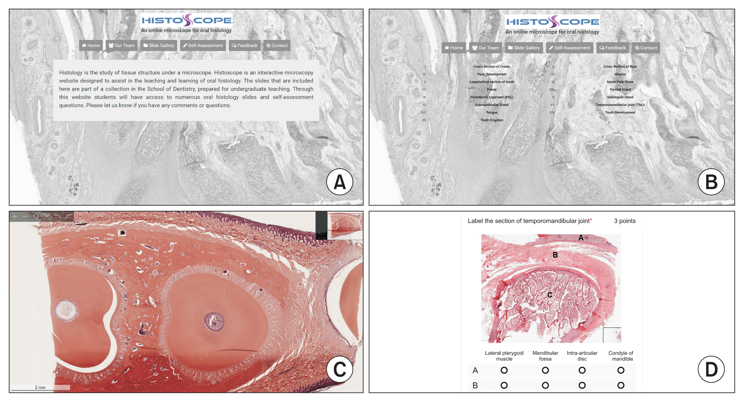
Digital scans of the slides were further checked for quality and resolution, organized in groups, annotated, and archived on the Histoscope website under the Slide Gallery tab (Figure 1). Currently, the website contains 76 digital slides representing teeth and surrounding oral structures.
We prepared a series of questions (Figure 1D) which were incorporated into the website under the Self-Assessment tab. The questions are grouped to correspond to the Slide Gallery organization, so students can choose to test their knowledge of a specific area of the oral cavity. Currently, we have 35 image-labeling and structure-identification questions on our website, which are randomized each time a user takes the test.
We aimed for a simple, responsive design with a minimum number of tabs and minimal text for easy navigation. For structural consistency, the title “Histoscope” and the background frame are kept visible on all pages (Figure 1A, 1B). The colors of the headings, tabs, and background were chosen for aesthetic harmony. The contents of the website are organized under six tabs. Digital slides are arranged in fifteen groups under the Slide Gallery tab (Figure 1A, 1B, Table 1). The Self-Assessment tab guides users to a series of questions to test their oral histology knowledge. The website also has an option for users to contact the authors and provide feedback for further improvement (Figure 1A).
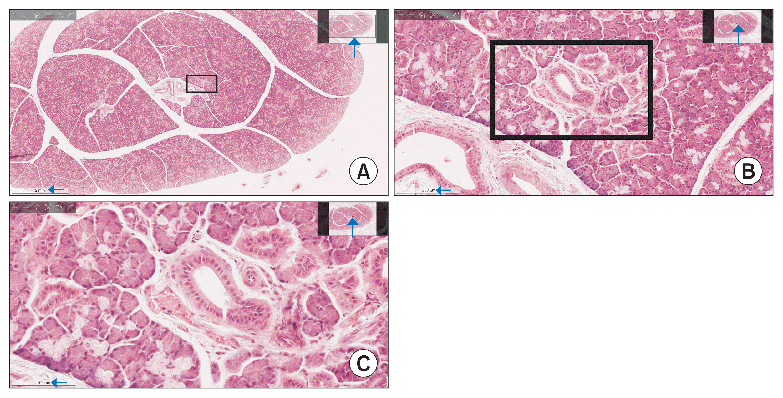
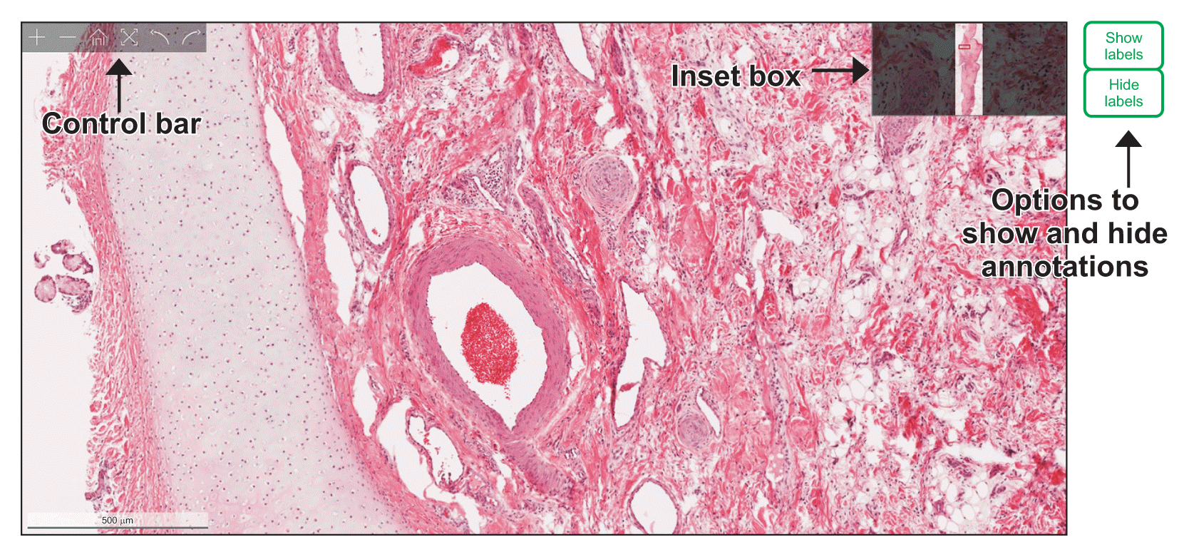
We designed Histoscope as an interactive microscopy tool. This interface’s primary goal is to mimic the experience of viewing slides through a real light microscope with the ability to manipulate and view various areas of the histological section, which is rare in the existing online histology databases. Our website was programmed with the Python programming language and uses Flask as a web framework, resulting in a dynamic platform capable of processing user requests and interacting with them accordingly. Users can load a digital slide of their choice in high resolution, rotate the slides as needed, toggle to full screen, and change the magnification in any mouse-pointed location. The digital slides on our website were scanned using the z-stacking technique to provide continuous z-axis information. Z-stacking allows for a greater depth of field and a unique ability to magnify a digital image at any given area and maintain a perfectly focused, high-resolution field-of-view (Figure 2). The slide viewer of the website provides an inset telescope box on the upper-right corner of the screen. When zooming in on fields of view, the inset shows a box outlined in red, delineating the exact position of the area being viewed on-screen with respect to the entire viewable field (Figure 2). After magnification, the user can navigate the magnified slide by grabbing the slide with a hand tool or by simply moving the red box in the inset (Figure 3). The slides are annotated by content experts to identify major structural components within a tissue section. Users can either “show” or “hide” the annotations, which allows them to self-test their learning (Figures 1C, 3).
The completed website includes 76 high-quality and rare sections of the tooth, oral structures, and development of tooth and facial regions, which are not readily available in any public online databases. A complete list of all the digital slides incorporated into our slide gallery is shown in Table 1. The digital images were scanned using a focus stacking technology to mimic the experience of focusing through a real light microscope (Figure 2). We have included an option to show or hide annotations to enable users to perform active memory recall (Figure 3). Self-assessment questions on our website allow learners to self-test their knowledge on oral histology (Figure 1).
Computer-based technologies to teach histology have increased over the years [3,12,13]. Virtual microscopy has improved learners’ performance in many training programs [3,5,14]. Some known limitations of this technology are low graphics resolution and lack of z-axis information [9,10]. With Histoscope, we aimed to overcome the shortcomings of previous virtual histology databases and to create an interactive and dynamic platform to mimic the experience of using an actual light microscope. The incorporation of our web-based microscope will stimulate active learning and student engagement. Educational theories suggest that adults learn proficiently when they are fully engaged in learning [15]. Active engagement also helps develop critical thinking skills in adults [16]. The use of web-based microscopy in conjunction with an actual light microscope is supported by Mayer’s cognitive theory of multimedia learning, which states that using two or more of each of the modalities of delivery, presentation, and sensory systems will support effective learning [17]. We believe that Histoscope will complement traditional teaching with the light microscope and allow students to study histology at their own pace.
Acknowledgments
The authors sincerely thank Rehabilitation Robotics Lab, University of Alberta for developing our website, Histoscope.
References
1. Garcia M, Victory N, Navarro-Sempere A, Segovia Y. Students’ views on difficulties in learning histology. Anat Sci Educ. 2019; 12(5):541–9.
2. Capela e Silva F, Rato LM, Lopes OS. Learning and teaching histology: traditional and computational methods. In : Proceedings of International Conference “Learning and Teaching in Higher Education”; 2010 Apr 15–16; Evora, Portugal.
3. Blake CA, Lavoie HA, Millette CF. Teaching medical histology at the University of South Carolina School of Medicine: transition to virtual slides and virtual microscopes. Anat Rec B New Anat. 2003; 275(1):196–206.
4. Pantanowitz L, Szymas J, Yagi Y, Wilbur D. Whole slide imaging for educational purposes. J Pathol Inform. 2012; 3:46.
5. Kumar RK, Velan GM, Korell SO, Kandara M, Dee FR, Wakefield D. Virtual microscopy for learning and assessment in pathology. J Pathol. 2004; 204(5):613–8.
6. Helle L, Nivala M, Kronqvist P. More technology, better learning resources, better learning? Lessons from adopting virtual microscopy in undergraduate medical education. Anat Sci Educ. 2013; 6(2):73–80.
7. Hortsch M. The SecondLook [Internet]. Ann Arbor (MI): University of Michigan;c2019. [cited at 2021 Apr 5]. Available from: https://secondlook.med.umich.edu/histology
.
8. MacPherson BR, Tieman JG. Oral histology: a digital laboratory and atlas [Internet]. Lexington (KY): University of Kentucky;n.d.. [cited at 2021 Apr 5]. Available from: http://www.uky.edu/~brmacp/oralhist/index.htm
.
9. Rinaldi VD, Lorr NA, Williams K. Evaluating a technology supported interactive response system during the laboratory section of a histology course. Anat Sci Educ. 2017; 10(4):328–38.
10. Van Es SL, Pryor WM, Belinson Z, Salisbury EL, Velan GM. Cytopathology whole slide images and virtual microscopy adaptive tutorials: a software pilot. J Pathol Inform. 2015; 6:54.
11. Ray SF. Applied photographic optics: lenses and optical systems for photography, film, video, electronic imaging and digital imaging. 3rd ed. Oxford, UK: Focal Press;2002.
12. Bloodgood RA, Ogilvie RW. Trends in histology laboratory teaching in United States medical schools. Anat Rec B New Anat. 2006; 289(5):169–75.
13. Cotter JR. Laboratory instruction in histology at the University at Buffalo: recent replacement of microscope exercises with computer applications. Anat Rec. 2001; 265(5):212–21.
14. McBride JM, Drake RL. National survey on anatomical sciences in medical education. Anat Sci Educ. 2018; 11(1):7–14.
15. Vella J. On teaching and learning: putting the principles and practices of dialogue education into action. San Francisco (CA): Jossey-Bass;2008.
16. Karge BD, Phillips KM, Jessee T, McCabe M. Effective strategies for engaging adult learners. J Coll Teach Learn. 2011; 8(12):53–6.
17. Mayer RE. Multimedia learning. 2nd ed. Cambridge, UK: Cambridge University Press;2009.