

 PDF
PDF Citation
 Print
Print


I. Introduction
Odontogenic cutaneous sinus tracts (OCSTs), or cutaneous fistulas of dental origin, are rare dermatoses that occur because of chronic dental draining infections
1
. It has been estimated that 50% of the individuals affected by OCSTs are subjected to inappropriate dermatological treatments and an array of antibiotic treatments before the correct diagnosis is established
2
. The diagnosis of OCST can be challenging because of the anatomic complexity of the oral cavity and the asymptomatic nature of chronic dental infections. Correct diagnosis often requires a variety of methods (e.g., radiological, microbiological, and/or pathological).
Patients with OCST often seek initial medical help from a general surgeon or a dermatologist instead of a dentist or a maxillofacial surgeon and undergo multiple surgical excisions, radiotherapy, biopsies, and antibiotic regimens with eventual recurrence because the primary odontogenic cause is not diagnosed
3
. Thus, a diagnosis made with suspicion of odontogenic origin, appropriate examination methods, and prompt treatment will reduce or eliminate complications such as asepsis, osteomyelitis, and patient discomfort
4
. In this article, we present our experience utilizing computed tomography (CT) with the soft tissue window setting to confirm suspected OCST and to visualize the sinus tract.
Go to : 
II. Cases Report
1. Case 1
A 27-year-old man presented with a chief complaint of a draining lesion on his left submandibular region with pus extrusion. Previously, the patient had undergone irrigation and drainage treatment by a general surgeon. The patient also disclosed that after the irrigation and the drainage treatment, intermittent pus discharge from the lesion was observed without pain. He had also visited a local dental clinic and extraction of his infected second molar tooth was recommended. The patient had no significant medical history with the exception of smoking one cigarette per day.
Clinical examination revealed a non-tender, erythematous nodule measuring about 0.7 cm in diameter on the left submandibular region.(Fig. 1. A) Upon gentle palpation pus discharge from the lesion was observed. Intra-oral examination was unremarkable. Panoramic radiogram revealed periapical rarefaction of the #31, #37, and #47 teeth.(Fig. 1. B) Coronal, sagittal, and axial CT optimized with 45 Hounsfield unit (HU) window length (WL) and 300 HU window width (WW) demonstrated a perforation of the cortical plate of the #37 tooth and a fistulous tract with high attenuation connected to the cutaneous layer.(Fig. 1. C-E)
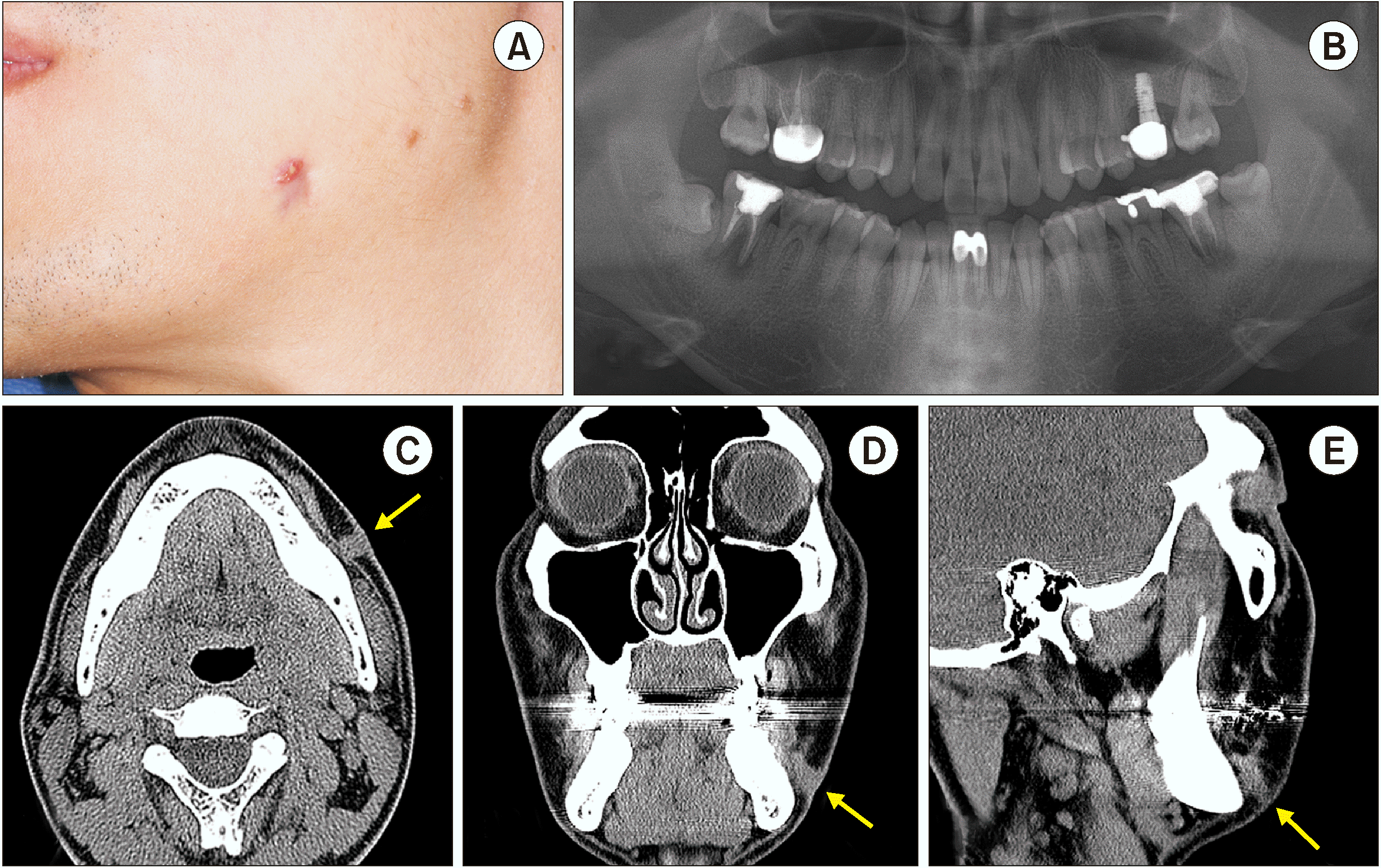 | Fig. 1A. Clinical examination revealed a non-tender, erythematous nodule measuring approximately 0.7 cm in diameter on the left submandibular region. B. Preoperative panoramic view revealed a periapical rarefaction of the #31, #37, #47 teeth and impacted #38, #48 third molar teeth. C-E. Axial, coronal, and sagittal computed tomography showing the exact path of the odontogenic sinus tract marked with arrows.
|
Based on these findings, the proposed treatment plan of fistulectomy of the left lower buccal cheek and mucosa, mass excision of the right and left molar region for biopsy and surgical extraction of the diseased #37, #47 teeth along with the impacted #38, #48 third molar teeth were carried out under conscious sedation with 4 mg midazolam. The biopsy results showed periapical granuloma from the periapical area of the #37 tooth and chronic periodontitis from the periapical area of the #47 tooth. Wound healing proceeded uneventfully. Significant improvements were observed at the one-year follow-up.
2. Case 2
In January 2018, a 59-year-old woman was referred to confirm the dental origin of a skin lesion. The patient had a chief complaint of the presence of a nonhealing extraoral nodule and a persistent abscess that had started three months prior. The patient was initially referred to a dermatologist and a general surgeon in a local hospital prior to the visit.
Extraoral examination revealed an erythematous, non-tender nodule with crusting approximately 1 cm in diameter slightly above the inferior border of the right mandible.(Fig. 2. A) Intra-oral examination revealed gold crown restorations on the #45, #46, and #47 teeth. The panoramic radiogram revealed a periapical rarefaction of the #45 tooth with an incomplete endodontic treatment status. Coronal, sagittal, and axial CT using the bone setting showed a perforation of the cortical bone of the #45 tooth and mild swelling of the adjoining buccal soft tissue.(Fig. 2. B-D) However, a cutaneous sinus tract could not be detected on the bone setting of the CT with a 3,000 HU WW and a 400 HU WL. After optimizing the window/level transformation to the soft tissue setting of 250 HU WW and 50 HU WL, an OCST connecting the affected tooth to the subcutaneous layer of the skin was delineated on the axial, coronal, and sagittal views of the scan.(Fig. 2. E-G)
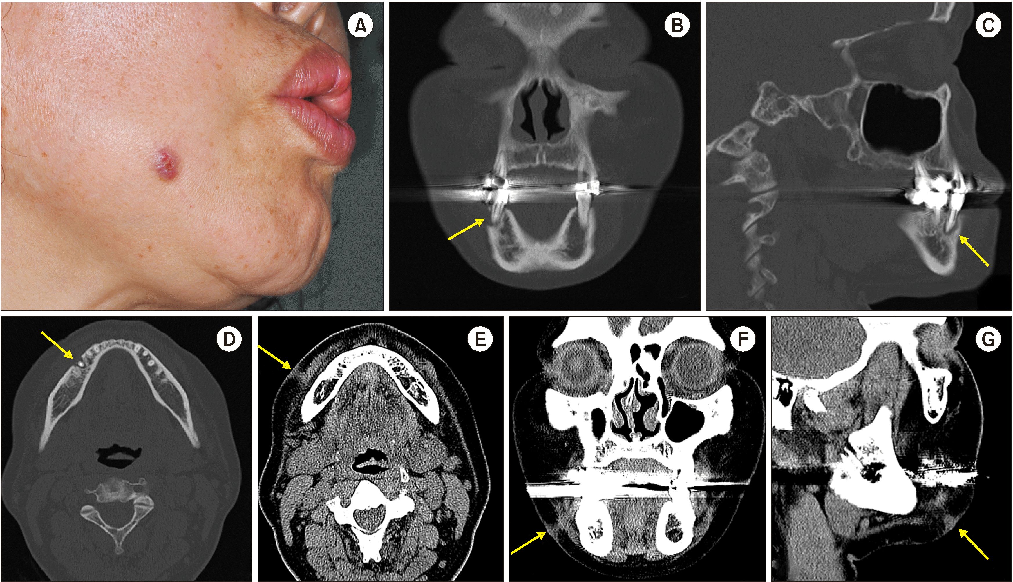 | Fig. 2A. Extraoral view of a patient with an orocutaneous fistula in the right mandible cheek. B-D. Preoperative axial, coronal, and sagittal computed tomography (CT) reveal lingual bone destruction of the #45 tooth and mild swelling of the adjoining buccal soft tissue marked with arrows. E-G. Axial, coronal, and sagittal CT with the soft tissue setting demonstrate the origin and the route of the fistula connected to the subcutaneous layer of the skin marked with arrows.
|
Cutaneous fistula closure on the right cheek and removal of the #45 tooth were performed without any complications. After ten months, the OCST on the right cheek healed completely and the scar was almost imperceptible.(Fig. 3. A) Dental implant installation for the edentulous area of the #45 tooth was carried out. A guided bone regeneration technique was applied to cover the bone defect using allogenic Oragraft (LifeNet Health, Virginia Beach, VA, USA). A 4.0×8.5-mm internal submerged-type Luna fixture (Shinhung, Seoul, Korea) was then inserted.
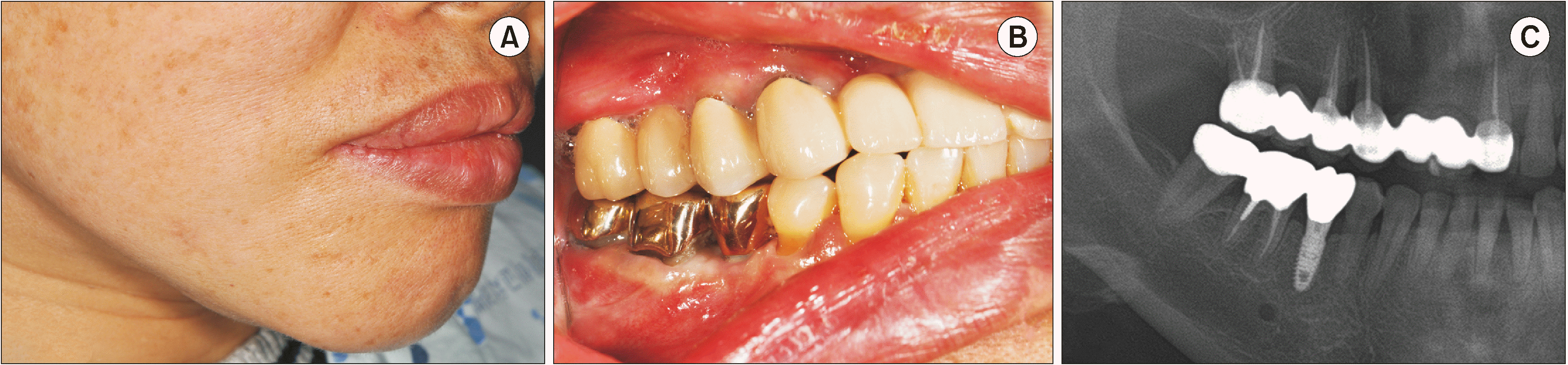
There were no remarkable findings at the four-month follow-up. Re-entry surgery was performed at that time under local anesthesia, and a limited incision was made to expose the implants for healing abutment placement. Three months after re-entry surgery, the final prosthesis was placed.(Fig. 3. B) Panoramic radiogram seven months after implant installation showed a well-osseointegrated implant placed in the right mandible.(Fig. 3. C)
Go to :
III. Discussion
A dental etiology should always be considered in the differential diagnosis of any orofacial skin lesion. A cutaneous sinus tract of the face and neck region is usually caused by dental infection. Other causes may include osteomyelitis, actinomycosis, pyogenic granuloma, neoplasms, salivary gland fistula, infected cyst, deep mycotic infection, and other local skin infections (e.g., carbuncles and infected epidermoid cysts)
3,5,6
. A delay in the treatment of such infections can cause the infection to spread into the fascial spaces of the orofacial area or into the deep spaces of the head and neck region (parapharyngeal space infection)
3
.
Most cutaneous sinus tracts have an intraoral opening, but in cases of chronic dental infection, the local inflammatory process progresses slowly as an alveolar bone abscess, following the path of least resistance through the maxillary or mandibular periosteum. The site of the opening depends on the location of the bony perforation in relation to the facial muscle attachments. In the mandible, if the osseous perforation occurs above the muscle attachment, the fistulae may occur in the attached mucosa, the buccal vestibule, or the floor of the mouth. If the osseous perforation is located below the muscle attachments, the sinus will open extraorally and appear in the chin, submental, or submandibular region
4
. In our first and the second case, the erythematous nodule presented slightly above the inferior border of the mandible.
Pulp sensitivity test, tracking the dental origin of the sinus tract with the gutta-percha point, and radiographic examination have been for cutaneous sinus tract evaluation
1,5,7
. However, the pulp sensitivity test may determine the condition of the teeth but fail to identify the path of the sinus tract. Furthermore, the gutta-percha technique may determine the direction and the depth of the sinus tract, the possible presence of a foreign body, and communication of fistula with the apices of teeth, but it is a blind procedure, and panoramic radiography may result in a false diagnosis due to overlapping. Panoramic radiography is often used for diagnosing cutaneous sinus fistulae of dental origin, but it cannot determine the exact extent of bone lesions and soft tissue pathology associated with the sinus tract. Cone-beam computed tomography (CBCT) has been the recommended imaging modality for evaluating the size and extent of bone destruction around the apices and locating the tooth involved in cutaneous sinus tract cases
5
. In our case, CT with the default bone window setting showed the exact extent of cortical bone destruction; by adjusting the window setting to soft tissue, the path of the sinus tract in the soft tissue could then be identified.
The sinus tracts found in the head and neck region are mainly caused by odontogenic infections, taking up 80%, while the remaining 20% is caused by non-odontogenic origin. The major causes of odontogenic sinus tracts include periapical and periodontal infection (63.5%), pericoronitis resulting from mandibular teeth (8.5%), fracture mandible (7%), infected dentigerous/keratocyst (6%)
8
. Therefore, it is important to have a high suspicion of odontogenic origin in the first place for cutaneous sinus tracts found in the head and region. Other major causes, which have been confused with the dermal origin is osteomyelitis and other congenital causes, eg, parotid, thyroglossal, or branchial
9
. For correct differential diagnosis, sinus tracts found in osteomyelitis will be identified with our method along with osteolysis, sequestra formation, and sclerosing lesions in CT findings
10
allow identify the obvious origin of the sinus tract. In parotid sinus tracts found in the head and neck region, the CT findings will also allow visualizing the abnormal congenital soft tissue findings such as ectopic parotid gland position allowing to differentiate from other origins
11
.
Furthermore, the developmental route of the odontogenic cutaneous sinus tract is also an important factor to consider for correct differential diagnosis. The inflammatory process causing periapical abscess progresses slowly resorbing the bone and spreads toward the cortical plate following the path of least resistance. If the location of perforation is located above the muscle attachment including the buccinator muscle in the maxilla and mentalis, mylohyoid, and buccinator muscles in the mandible, then it is most likely that the sinus tract will drain extra-orally. If the perforation is located below the mentioned muscle attachments, the sinus tract is most likely to drain intra-orally
7
. Thus, in clinical examination, if the orifice is identified externally above the muscles, the dermal origin should be considered and CT analysis with soft tissue setting will help to verify the diagnosis.
The primary objective for managing OCST is to remove the source of infection which may be eliminated by removal of the infected pulp tissue through endodontic treatment or, if there is no indication for endodontic treatment or apical resection, by extraction of the infected tooth and curettage
1
. Once the odontogenic cause is removed, the OCST will heal without further treatment within a few weeks as our cases’ healing uneventfully
12
. Accordingly, our cutaneous sinus tract cases resolved uneventfully after elimination of the source of infection.
Coronal, sagittal, and axial CT adjusted with abdomen setting, such as 45 HU WL and 300 HU WW in the INFINITT PACS software (INFINITT Healthcare, Seoul, Korea), demonstrated a perforation of the cortical plate of the #37 and #45 tooth with a high attenuated fistulous tract connected to the cutaneous layer in each case.(Fig. 3) After optimizing the WL/WW transformation to the abdomen setting of 45-50 HU WL and 250-300 HU WW, any OCST connecting the affected tooth to the subcutaneous layer of the skin was delineated clearly with mild swelling of the adjoining buccal soft tissue on the axial, coronal, and sagittal views. Panoramic radiography and CBCT were often used to visualize the exact location of spatial periapical radiolucency and the cortical plate perforation, but it demonstrates a poor diagnostic potential with soft tissue lesions due to limited contrast resolution
5,13-15
. This proposed method can be implemented with any other types of commercially available picture archiving and communications systems (PACS) such as the M-view (Marosis M-view 4.5; Marotech, Seoul, Korea), Philips DiagNET 2.2 software (Philips, Amsterdam, The Netherlands)
16
, PACSPLUS viewer 3.2 (Pacsplus, Orange, CA, USA)
17
, dicomPACS DX-R (OR Technology, Rostock, Germany)
18
or Digital Imaging and Communications in Medicine (DICOM) viewing software RadiAnt DICOM 2.2.9 viewer (Medixant Company, Poznań, Poland)
19
. The application system of these programs allows medical practitioners to adjust WL/WW values manually, focusing on the visibility of tissues relevant to the pathology. Recently, any PACS programs used in the clinical practice has the pre-set WL/WW values such as the “abdomen”, “angio”, “bone”, “brain”, “chest”, and “lungs” configurated for analyzing the relevant tissues. Therefore, this method can be used universally in any clinical practice with PACS system, which will allow an easy and convenient way to validate the exact route and the dental origin of the OCSTs.
In conclusion, the use of CT with the WL/WW values adjusted for bone and soft tissue evaluation, allows clinicians to determine the exact source of infection, the route of medullary and cortical bone perforation, and soft tissue changes of OCSTs. Thus, use of different WL/WW values make timely and accurate diagnosis, avoiding time-consuming, unnecessary additional diagnostic techniques in the facial dermatologic management.
Go to :
 XML Download