

 PDF
PDF Citation
 Print
Print


I. Introduction
The increasing demand for an attractive smile in the last 40 years has resulted in development of various techniques to respond to patient concerns. Some individuals exhibit excessive gingival display (EGD), which is reported in 7% of in males and 14% of in females, and can impose esthetic issues1. EGD can be classified into four distinct types: continuous band of EGD (type 1), which is the most common type; excessive display of the posterior gingiva (type 2); unilateral (type 3); and EGD in the anterior area (type 4)2. In addition to a high smile line, which is prevalent among 10.57% to 38.9% of individuals1,3, factors including delayed tooth eruption or excessive tooth coverage with gingival tissue, inadequate upper lip movement, upper lip length, and skeletal issues can contribute to this condition4.
In general, the underlying etiology of a gummy smile dictates the primary treatment approach. Such techniques include crown lengthening procedures5, orthodontic leveling of the gingival margins6, maxillary tooth intrusion7, lip repositioning8, orthognathic surgery9, and nonsurgical procedures such as administration of botulinum toxin A10.
Rubinstein and Kostianovsky11 in 1973 described lip repositioning surgery without muscular intervention. This technique is conducted to limit retraction of the upper lip elevator muscles through removal of a strip of mucosa from the maxillary buccal vestibule. More aggressive procedures to increase predictability have been reported with short-term follow-up periods including detachment of labial muscles12, applying a silicone spacer13, lip elongation with rhinoplasty14, and myotomy of the levator labii superioris along with frenectomy8. However, these techniques are not indicated in all cases suffering from gummy smile. Contraindications consist of severe vertical maxillary excess (>8 mm) and the presence of a minimal zone of attached gingiva, which can create difficulties in flap design, stabilization, and suturing15.
Regardless of its gain in popularity, lip repositioning procedures are mainly based on case reports and case series and do not seem to address any points beyond short-term improved outcomes. Furthermore, there has been greater emphasis on the importance of myotomy for long-term stability. Therefore, the aim of this study was to evaluate the available scientific evidence regarding the outcomes and long-term stability of lip repositioning surgery with or without myotomy.
Go to : 
II. Methods
A detailed protocol was developed and followed according to the PRISMA (Preferred Reporting Items for Systematic Reviews and Meta-Analyses) statement16.
1. Focused questions
PICO question (Participant, Intervention, Comparison, and Outcome):
(1) Participants (P): Patients with EGD
(2) Intervention (I): Lip repositioning surgery along with myotomy or muscle containment
(3) Comparison (C): Lip repositioning surgery alone
(4) Outcomes (O): Amount of improvement in gingival display and stability of the results
The literature was reviewed to answer the following questions:
(1) Does lip repositioning improve esthetics?
(2) Are the results achieved by these techniques stable?
(3) Is there any difference among the various techniques?
2. Search strategy
Two researchers (A.M. and N.K.V.) searched MEDLINE (through PubMed), Embase, and the Cochrane Library (including the Cochrane Central Register of Controlled Trials [CENTRAL]) up to October 2019. The search term ‘lip repositioning’ was utilized to retrieve all relevant studies. We did not limit our search strategy regarding study design, as doing so could have excluded pertinent publications17. No publication status, language, or time restrictions were applied. The electronic search was complemented by a manual search of the reference lists of all relevant articles.
4. Exclusion criteria
Studies with the following criteria were excluded from the final assessment:
(1) Follow-up duration less than 6 months
(2) Lack of data on preoperative and/or postoperative measurements
5. Study selection and data synthesis
Two authors (M.T.A. and A.M.) independently screened the titles and abstracts based on the inclusion criteria. Full-text articles were obtained in case supplementary data were needed. The extracted data included patient information, preoperative and postoperative gingival display in millimeters at maximum smile, surgical technique, EGD etiology, follow-up period, and complications. Any disagreements during the process were resolved by discussion.
6. Statistical analysis
Standard methods were used to obtain the estimated overall effect size and the corresponding forest plot. All calculations were carried out using Comprehensive Meta-Analysis 2.2.064.
Go to :
III. Results
The initial search yielded 580 articles: 273 articles through PubMed, 290 through Embase, and 17 articles through the Cochrane Library.(Fig. 1) Manual search resulted in no additional articles. Three hundred thirty-eight studies were screened by title (after elimination of duplicate entries) and 44 articles were considered for full-text assessment. Excluding 28 articles (Table 1)15,20-46, 16 studies remained for data extraction.(Fig. 1) Table 28,47-61 shows the data extracted from these remaining studies.
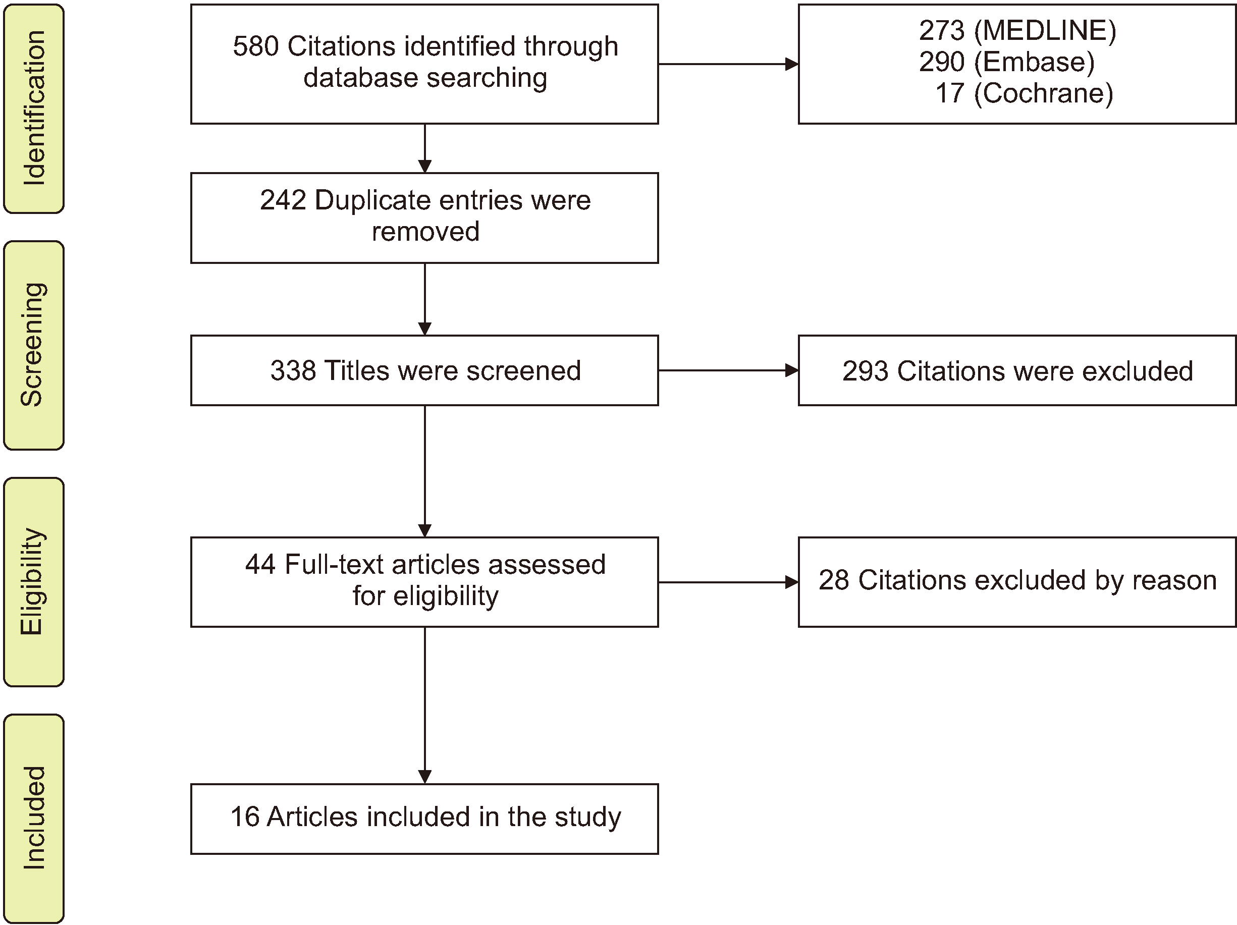
Table 1
Articles excluded following full-text assessment
| Study |
Follow-up less than 6 months |
Lack of data on preoperative and/or postoperative measurements |
Lack of data on etiology |
Conflicting data |
Study design |
No surgical intervention |
|---|---|---|---|---|---|---|
| Ramesh et al.20 (2019) | * | |||||
| Foudah21 (2019) | * | |||||
| K et al.22 (2018) | * | * | ||||
| Faus-Matoses et al.23 (2018) | * | * | ||||
| Sharma et al.24 (2017) | * | * | ||||
| Sánchez et al.25 (2017) | * | |||||
| Littuma et al.26 (2017) | * | |||||
| Khan et al.27 (2017) | * | |||||
| Gibson and Tatakis28 (2017) | * | |||||
| Farista et al.29 (2017) | * | |||||
| Mahn30 (2016) | * | |||||
| Aly and Hammouda31 (2016) | * | |||||
| Rao et al.32 (2015) | * | |||||
| Muthukumar et al.33 (2015) | * | * | ||||
| Bhola et al.34 (2015) | * | |||||
| Storrer et al.35 (2014) | * | * | ||||
| Grover et al.36 (2014) | * | |||||
| Gaddale et al.37 (2014) | * | |||||
| Sheth et al.38 (2013) | * | |||||
| Humayun et al.39 (2010) | * | |||||
| Gupta et al.40 (2010) | * | * | ||||
| Simon et al.15 (2007) | * | |||||
| Rosenblatt and Simon41 (2006) | * | * | ||||
| Ambrosio et al.42 (2018) | * | |||||
| Assenza et al.43 (2011) | * | |||||
| Ergezen et al.44 (2017) | * | |||||
| Mangano and Mangano45 (2013) | * | |||||
| Polo46 (2011) | * |
![]()
Table 2
Characteristics of the included studies
| Study | Study design | No. of patients | Sex | Age (yr) | Etiology | Surgical technique | Additional intervention | Follow-up duration (mo) | Preoperative display (mm) | Postoperative display (mm) | Complications |
|---|---|---|---|---|---|---|---|---|---|---|---|
| Ganesh et al.47 (2019) | Case report | 1 | F | 25 |
1. APE 2. HUL |
Type of instrument: diode laser (940 nm), 400 µm laser tip/continuous mode at 0.8 W Lower incision: MGJ Upper incision: 10 mm Type of incision: partial thickness MD extension: maxillary premolar Suturing: resorbable (muscle containment)/4-0 silk interrupted Removed after 14 days |
1. Muscle containment 2. Crown lengthening: laser gingivectomy |
12 | 7 | 3 |
1. Mild pain and tension 1st week 2. Minor scar |
| Torabi et al.48 (2018) | Case report | 3 |
2 F 1 M |
41-54 |
1. VME 2. HUL 3. APE |
Type of instrument: blade Lower incision: MGJ Upper incision: labial vestibule Type of incision: partial thickness (periosteal fenestration) MD extension: maxillary 1st molar Suturing: 4-0 silk, 4-0 Vicryl/ suspensory triangular+extraoral stabilization tape |
N/A | 13-16 | 6.3±4.06 |
2 months: 0.96±0.73 6 months: 0.76±0.87 13-16 months: 0.72±0.96 |
N/A |
| Tawfik et al.49 (2018) | RCT |
Group 1: 20 Group 2: 20 |
18 F 2 M |
N/A | Various etiologies |
Type of instrument: #15 blade Lower incision: MGJ Upper incision: 2× the display Type of incision: partial thickness MD extension: maxillary 1st molar Suturing: 5-0 polyglycolic sutures/continuous & interrupted Removed after 14 days |
Myotomy: Group 1 | 12 |
Group 1: 6.29±2.6 Group 2: 4.31±1.12 |
Group 1: 3 months: 3±1.53 6 months: 3.42±1.23 12 months: 3.57±1.62 Group 2: 3 months: 1.65±0.90 6 months: 2.21±1.0 12 months: 2.73±1.28 |
1. Pain 2. Swelling 3. Numbness |
| Alammar and Heshmeh50 (2018) | Case series | 14 | N/A | 18-38 |
1. SUL 2. HUL |
Type of instrument: blade Lower incision: 1 mm coronal to MGJ Upper incision: 10-12 mm Type of incision: full thickness MD extension: maxillary 1st molar Suturing: 4-0 Vicryl (muscle), 3-0 silk (mucosa)/interrupted sutures Removed after 14 days |
1. Myotomy 2. Crown lengthening |
6 | 6.36±1.12 |
1 month: 0.91±1.22 3 months: 2.27±1.27 6 months: 2.45±1.13 |
1. Scar 2. Tension in the upper lip 3. Minimal discomfort 4. Ecchymosis 5. Edema 6. Flap dehiscence 7. Numbness |
| Alammar et al.51 (2018) | PCT | 22 |
19 F 3 M |
18-38 |
1. SUL 2. HUL (lip mobility >8 mm) |
Type of instrument: blade Lower incision: 1 mm coronal to MGJ Upper incision: 10-12 mm Type of incision: partial thickness (Group 1)full thickness (Group 2) MD extension: maxillary 1st molar Suturing: 3-0 silk/interrupted (Group 1) 4-0 Vicryl (muscle), 3-0 silk (mucosa)/ interrupted (Group 2) Removed after 14 days |
1. Myotomy (Group 2) 2. Crown lengthening |
6 |
5.82±0.87 (Group 1) 6.36±1.12(Group 2) |
Group 1: 1 month: 2.18±0.75 3 months: 2.55±0.93 6 months: 3.27±0.79 Group 2: 1 month: 0.91±1.22 3 months: 2.27±1.27 6 months: 2.45±1.13 |
1. Scar 2. Tension in the upper lip 3. Minimal discomfort 4. Ecchymosis 5. Edema 6. Flap dehiscence 7. Numbness |
| Storrer et al.52 (2017) | Case report | 1 | F | 23 | HUL |
Type of instrument: #15C blade Lower incision: MGJ Upper incision: N/A Type of incision: partial thickness MD extension: maxillary canines Suturing: 5-0 nylon/4-0 silk/interrupted external sutures removed after 10 days |
1. Muscle containment 2. Frenectomy 3. Crown lengthening |
24 | 8 |
2 mm midline 4 mm lateral incisors |
Tension |
| Mantovani et al.53 (2016) | Case report | 1 | F | 23 |
1. HUL 2. APE |
Type of instrument: N/A Lower incision: 1 mm coronal to MGJ Upper incision: 10-12 mm Type of incision: partial thickness MD extension: maxillary 1st molars Suturing: 5-0 polygalactin/continuous interlocking sutures |
Crown lengthening: 2 mm after lip repositioning | 9 | 5 | 6 months: 0-1 | N/A |
| Abdullah et al.54 (2014) | Case series | 12 |
10 F 2 M |
20-29 | N/A |
Type of instrument: blade Lower incision: 4-5 mm from gingival margin Upper incision: 8-10 mm Type of incision: full thickness MD extension: maxillary 2nd premolar Suturing: 3-0 Vicryl/interrupted Removed after 14 days |
1. Myotomy 2. Frenectomy |
12 | 5±0.95 |
1 month: 2.66±0.77 3 months: 3.08±1.16 6 months: 3.08±1.16 12 months:3.08±1.16 |
1. Feeling of tension 2. Mild pain 3. Ecchymosis 4. Edema 5. Complete relapse in 1 patient after 3 months |
| Ozturan et al.55 (2014) | Case series | 10 | F | 22-34 | HUL |
Type of instrument: diode laser (940 nm, 4 W, continuous wave) Lower incision: MGJ Upper incision: 10-12 mm Type of incision: partial thickness Procedure was done in two stages left/right. MD extension: maxillary 1st molars Suturing: 5-0 monofilament/continuous interlocking |
N/A | 12 | 4.3±1.8 |
6 months: 1.1±1.0 12 months: 1.2±1.5 |
N/A |
| Jananni et al.56 (2014) | Case report | 1 | F | 18 |
1. VME 2. HUL |
Type of instrument: blade #15 Lower incision: MGJ Upper incision: 2× the display Type of incision: partial thickness MD extension: maxillary 2nd premolars Suturing: 4-0 silk/continuous interlocking sutures/periodontal pack Removed after 14 days |
N/A | 18 | 6 | 4 | N/A |
| Gabrić Pandurić et al.57 (2014) | Case report | 1 | F | 27 |
1. VME 2. HUL 3. APE |
Type of instrument: diode laser (975 μm, 4 W, CW) Lower incision: MGJ Upper incision: 1.5× display Type of incision: partial thickness MD extension: maxillary 1st molars Suturing: 3-0 silk/interrupted sutures Removed after 10 days |
1. Frenectomy 2. Crown lengthening: laser gingivectomy |
6 | 5.5-10 | 0-2 |
1. Tension 2. Slight pain 3. Numbness 1st week 4. Scar |
| Dayakar et al.58 (2014) | Case report | 1 | F | 22 | N/A |
Type of instrument: blade Lower incision: MGJ Upper incision: 10-12 mm Type of incision: partial thickness MD extension: maxillary 1st molars Suturing: continuous interlocking Removed after 14 days |
N/A | 12 | 5-6 |
3 months: 3 6 months: 3 12 months: complete relapse |
1. Mild pain and tension 1st week 2. Scar |
| Silva et al.59 (2013) | PCT | 13 |
11 F 2 M |
19-49 | HUL |
Type of instrument: N/A Lower incision: 1 mm coronal to MGJ Upper incision: 10-12 mm Type of incision: partial thickness MD extension: maxillary 1st molar Suturing: 4-0 polygalactin/continuous interlocking sutures |
N/A | 6 | 4-10 (5.8±2.1) |
3 months: 0-3 (1.4±1.0) 6 months: 0-5 (1.3±1.6) |
1. Tension 2. Scar |
| Ribeiro-Júnior et al.60 (2013) | Case report | 2 | F | 20-22 |
1. HUL 2. APE(1 patient) |
Type of instrument: N/A Lower incision: 1 mm coronal to MGJ Upper incision: 10-12 mm Type of incision: partial thickness MD extension: maxillary 1st molars Suturing: 4-0 poligalactin/continuous interlocking sutures Removed after 14 days |
Crown lengthening: 1 patient | 6 |
7 6 |
1 |
1. Tension 1st week 2. Scar |
| Jacobs and Jacobs61 (2013) | Case series | 7 | F | 21-59 | N/A |
Type of instrument: diode laser/#15 blade Lower incision: MGJ Upper incision: 2× display Type of incision: partial thickness MD extension: maxillary 1st molars Suturing: 3-0 chromic or silk/interrupted or continuous interlocking sutures |
N/A | 1-36 | 5.36±1.5 | 1.1±2.5 |
1. Scar 2. Pain, swelling and tension 1st week |
| Ishida et al.8 (2010) | Case series | 14 | F | 15-35 | N/A |
Type of instrument: #15 blade Lower incision: N/A Upper incision: N/A Type of incision: N/A MD extension: N/A Suturing: 5-0 resorbable/interrupted |
1. Myotomy 2. Frenectomy |
6-18 | 5.22±1.48 | 6 months: 1.91±1.50 | N/A |
![]()
The risk of bias assessment for non-comparative studies is presented in Table 38,47,48,50,52-61. The risk of bias for one non-randomized comparative study51 was estimated at 23 (of 24) based on the MINORS scoring criteria. The only randomized clinical trial49 was evaluated as “low risk” of bias based on the Cochrane risk of bias tool for randomized trials.
Table 3
Quality assessment for non-comparative studies
| Study | Aim | Inclusion | Data collection | Endpoint | Evaluation (bias) | Follow-up period | Loss to follow-up |
Sample size |
Total score |
|---|---|---|---|---|---|---|---|---|---|
| Ganesh et al.47 (2019) | 2 | 1 | 1 | 2 | 1 | 2 | 2 | 0 | 11 |
| Torabi et al.48 (2018) | 1 | 2 | 1 | 2 | 2 | 2 | 2 | 1 | 13 |
| Alammar and Heshmeh50 (2018) | 2 | 2 | 2 | 2 | 1 | 1 | 2 | 1 | 13 |
| Storrer et al.52 (2017) | 1 | 1 | 1 | 1 | 1 | 1 | 2 | 0 | 8 |
| Mantovani et al.53 (2016) | 2 | 1 | 1 | 1 | 0 | 1 | 2 | 0 | 8 |
| Abdullah et al.54 (2014) | 2 | 2 | 2 | 2 | 2 | 2 | 2 | 1 | 15 |
| Ozturan et al.55 (2014) | 2 | 2 | 2 | 2 | 2 | 2 | 2 | 1 | 15 |
| Jananni et al.56 (2014) | 0 | 1 | 1 | 1 | 1 | 2 | 2 | 0 | 8 |
| Gabrić Pandurić et al.57 (2014) | 2 | 1 | 2 | 2 | 2 | 2 | 2 | 0 | 13 |
| Dayakar et al.58 (2014) | 1 | 1 | 1 | 1 | 1 | 2 | 2 | 0 | 9 |
| Silva et al.59 (2013) | 2 | 2 | 2 | 2 | 2 | 1 | 2 | 1 | 14 |
| Ribeiro-Júnior et al.60 (2013) | 2 | 1 | 1 | 1 | 1 | 1 | 2 | 1 | 10 |
| Jacobs and Jacobs61 (2013) | 2 | 2 | 2 | 2 | 2 | 2 | 2 | 1 | 15 |
| Ishida et al.8 (2010) | 2 | 2 | 2 | 2 | 2 | 2 | 2 | 1 | 15 |
![]()
The included studies assessed lip repositioning procedures in 144 patients aged between 15-59 years (134 females and 10 males, not reported in one study with 14 patients50). The most prevalent reported etiology was hypermobile upper lip47,48,50-53,55-57,59,60, followed by altered passive eruption25,28,30,39,47,48,53,57,60, vertical maxillary excess48,56,57, and short upper lip50,51.
All but two studies50,54 used a partial-thickness flap. However, one comparative study reported better results with a full-thickness approach51. Some authors used additional interventions including crown lengthening47,50-53,57,60 and frenectomy8,23,33,37,52,54,57 to achieve more favorable results with higher stability. Alammar et al.51 mentioned two cases with complete relapse in their conventional surgical group. In addition, Dayakar et al.58 reported complete relapse after 12 months, but they did not report additional details.
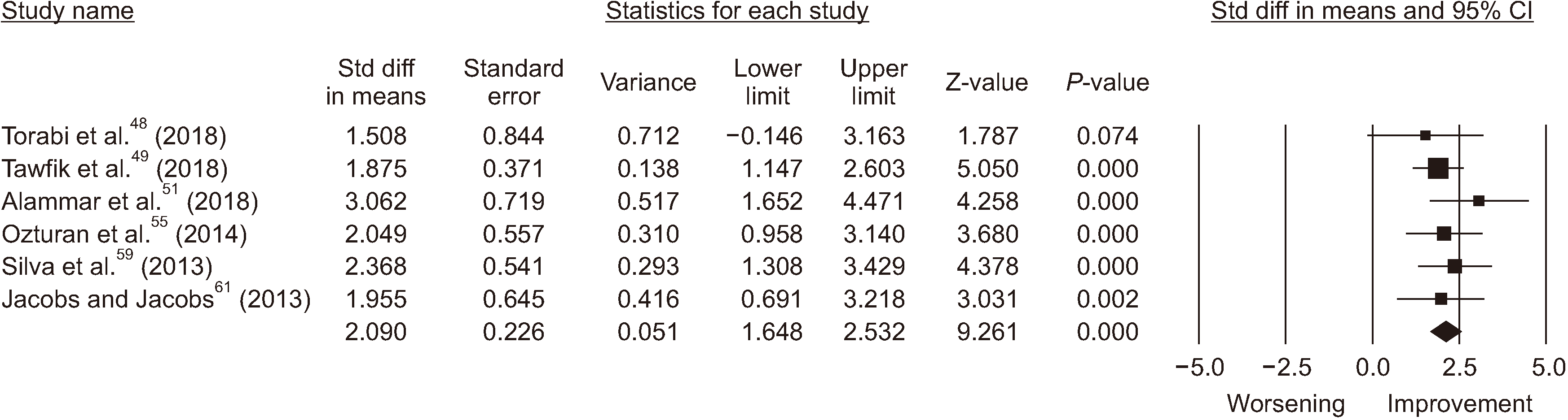
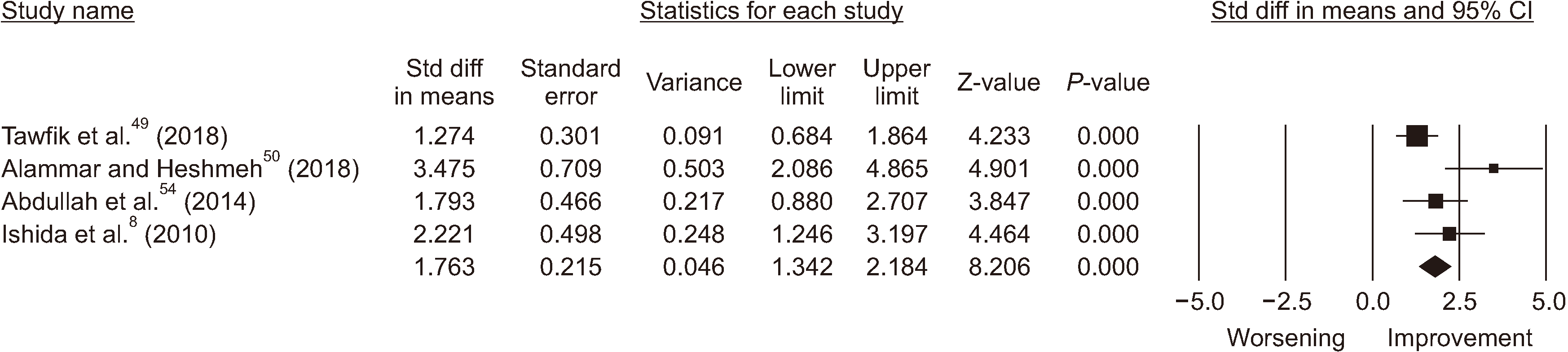
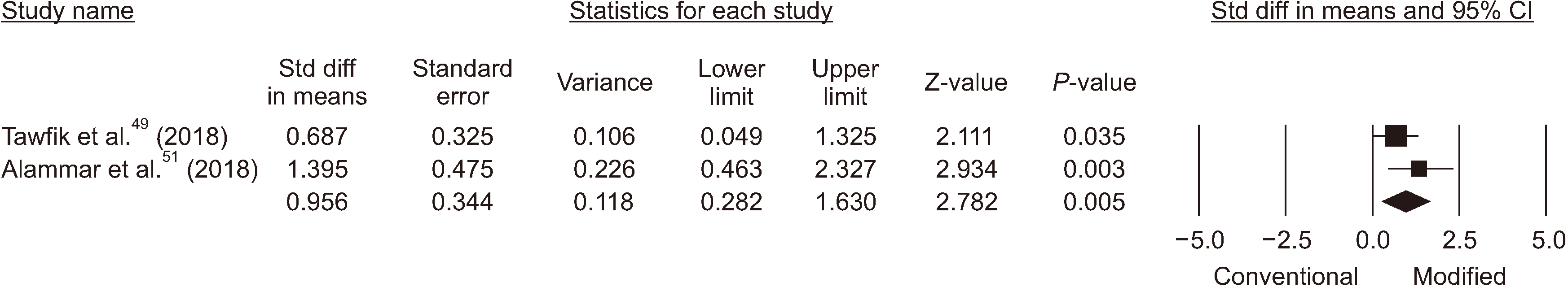
Six studies provided sufficient data (sample size, mean, and either standard deviation or standard error) to contribute to estimation of the overall effect size for improvement after lip repositioning surgery.(Fig. 2) In five studies, myotomy was performed along with lip repositioning surgery8,49-51,54, while two investigations used sutures to confine the elevator muscles47,52. The analyzed data indicate an improvement of 1.76 mm (95% confidence interval, 1.34-2.18 mm) with myotomy/muscle containment.(Fig. 3) Tawfik et al.49 reported that lip repositioning with myotomy resulted in greater improvement and higher patient satisfaction compared to that without myotomy, which was consistent with the results of another comparative study51.(Fig. 4)
 | Fig. 2Forest plot of effect size for lip repositioning without myotomy/muscle containment after 6 months. (Std diff: standard difference, CI: confidence interval)
|
Go to :
IV. Discussion
Among the procedures used to improve EGD, lip repositioning is a promising alternative. This procedure has been suggested for patients with minor discrepancies requesting a less invasive procedure compared with orthognathic surgery. It also satisfies patients who do not desire to undergo orthodontic therapy or botulinum toxin A administration. This study was conducted to evaluate the current literature regarding the efficacy of myotomy/muscle containment on outcome and long-term stability of lip repositioning surgery.
Most retrieved articles were case series or case reports. The ideal score for non-comparative studies according to MINORS scoring criteria is 16, though none of the chosen studies obtained this. Therefore, the findings should be interpreted with caution.
Among 7 studies involving myotomy/muscle containment, 6 studies8,47,50-52,54 used additional modalities such as crown lengthening and frenectomy. Therefore, it is unclear how much of the improvement is related to lip repositioning surgery alone. One comparative study49 that evaluated the effect of myotomy without other interventions reported better results with the modified technique. However, they did not specify the etiologic contributors for gummy smile in their patients and concluded no clear correlation between etiology and achieved improvement. Studies reported various etiologies for their study population, limiting the ability to draw a conclusion, as vertical maxillary excess48,56,57 can compromise long-term stability15.
Some studies reported subjective improvement without an exact measurement of pre/postoperative gingival display15,20,23,28,37,40. Success must be reported based on measurement of all parameters before and after the procedure to allow exact assessment of the improvement. Consequently, we included studies that reported pre- and postoperative measurements to assess treatment success more accurately. This also applies to patient-related outcome measures (PROMs). Therefore, standardization of the measurement protocol, techniques, outcomes, and PROMs is essential to draw meaningful conclusions.
Tawfik et al.62 published a systematic review in 2018, in which they concluded a persuasive need for randomized and comparative trials to assess the influence of different factors on outcomes, complications, and patient satisfaction. They carried out a randomized clinical trial49 to evaluate the effect of myotomy on lip repositioning results, reporting EGD reduction by 2.73±1.281 mm with classic lip repositioning and greater reduction of 3.57±1.62 mm with myotomy. In another study, Alammar et al.51 compared the conventional technique with the modified approach and demonstrated greater sustainability and less relapse in the modified surgical technique including myotomy. However, they conducted crown lengthening in addition to lip repositioning surgery, which must be considered when interpreting the results.
Based on the available data, lip repositioning can be a successful approach for EGD treatment in carefully selected cases, though it has been shown to have a higher chance of unfavorable results in cases of severe maxillary excess (EGD >8 mm)15. It seems that myotomy/muscle containment can result in better outcomes and stability of the achieved improvement. However, due to the limited number of studies and the discrepancy in the current evidence, further well-organized comparative clinical trials are needed to derive a conclusion regarding the effectiveness of myotomy/muscle containment compared with the conventional approach.
Go to :
 XML Download