

 PDF
PDF Citation
 Print
Print


INTRODUCTION
MATERIALS AND METHODS
Study population
Parameters collected
Radiographic scoring
Statistical analysis
RESULTS
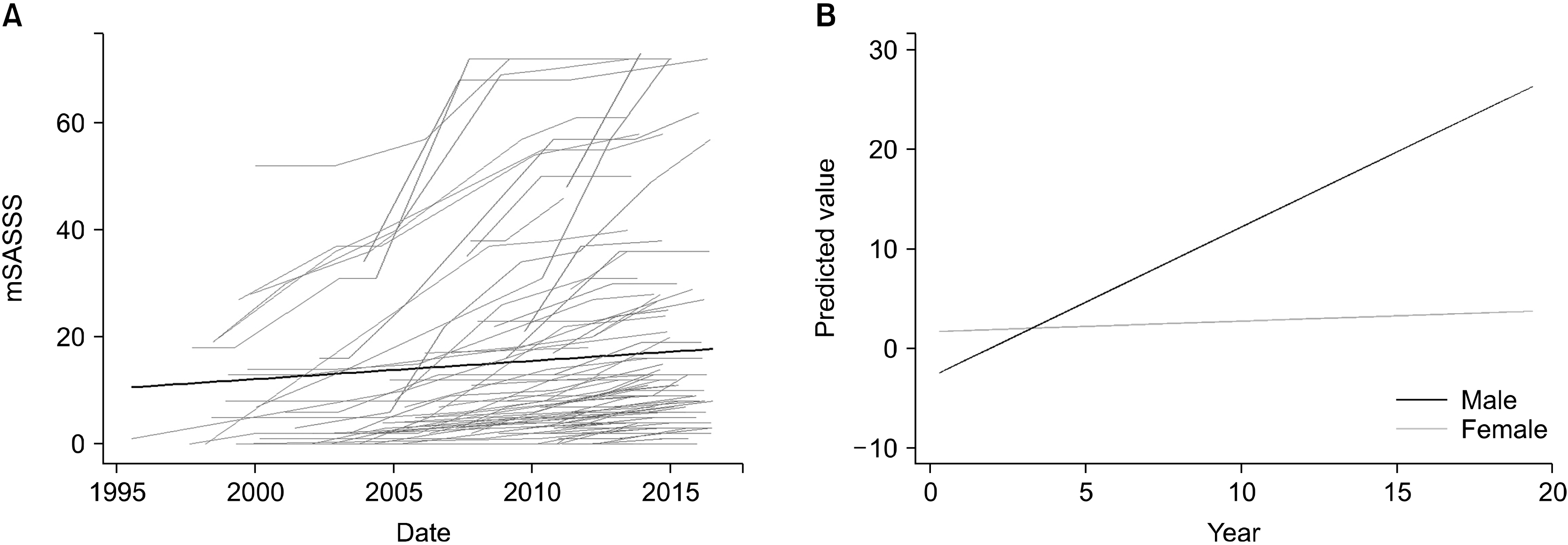 | Figure 1(A) The natural course of radiographic progression. The black line indicates the mean radiographic progression of the entire patient cohort. (B) Predicted value of radiographic progression over time in multivariable analysis according to sex. mSASSS: modified Stoke Ankylosing Spondylitis Spine Score.
|
Table 1
| Variable | Male (n=85) | Female (n=16) | p-value |
|---|---|---|---|
| Age at disease onset (yr) | 26.6±10.8 | 31.4±9.7 | 0.100 |
| Age at estrogen sampling (yr) | 38.3±11.4 | 39.3±8.8 | 0.739 |
| Disease duration (mo) | 55.6±66.7 | 29.3±29.0 | 0.126 |
| Peripheral arthritis | 38 (44.7) | 7 (43.8) | 0.944 |
| Hip involvement | 19 (22.4) | 2 (12.5) | 0.512 |
| Uveitis | 23 (27.1) | 4 (25.0) | 1.000 |
| Enthesitis | 12 (14.1) | 2 (12.5) | 1.000 |
| mSASSS, initial | 8.9±11.2 | 4.0±3.2 | 0.001 |
| mSASSS, final | 20.3±21.9 | 10.9±9.3 | 0.188 |
| Sacroiliitis grade* | 5.5±1.6 | 5.1±1.5 | 0.337 |
| E2 (pg/mL)† | 114.1±35.9 | 108.8±45.7 | 0.552 |
| Dkk1 (ng/mL) | 1,243.5±760.4 | 962.0±521.7 | 0.160 |
| Leptin (pg/mL) | 1,896.1±1,847.5 | 2,968.5±2,361.3 | 0.045 |
| ESR, initial (mm/h) | 51.9±31.9 | 60.2±39.7 | 0.361 |
| CRP, initial (mg/dL) | 3.17±3.68 | 1.93±2.07 | 0.196 |
| BMI (kg/m2) | 24.6±3.8 | 22.2±2.7 | 0.018 |
| Anti-TNF use | 74 (87.1) | 12 (75.0) | 0.250 |
| Current smoker | 31 (36.5) | 0 (0.0) | 0.002 |
| X-ray follow-up duration (mo) | 97.2±60.9 | 58.5±54.3 | 0.020 |
Data are expressed as mean±standard deviation or number (%). mSASSS: modified Stoke Ankylosing Spondylitis Spine Score, E2: 17β-estradiol, Dkk1: dickkopf-1, ESR: erythrocyte sedimentation rate, CRP: C-reactive protein, BMI: body mass index, TNF: tumor necrosis factor. *Sacroiliitis grade refers to the sum of sacroiliitis grades of each side according to the modified New York criteria. †Reference range for E2 are 0∼400 pg/mL.
![]()
Table 2
| Variable | Non-progressor (n=66) | Progressor (n=35) | p-value |
|---|---|---|---|
| Age at disease onset (yr) | 26.8±11.5 | 28.3±9.2 | 0.528 |
| Age at estrogen sampling (yr) | 36.3±11.6 | 42.6±8.5 | 0.006 |
| Disease duration (mo) | 37.1±38.4 | 78.4±87.5 | 0.011 |
| Female | 14 (21.2) | 2 (5.7) | 0.048 |
| Peripheral arthritis | 32 (48.5) | 13 (37.1) | 0.275 |
| Hip involvement | 17 (25.8) | 4 (11.4) | 0.123 |
| Uveitis | 18 (27.3) | 9 (25.7) | 0.866 |
| Enthesitis | 9 (13.6) | 5 (14.3) | 0.928 |
| mSASSS, initial | 3.8±4.5 | 16.3±13.4 | <0.001 |
| mSASSS, final | 17.9±20.2 | 21.6±22.7 | 0.445 |
| Sacroiliitis grade* | 4.94±1.37 | 6.31±1.62 | <0.001 |
| Estrogen (pg/mL)† | 107.7±37.5 | 123.6±35.6 | 0.042 |
| Dkk1 (ng/mL) | 1,303.7±778.9 | 1,001.2±598.7 | 0.048 |
| Leptin (pg/mL) | 1,939.0±1,838.8 | 2,305.4±2,189.4 | 0.375 |
| ESR, initial (mm/h) | 49.1±35.4 | 61.0±27.3 | 0.087 |
| CRP, initial (mg/dL) | 2.31±2.75 | 4.21±4.37 | 0.024 |
| BMI (kg/m2) | 23.1±3.2 | 26.3±3.8 | <0.001 |
| Anti-TNF use | 53 (80.3) | 33 (94.3) | 0.079 |
| Current smoker | 17 (25.8) | 14 (40.0) | 0.140 |
| X-ray follow-up duration (mo) | 93.3±58.3 | 119.1±66.9 | 0.080 |
Data are expressed as mean±standard deviation or number (%). mSASSS: modified Stoke Ankylosing Spondylitis Spine Score, Dkk1: dickkopf-1, ESR: erythrocyte sedimentation rate, CRP: C-reactive protein, BMI: body mass index, TNF: tumor necrosis factor, E2: 17β-estradiol. *Sacroiliitis grade refers to the sum of sacroiliitis grades of the each sides according to the modified New York criteria. †Reference ranges for E2 are 0∼400 pg/mL.
![]()
Table 3
| Variable | Non-progressor (n=52) | Progressor (n=33) | p-value |
|---|---|---|---|
| Age at disease onset (yr) | 25.9±11.8 | 27.5±8.9 | 0.507 |
| Age at estrogen sampling (yr) | 35.6±12.1 | 42.6±8.7 | 0.005 |
| Disease duration (mo) | 38.5±40.4 | 82.4±88.6 | 0.003 |
| Peripheral arthritis | 26 (50.0) | 12 (36.4) | 0.218 |
| Hip involvement | 15 (28.8) | 4 (12.1) | 0.108 |
| Uveitis | 14 (26.9) | 9 (27.3) | 0.972 |
| Enthesitis | 7 (13.5) | 5 (15.2) | 0.827 |
| mSASSS, initial | 3.9±4.8 | 16.8±13.7 | <0.001 |
| mSASSS, final | 19.2±21.5 | 22.0±23.0 | 0.601 |
| Sacroiliitis grade* | 4.90±1.33 | 6.39±1.61 | <0.001 |
| Estrogen (pg/mL)† | 109.6±36.7 | 121.1±34.1 | 0.153 |
| Dkk1 (ng/mL) | 1,384.0±823.6 | 1,022.1±595.0 | 0.032 |
| Leptin (pg/mL) | 1,591.7±1,497.3 | 2,375.7±2,235.8 | 0.082 |
| ESR, initial (mm/h) | 47.2±34.3 | 59.3±26.6 | 0.088 |
| CRP, initial (mg/dL) | 2.51±2.96 | 4.19±4.44 | 0.039 |
| BMI (kg/m2) | 23.4±3.3 | 26.5±3.7 | <0.001 |
| Anti-TNF use | 43 (82.7) | 31 (93.9) | 0.132 |
| Current smoker | 17 (32.7) | 14 (42.4) | 0.364 |
| X-ray follow-up duration (mo) | 88.7±58.3 | 110.6±63.4 | 0.106 |
Data are expressed as mean±standard deviation or number (%). mSASSS: modified Stoke Ankylosing Spondylitis Spine Score, Dkk1: dickkopf-1, ESR: erythrocyte sedimentation rate, CRP: C-reactive protein, BMI: body mass index, TNF: tumor necrosis factor, E2: 17β-estradiol. *Sacroiliitis grade refers to the sum of sacroiliitis grades of the each sides according to the modified New York criteria. †Reference ranges for E2 are 0∼400 pg/mL.
![]()
Table 4
![]()
Table 5
![]()
 XML Download