

 PDF
PDF Citation
 Print
Print


INTRODUCTION
Crowding can be managed in a number of different ways, including arch expansion, flaring of the anterior teeth, and distalization. In the absence of significant anteroposterior discrepancies such as in Class II or Class III molar relationships, a transverse deficiency may be suspected to be a major cause of crowding. Transverse expansion can be achieved by dentoalveolar displacement using a transpalatal arch (TPA) or expansion archwire. Since TPAs and expansion archwires largely result in buccal tipping of the posterior teeth,1 they are not readily used unless the molars are lingually tipped. Furthermore, dentoalveolar expansion can cause undesirable periodontal side effects. The reduced periodontal support can be functionally and esthetically detrimental.
Displacement of the premolars and/or molars to the buccal side is mostly contraindicated, especially in aged patients, due to the risk of gingival recession and bony dehiscence. Even in the absence of active periodontal lesions, alveolar bone loss is a natural consequence of aging.2,3 Therefore, studies reporting the use of active expansion in aged patients are scarce in orthodontic literature.
In patients with maxillary transverse deficiency, skeletal expansion offers a good therapeutic option without the need for periodontal compromise, while maintaining the buccolingual axis of the posterior segment. Angell4 first introduced the idea of rapid palatal expansion (RPE) in 1860, which was again demonstrated by Haas5 in the 1960s mainly in around the pubertal stage. Moreover, successful orthopedic expansion using miniscrew-assisted RPE (MARPE) has been shown in young adults.6,7 Nonetheless, skeletal maturity and the increasing interdigitation of the facial sutures are known to be the main limiting factors for RPE.8,9 Accordingly, surgically assisted RPE (SARPE) has been suggested as an alternative to overcome the mechanical resistance to suture opening in adult patients.10,11 Meanwhile, the age range for successful orthopedic expansion has remained a topic of debate, possibly due to the underlying individual variations in suture maturity.
Unlike calvarial sutures, which are known to fuse at around the 20s, the facial sutures have been shown to remain patent after cessation of active growth.12 This indicates the possibility of successful orthopedic expansion even in skeletally mature and old patients. The following case describes the treatment of a 60-year old patient who showed crowding accompanied by the risk of gingival recession and alveolar bone loss via nonsurgical orthopedic expansion.
DIAGNOSIS AND ETIOLOGY
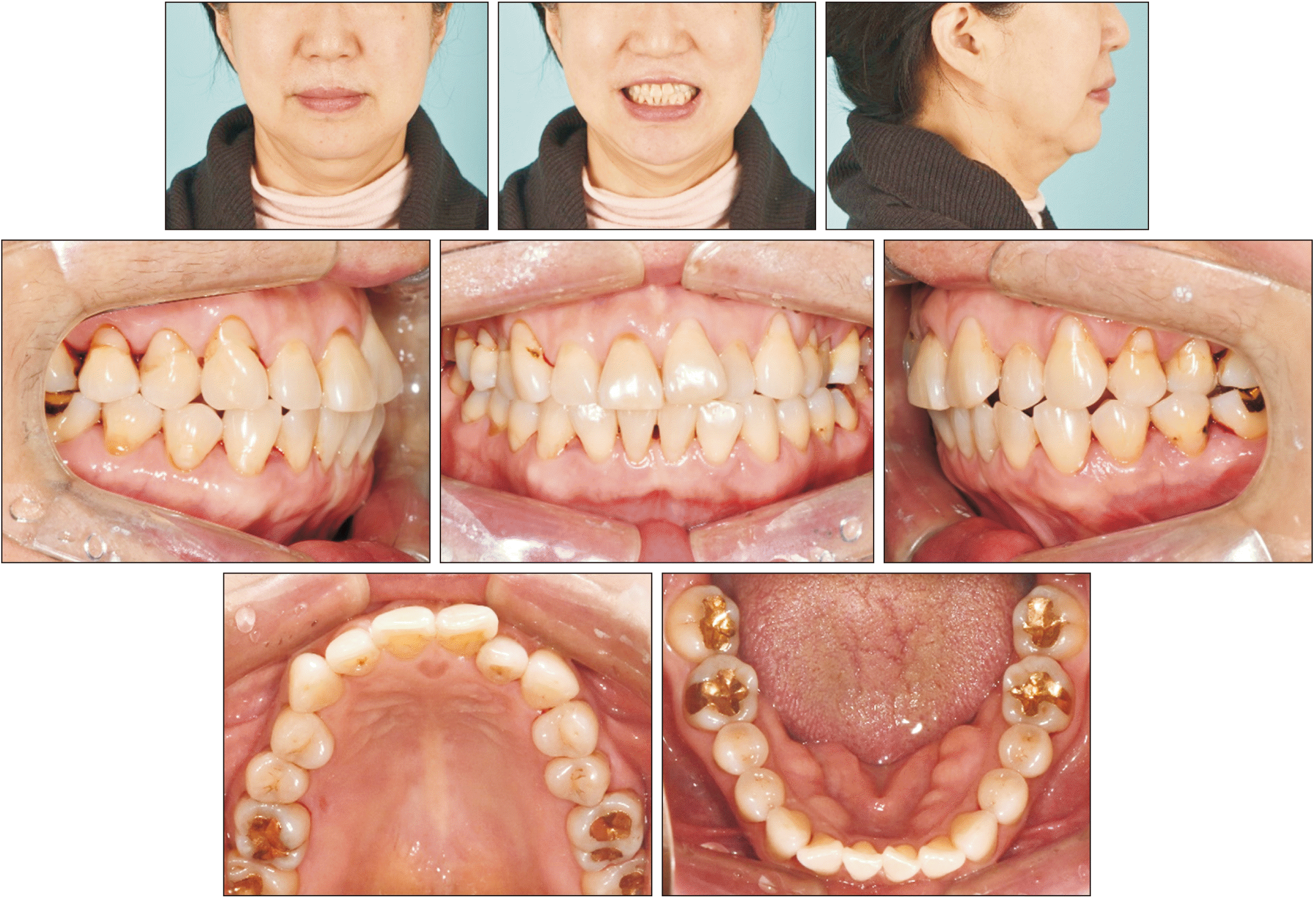
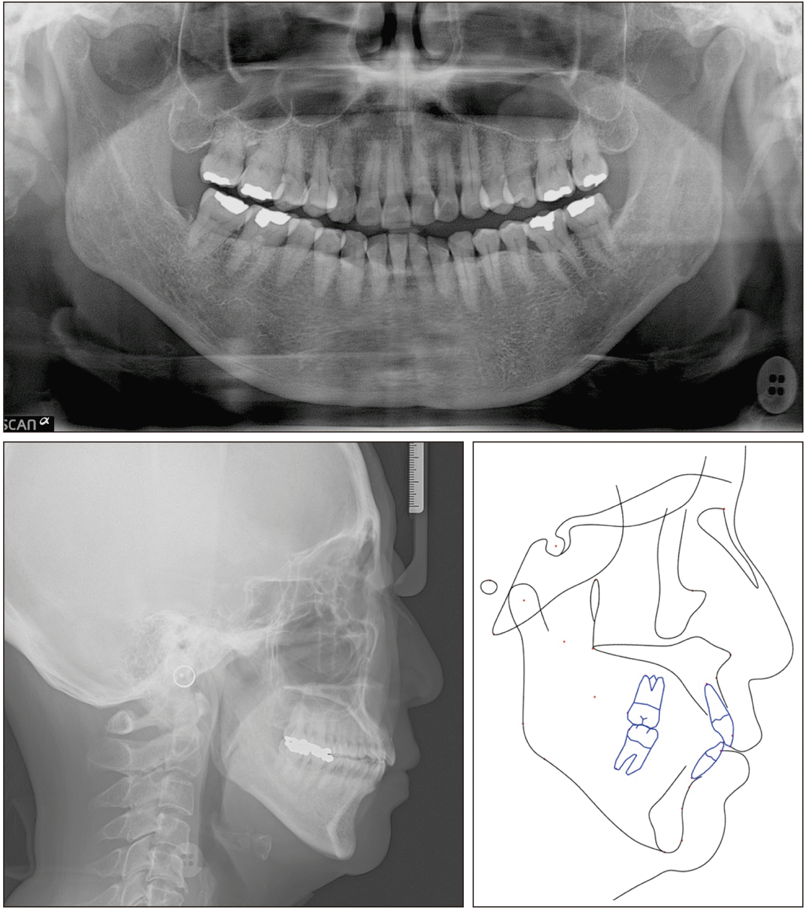
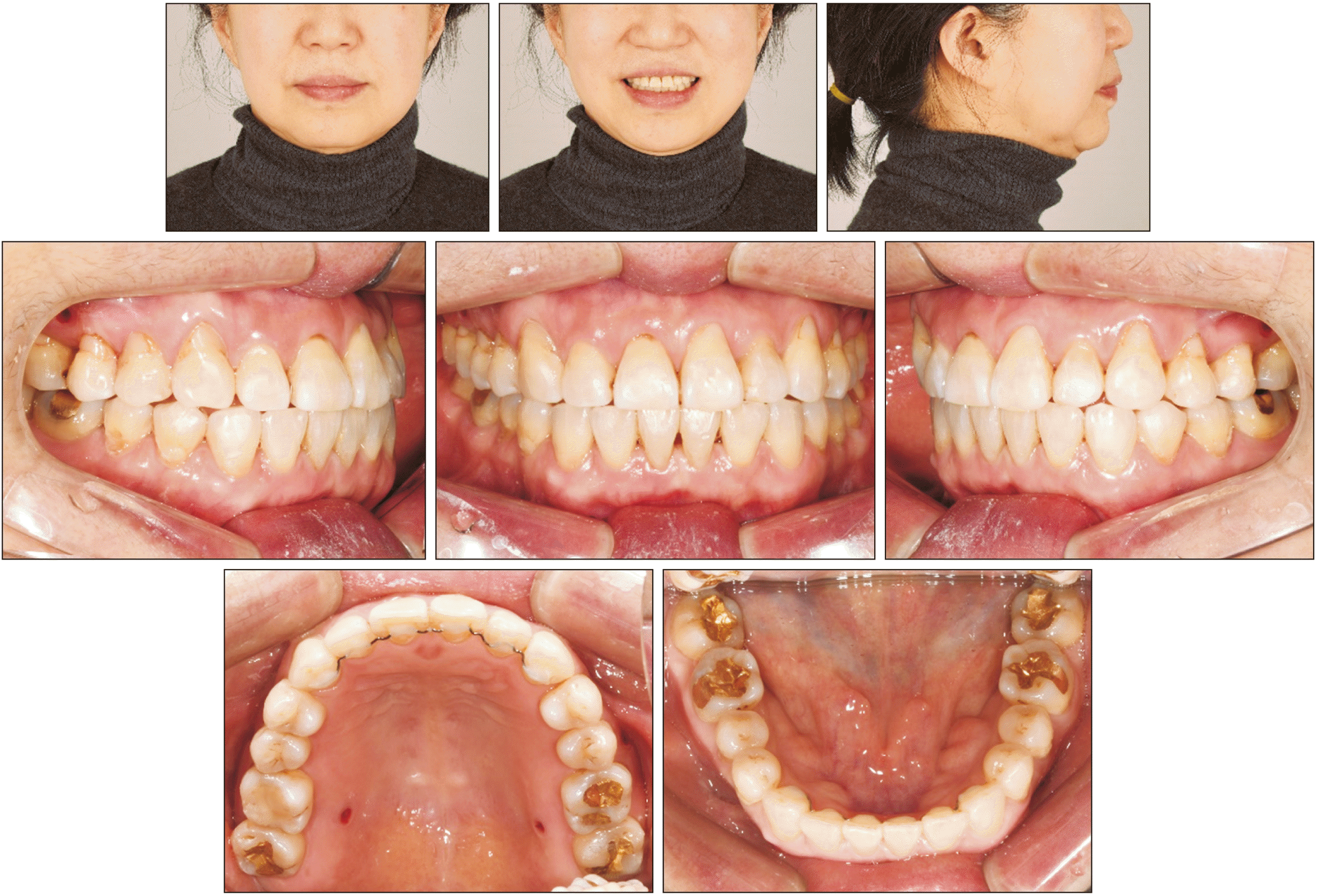
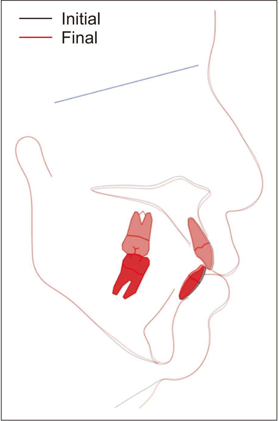
A 60-year old female patient presented with the chief complaint of unaesthetic smile involving a crossbite and crowding of the anterior teeth. She had a history of previous orthodontic treatment due to crowding approximately 50 years ago. The intraoral examination indicated an anterior crossbite of the left lateral incisors and a posterior crossbite of the left second premolars, as well as an edge-to-edge bite of the left first premolars. She had a symmetrical Class I molar relationship. The dental midline deviated 2 mm to the left in the maxilla and 1 mm to the left in the mandible relative to the facial midline (Figure 1). The intermolar width, which was measured as the distance between the central fossa of the right and left first molars, was 47.0 mm in the maxilla and 43.0 mm in the mandible, whereas the normal intermolar width difference ranged from 5 to 8 mm.13,14 The interpremolar width was 34.0 mm in the maxilla and 32.6 mm in the mandible. The arch widths were also measured at the estimated centers of resistance (middle point of furcation).14 The difference was –4.0 mm, with the widths being 44.1 mm in the maxilla and 48.1 mm in the mandible. This was in contrast to the reported mean value in normal occlusion, which is –0.39 ± 1.87 mm.14 Accordingly, the basal arch width of the maxilla was considered as relatively narrow. Further assessment of the diagnostic data revealed a mild hyperdivergence (skeletal Class II) with maxillary and mandibular arch length discrepancies amounting to 3 and 2 mm, respectively (Figure 2, Table 1). The patient showed some generalized horizontal alveolar bone loss and non-inflammatory gingival recession, especially on the facial surfaces of the upper left central incisor, canine, and premolars. Furthermore, a predisposition to cervical abfraction could be inferred from the presence of multiple cervical fillings (Figure 1).
Informed consent of the patient was obtained for release of the patient’s records.
TREATMENT OBJECTIVES
The treatment objectives were (1) to correct the anterior and posterior crossbites, (2) to relieve the maxillary and mandibular crowding without any change in the profile, (3) to maintain the bilateral Class I molar relationship, (4) to maintain periodontal health by avoiding further buccal bone loss and gingival recession, and (5) to improve the smile esthetics. Considering the advanced age of the patient and the presence of periodontal issues, the treatment was kept as minimally invasive and short as possible.
TREATMENT ALTERNATIVES
Based on the diagnosis, several treatment options were suggested. Flaring of the anterior teeth provides space for crowding relief; however it results in increasing convexity of the facial profile. Since the patient did not want any change in the profile, this treatment option was discarded.
Molar distalization is an effective treatment approach to relieve crowding, especially to correct Class II or III molar relationships without affecting the position of the incisors.15-17 However, the present patient had a Class I molar relationship, indicating the possibility of slight transverse maxillary arch constriction, shown as the crossbite of the left second premolars. Hence, molar distalization was not considered as the primary treatment option.
Extraction was not regarded as a reasonable treatment approach, because the patient showed only mild to moderate crowding. For extraction, closure of excessive space was expected to be challenging with prolonged treatment time.
Correction of the maxillary transverse deficiency by arbitrary arch expansion was seen as risky due to the potential periodontal adverse effects such as alveolar bone dehiscence and gingival recession, especially in the upper left central incisor.18 Therefore, expansion by RPE was suggested. Ideal alignment without periodontal compromise can be achieved through orthopedic expansion of the maxilla and relocation of the teeth within the expanded basal bone. Furthermore, RPE allows correction of crossbite and crowding without any considerable effect on the anteroposterior relationship, as desired in this patient.
Although SARPE provides higher predictability, it involves surgical procedures with additional expenses and general risks related to surgery. The patient wished to avoid an invasive treatment. An approach with nonsurgical MARPE was chosen to minimize the undesirable effects of conventional tooth-borne RPE.19
The patient was informed of the uncertainty of suture opening and the eventual need for an alternative treatment method and agreed to attempt nonsurgical MARPE.
TREATMENT PROGRESS
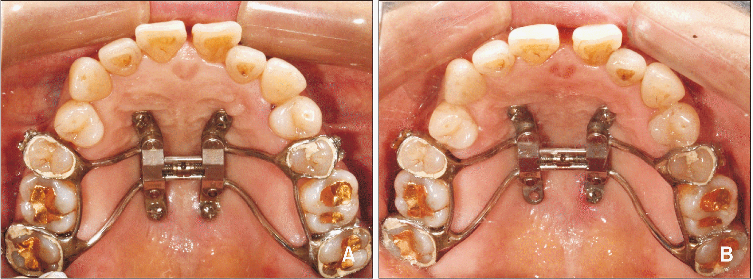
After evaluation of the periodontal status at the periodontal department, a customized MARPE appliance using an expander screw with extension plates (KBE; BioMaterials Korea Inc., Seoul, Korea) was fabricated. The MARPE consisted of four bands, connecting arms, an expander body with an expansion screw and four bendable extension plates with insertion holes for the miniscrews. The preliminary assessment of the insertion sites was conducted in the initial cone-beam computed tomography (CBCT) images to determine the anteroposterior position of the expander body to ensure that the palatal bone accommodated the bone screws. The positions of the expander body and the screw holes were marked on the pickup model with four bands. Bands were placed on the second premolars and second molars to avoid injury to the first molars, which had hypersensitivity. The appliance was fabricated by soldering the connecting arms to the bands and contouring the extension plates according to the depth of the palatal vault. The posterior plates were converged toward the midpalatal suture to allow the use of the thickness of the nasal crest.20 The MARPE was then cemented on the second premolars and the second molars, and four miniscrews (length, 9.0 mm; diameter, 2.0 mm for the anterior holes, and length, 7.0 mm and diameter, 2.0 mm for the posterior holes; BioMaterials Korea Inc.) were inserted (Figure 3). The selection of the insertion sites and miniscrew dimensions were also based on the norm values of the palatal bone and soft tissue thickness reported in the literature.20,21 The anterior miniscrews were inserted in the rugae region, where the palatal bone thickness is known to be sufficient (more than 6 mm on average). The posterior miniscrews were inserted within 3 mm from the midpalatal suture, because the bone thickness is known to decrease laterally.21
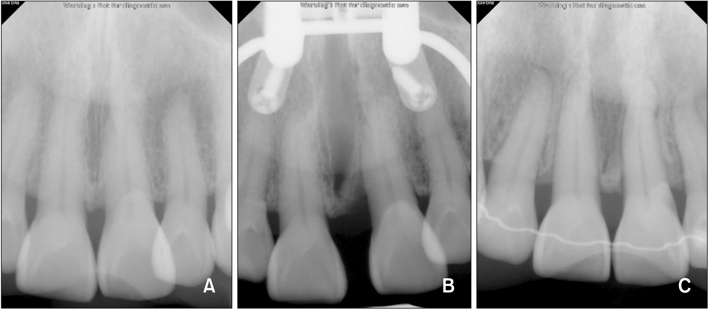
The activation was initiated at a rate of one turn a day (semi-rapid expansion protocol, 0.2 mm/day). After four weeks of activation, a median diastema was noted clinically (Figure 3). The separation of the midpalatal suture could be confirmed radiographically on a series of periapical views (Figure 4). The activation was completed after a total of 38 turns and 7.6 mm of MARPE. After completion of expansion, a CBCT scan was obtained.
On the periapical radiographs and CBCT images, a structure resembling a bone-bridge could be observed at the crestal level in the suture gap, indicating a somewhat uneven orthopedic expansion pattern. This structure, however, did not seem to have hindered the suture separation. The post-expansion periapical radiograph showed some vertical alveolar bone loss on the mesial side of the left central incisor. This defect had fully recovered by the end of the active treatment (Figure 4).
After the expansion, 0.018-inch slot self-ligation brackets (Clippy-C; Tomy, Tokyo, Japan) were bonded. The alignment in the mandible was performed directly, while the maxillary alignment was first initiated after two months of consolidation. For the patient’s comfort and to secure the transverse dimension of the basal bone during alignment, the MARPE appliance was transformed into a passive bone-borne retainer by cutting the arms and removing the bands. In order to further upright the second molars to the lingual side, additional palatal miniscrews were used. All fixed appliances were removed after 12 months of active treatment.
TREATMENT RESULTS
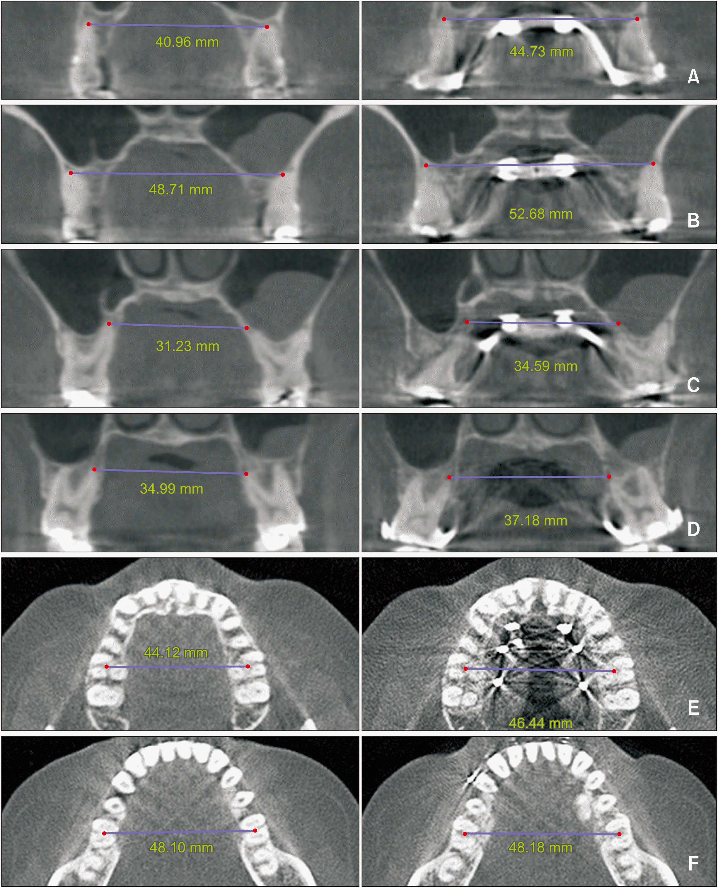
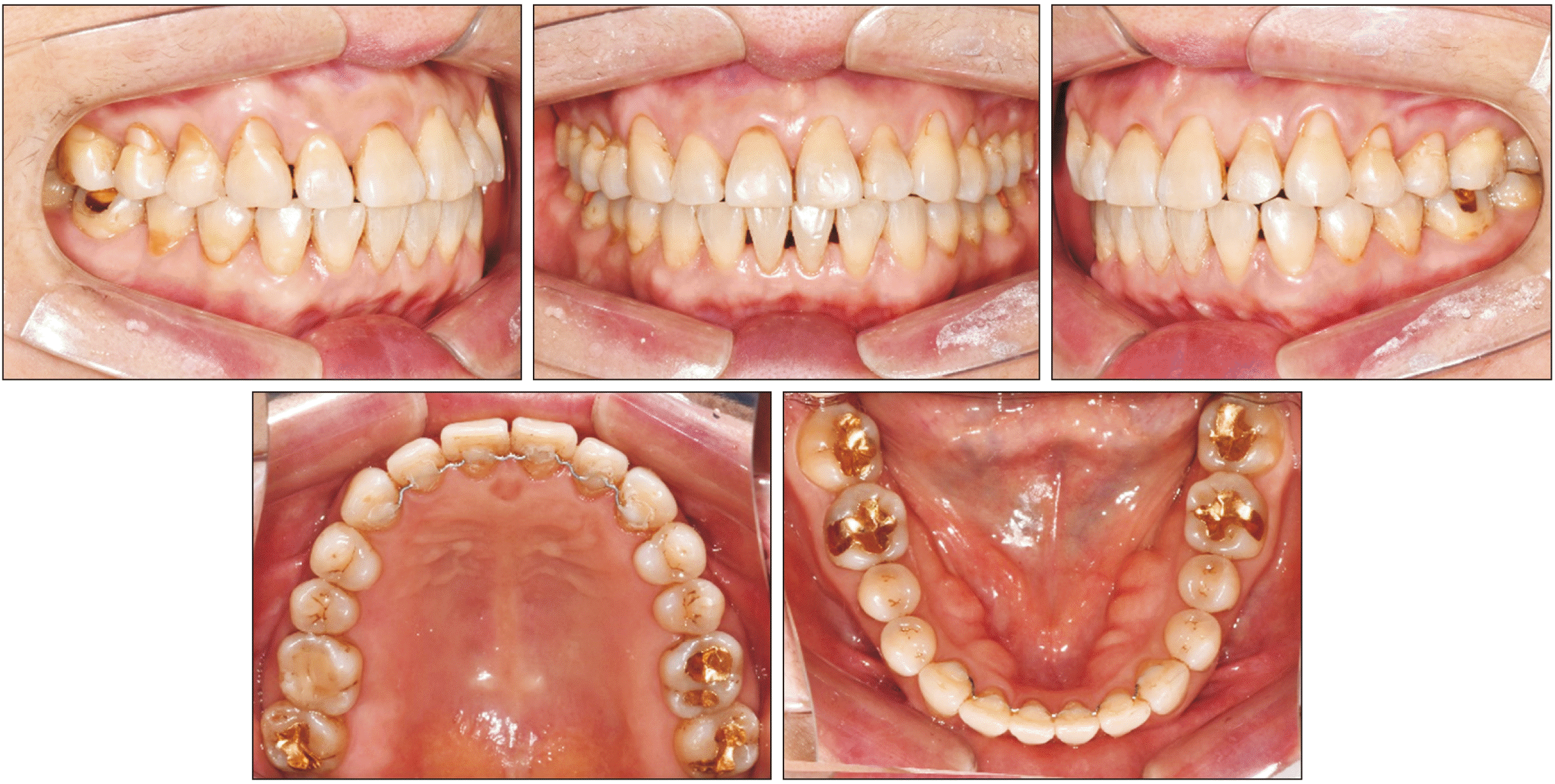
At the completion of treatment, crossbites were corrected, and acceptable overbite and overjet were established (Figure 5). A Class I molar relationship was maintained on both sides, and the maxillary and mandibular crowding were relieved without any considerable change in the profile (Figure 6). The transverse width, measured on CBCT images at the palatal root apex of the maxillary first molars, increased from 31.23 mm to 34.59 mm after MARPE (Figure 7). Additionally, the Yonsei Transverse Index was measured in order to demonstrate the skeletal change achieved by the expansion. It increased by 2.3 mm in the maxilla and remained unchanged in the mandible (Figure 7).
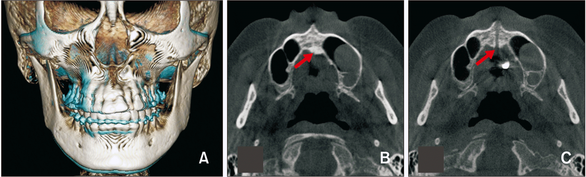
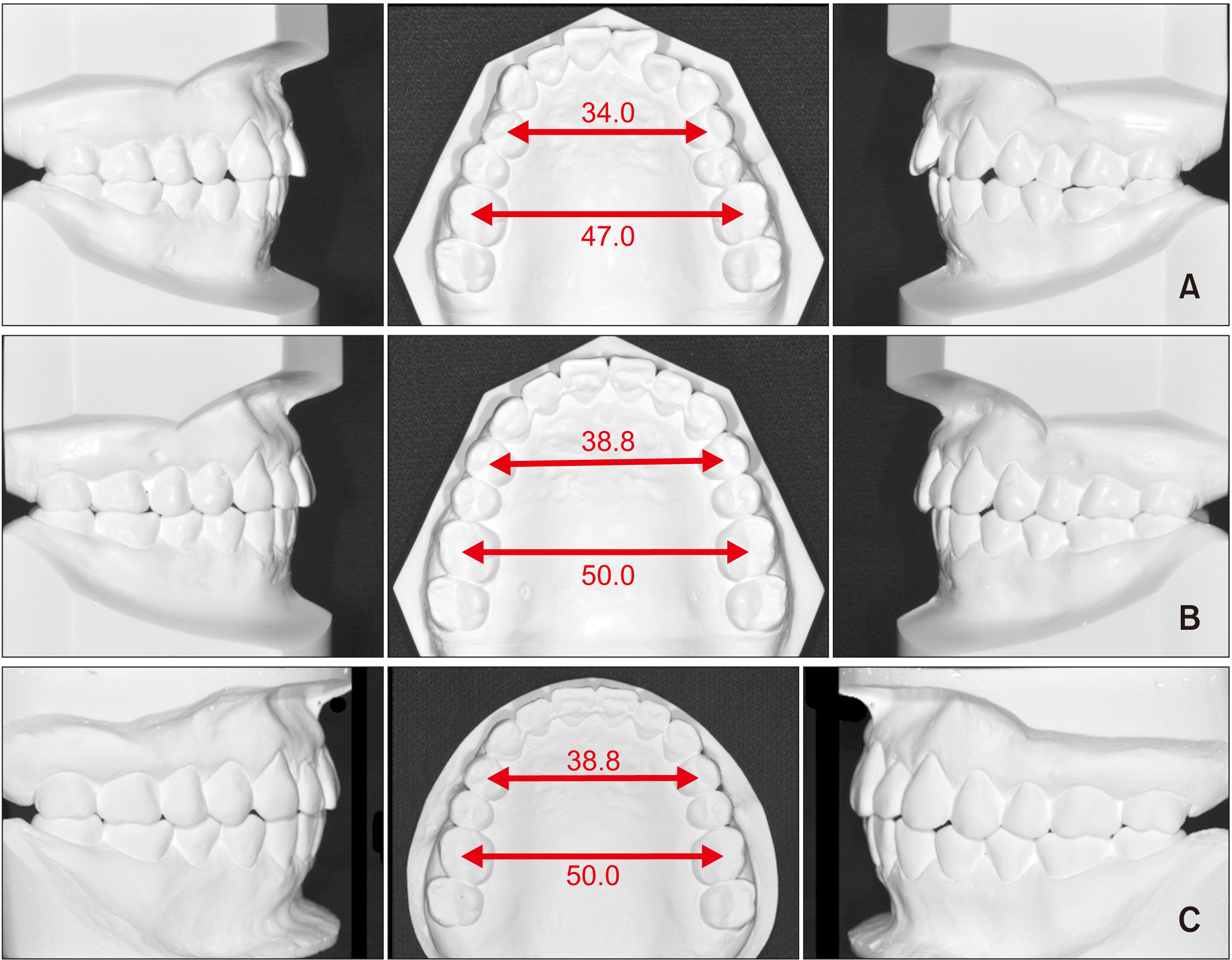
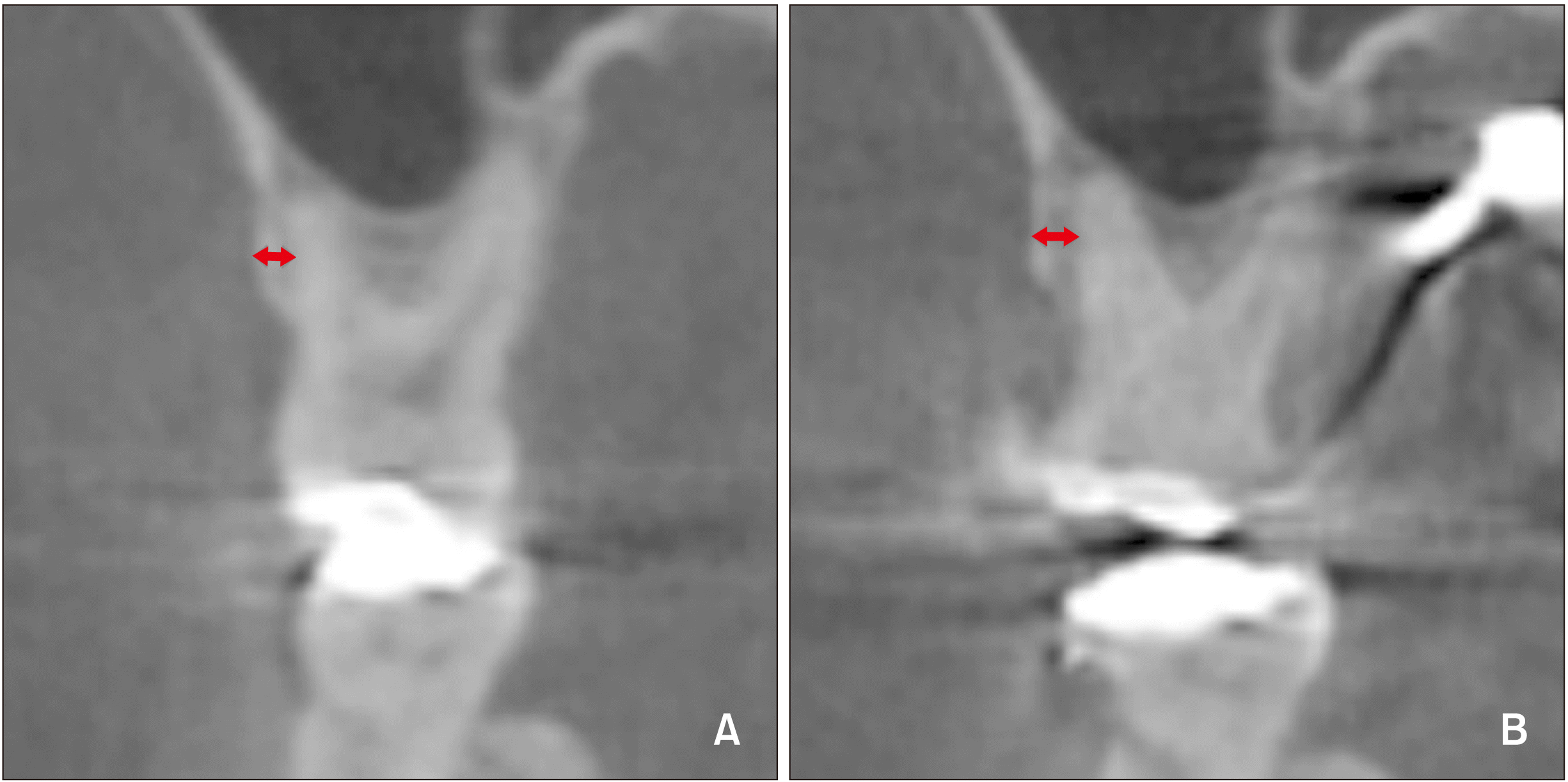
Moreover, the opening of the midpalatal suture could be confirmed on the axial palatal section of post-expansion CBCT (Figure 8). A comparison of the pre- and posttreatment dental casts indicated that the maxillary intermolar width increased by 3.0 mm, and the interpremolar width by 4.8 mm (Figure 9). Despite the adequate amount of expansion, there was no significant change in the clinical crown height of the anchor teeth and the teeth with pre-existing gingival recession (Table 2). Furthermore, comparison of the buccal bone plate at the level of the maxillary first molar on CBCT images before and after expansion did not show any considerable reduction in thickness (Figure 10). The smile esthetics improved, and the patient was satisfied with the outcome.
DISCUSSION
Adult patients with transverse discrepancies often present conditions unfavorable to periodontal health. Maxillary transverse deficiency is associated with anterior and posterior crossbites and crowding.22 Nonaxial loading of the teeth, as a consequence of dental compensation, may result in cervical abfraction.23 Such cervical defects can cause apical migration of the gingival margins. Considering these factors, the multiple cervical restorations and gingival recessions found in the present patient may be related to the underlying transverse problems.
The initial alveolar support and clinical gingival margins were not significantly detrimental. However, considering the age of the patient, a safe treatment modality was needed. In order to ensure periodontal health, transverse correction by orthopedic expansion was suggested, despite the uncertainty of successful suture opening.
The present case describes, to the best of our knowledge, nonsurgical palatal expansion in the oldest patient reported in the literature.24 The general consensus has been that orthopedic palatal expansion by nonsurgical means is feasible only until adolescence.10,25 The mechanical resistance resulting from increasing interdigitation of the facial sutures is commonly believed to limit orthopedic expansion in mature patients.8-11 On the other hand, the regulatory mechanism behind the fusion of facial sutures differs from that of calvarial sutures.12,26 According to a previous study, cranial sutures are obliterated in the absence of dura mater.27 The lack of such biochemical modulation by dura mater in the facial sutures may enable nonsurgical expansion even in elderly patients with interdigitated sutures, as long as they are not fused at the histologic level.
Unfortunately, no reliable guidelines have been proposed to predicting the success of skeletal expansion in nongrowing patients. According to Wehrbein and Yildizhan,28 besides the factors related to the midpalatal suture itself, the complexity and patency of pterygomaxillary junction and other circumaxillary sutures may affect the outcome. Because it is technically not possible to examine all affecting sutures, the only reliable method to confirm suture patency is an actual clinical trial of orthopedic expansion.
For these reasons, nonsurgical orthopedic expansion was attempted in the present patient. In order to reduce the adverse effects such as buccal tipping of the anchor teeth and thinning of the buccal bone plate, a MARPE appliance was used. Previous studies have shown that the incorporation of miniscrews in palatal expanders reduces dental side effects, but does not completely eliminate them.19 Moreover, in the present case, the second molars showed some dental tipping, which caused an intermediate occlusal interference and overall open bite. However, the open bite spontaneously subsided following the uprighting movement of the molars, and desirable transverse occlusion was obtained after treatment (Figure 5). Eventually there was no significant change in the overall facial vertical dimension (Figure 6).
Regarding the frequency of activation, a semi-rapid expansion protocol (0.2 mm/day) was used to allow enough time for remodeling of the interdigitated suture structure and to monitor the periodontal changes in the buccal plates of the anchor teeth. Considering the inherent complexity of the interdigitation along the suture and the uncertainty in orthopedic expansion due to the possible synostosis in adults,26 careful monitoring is crucial. Abrupt expansion may lead to unfavorable fracture or bony dehiscence, so a modest expansion rate was supposed to be reliable. Moreover, studies with experimental animal models have demonstrated that constant pressure to the suture may lead to compression osteogenesis, which causes changes in the suture structure.29 To allow this process to occur, it is presumably important to provide enough time for bone remodeling, rather than applying greater force by activating the screw with shorter intervals. Overall, orthopedic palatal expansion in aged individuals requires careful observation of the changes in the buccal plates, midpalatal suture, dental tipping, and general occlusion as well as the patient response. Considering these aspects, a semi-rapid protocol may be relatively safer in older patients with highly interdigitated facial sutures.
The use of MARPE in the present case yielded satisfactory outcomes with good maintenance of gingival health. The bone-bridge observed after the expansion is possibly due to the tight interdigitation in the incisal interproximal crestal area, which seemed to have caused some bone bending on the mesial side of the left central incisor. Nevertheless, the opening of the suture was relatively symmetric and constant along the midpalatal suture, indicating an overall orthopedic expansion of the maxilla. In relation to this orthopedic expansion, the intermolar width increased by 3.0 mm and the interpremolar width increased by 4.8 mm without any significant buccal tipping. The treatment change, although relatively small, approximated the average amount of expansion reported by Choi et al.6 and is considered meaningful since the movement pattern can be defined as bodily displacement of the posterior segment against the thin buccal plate. Since an increase of more than 4 mm in the interpremolar width is possibly associated with gingival recession,30 the use of MARPE in the present case can be justified.
The occlusion and transverse width remained stable, and gingival health was maintained during the follow-up period of six months (Figure 11). According to Reitan,31 the periodontal fibers under orthodontic force would remain stretched after treatment, which may lead to clinical changes in the periodontal tissue. Therefore, we presumed that observation of the clinical changes in the gingival tissue within a relatively short term would be crucial to detect some adverse effects of the expansion in the present case. A longer follow-up period is necessary to evaluate the long-term stability of the occlusion. However, long-term gingival changes may also reflect individual lifestyle factors such as brushing and mastication habits.32 Thus, proper orthopedic expansion in aged individuals may be an option for preservation of the periodontal structures on the buccal side.
CONCLUSION
Nonsurgical orthopedic expansion of the maxilla was achieved in a 60-year-old patient with the help of a MARPE appliance. This approach allowed the correction of crossbites and crowding without negatively affecting the pre-existing gingival recession. This case does not represent the general clinical observations. However, it is a good example demonstrating that advanced age is not an absolute contraindication for nonsurgical RPE. Even in elderly patients, orthopedic expansion can be a reasonable option and may eventually deliver satisfactory results, as shown in the present case.
 XML Download