

 PDF
PDF Citation
 Print
Print


INTRODUCTION
External apical root resorption (EARR) is one of the iatrogenic effects of fixed appliance treatment. Resorption activity during orthodontic tooth movement (OTM) is associated with a wide range of cytokines and molecular factors that are involved in clastic cell fusion and activation, including receptor activator of nuclear factor kappa-B ligand (RANKL)/RANK/osteoprotegerin (OPG) and interleukin (IL)-1-related pathways.1 The OPG:RANKL ratio in the crevicular fluid varies according to the magnitude of the compressive force. Root resorption is initiated by macrophage-like cells from the periodontal ligament blood supply, causing damage to the nearby cementoblast layer overlying the cementoid. This leads to cementum exposure, causing the denuded root surface to become more susceptible to resorption by scavenger cells and osteoclasts.1
Many factors contribute to EARR, and they can be categorized as patient- and treatment-related factors. Biological elements include genetic susceptibility (IL-Iβ gene), systemic disease, age, sex, previous resorption due to trauma, personal habits, and tooth structure.2 However, a recent meta-analysis suggested that the IL-1β polymorphism is not correlated with a predilection to EARR.3 Earlier literature showed that the incidence and severity of root resorption was connected with numerous mechanical factors, including force magnitude, the amount or distance of tooth movement, force type, and treatment appliance.4,5 Increased treatment time or prolonged treatment duration positively correlated with increased EARR4; thus, shortening the treatment duration is a worthwhile goal as it could decrease the risk of EARR.
Various surgical techniques ranging from total block osteotomies to flapless partial corticotomy have been reported to accelerate OTM with different degrees of invasiveness.6 Corticotomy involves superficial punctures or cuts on the cortical bone. It is considered invasive since it would necessitate the use of full-thickness mucoperiosteal flaps either buccally or lingually, followed by suturing.7 Al-Naoum et al.7 reported in their split-mouth study that alveolar corticotomy increases canine retraction by 2- to 4-fold on the experimental side than on the control side. However, this procedure compromises the dental periodontium and is associated with surgical risks such as an increase in postoperative pain, discomfort, and swelling.8 Due to its high morbidity, this technique has low acceptance among patients. Since then, corticotomy surgical procedures have evolved into flapless techniques.
Piezocision is a flapless corticotomy technique in which a microsurgical blade is used to make gingival microincisions, which are followed by piezoelectric incisions of the cortical alveolar bone made with an ultrasonic piezosurgical knife.9,10 Charavet et al.11 reported that treatment length was significantly shortened by 43% in the piezocision group compared to that in the conventional orthodontic treatment, without affecting gingival recession, bone dehiscence, and root resorption. Another part of their study reported that this technique was associated with a higher degree of apprehension and pain in the trial group.10 Even though piezocision is less invasive and has been reported to be effective in accelerating OTM,12 the procedure involves the potential risk of scarring.11,13
The use of micro-osteoperforations (MOPs) is the most current minimally invasive modality to accelerate OTM; MOPs have better acceptance among patients, and their application is more favorable than other invasive adjunctive methods in routine practice. However, a single application of MOPs does not accelerate OTM, considering the rate of canine or en masse retraction.14 The first study of the effect of MOPs on the rate of OTM in humans by Alikhani et al.15 did not investigate the associated root resorption due to the short duration of only one month. In their study, dental panoramic tomography (DPT) showed no indisputable evidence of root resorption or alveolar bone loss. However, this technique was considered to be not precise enough for measuring the magnitude of root resorption; thus, further studies are warranted. Subsequently, several randomized controlled trials (RCTs) have been conducted to investigate the effects of MOPs on EARR. Sivarajan et al.14 included eight RCTs in their systematic review, and only two RCTs reported EARR as the secondary outcome while evaluating the effects of MOPs on canine retraction. The authors concluded that MOPs do not have an effect on root resorption.
Thus, root resorption related to MOPs has not been studied extensively, with the reported studies primarily focusing on root resorption during space closure. As a result, additional studies investigating the association between MOPs and root resorption are warranted. This study aimed to investigate the effect of MOPs on EARR during the six-month initial orthodontic alignment phase of anterior maxillary crowding. The secondary outcome was to assess the frequency of EARR according to severity in relation to the MOP procedure.
Go to : 
MATERIALS AND METHODS
Ethics approval
Ethical approval for this study was obtained by the Universiti Teknologi MARA (UiTM) Research Ethics Committee on September 29, 2017 (Reference: 600-IRMI [5/1/6]; REC/297/17). This trial was also registered retrospectively at the International Standard Randomised Controlled Trial Number (ISRCTN) registry with the study identification (ID) ISRCTN15080404. Participation was voluntary, and withdrawal from the trial was allowed without jeopardizing the patient’s orthodontic treatment.
Trial design
This study was a prospective, randomized, single-center, two-arm parallel clinical trial to compare the effects of MOPs on EARR during the initial alignment stage of moderate maxillary crowding in comparison with conventional orthodontic treatment alone. The trial was conducted at the Orthodontic Clinic, Faculty of Dentistry, UiTM.
Participants
Inclusion criteria included age ranging from 18 to 45 years, moderate crowding of 5–8 mm in the maxillary arch, full dentition up to the second molars prior to extraction of the upper first premolars with or without anchorage reinforcement, healthy periodontal status with probing depth less than or equal to 3 mm, full-mouth plaque score of 20% or lower, and full-mouth bleeding score of 20% or lower. Exclusion criteria included previous orthodontic treatment, systemic diseases, smoking, pregnancy, and compromised periodontal oral health with evidence of bone loss and root resorption on DPT, as well as history of dental trauma. Patients with dental (e.g., hyperdontia, hypodontia) or craniofacial anomalies (e.g., cleft lip and palate) were also excluded. Patients receiving medications that could interfere with tooth movement, such as anti-inflammatory drugs, systemic corticosteroids, or calcium channel blockers were not taken into consideration. The period of recruitment was from October 2017 until February 2019.
Orthodontic treatment
Standardized conventional pre-adjusted edgewise McLaughlin, Bennett, and Trevisi brackets with a 0.022 × 0.028-inch (in) slot (Victory Series; 3M Unitek, Monrovia, CA, USA) were used for all participants. The archwire sequence was similar for both groups, which involved the use of 0.014-in nickel titanium (NiTi) archwires followed by 0.018-in NiTi archwires (TruFlex NiTi archwires; Ortho Technology, Lutz, FL, USA) until alignment was achieved.
Micro-osteoperforation procedure
A single operator (A.A.S.) performed MOPs under local anesthesia infiltration (Lignocaine with 1:100,000 adrenaline). The patients in the MOP group received two perforations 3 mm apart within the attached gingiva to 1 mm apical to the mucogingival junction, with the MOPs placed equidistant between the anterior teeth from the upper right canine to the upper left canine, except in the midline alveolar bone. The perforations were 1.5-mm wide and 3-mm deep and were made using a disposable MOP device (PROPEL Ortho Singapore, Kallang, Singapore). All participants underwent follow-up assessments at four-week intervals, and MOPs were repeated at every visit until completion of the alignment stage. Postoperatively, local measures with cotton roll pressure were applied to arrest bleeding after MOPs. Participants in the MOP group were prescribed 0.12% chlorhexidine digluconate (Oradex; CAVICO, Selangor, Malaysia) mouthwash to be used twice a day for one week. In addition, proper oral hygiene maintenance was reinforced, and the participants were advised to avoid modifying their flossing and brushing routines in the MOP region.
Measurement of root resorption
Long-cone intraoral periapical (IOPA) radiographs were used to assess the occurrence of EARR. All images were taken by a standardized paralleling technique using a single digital dental intraoral machine (Satelec X-Mind-AC/DC; Acteon, VA, Italy). IOPA radiographs involving six anterior maxillary teeth were taken before bracket placement, on initiation of MOPs (T1), and after six months into treatment (T2) (Figure 1). Crown length and root length were measured using a virtual ruler of the EasyDent V4 viewer software (Vatech, Hwaseong, Korea) by identifying these landmarks in all three radiographs at each stage (Figure 2). The crown length was gauged from the mid-incisal point of the crown to the median cementoenamel junction (CEJ) point, which was the midpoint between the mesial and distal CEJ. The root length was calculated from the median CEJ point to the tipping point of the root. Apical root resorption was measured as the difference between the length of the root at T1 and the length of the root at T2 in millimeters.
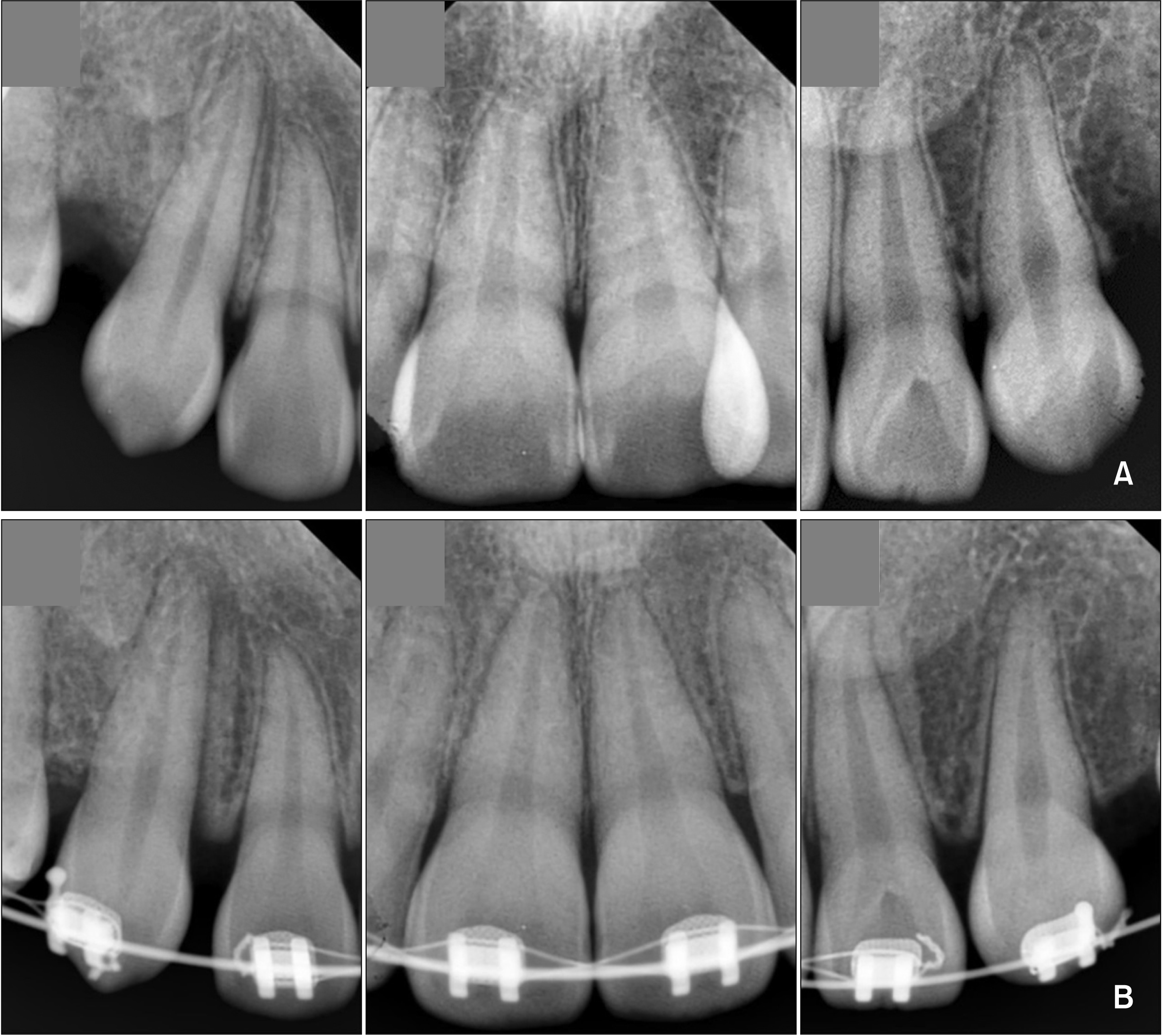 | Figure 1Intraoral periapical (IOPA) radiographs. A, Pre-IOPA taken before orthodontic treatment. B, Post-IOPA repeated after 6 months of treatment.
|
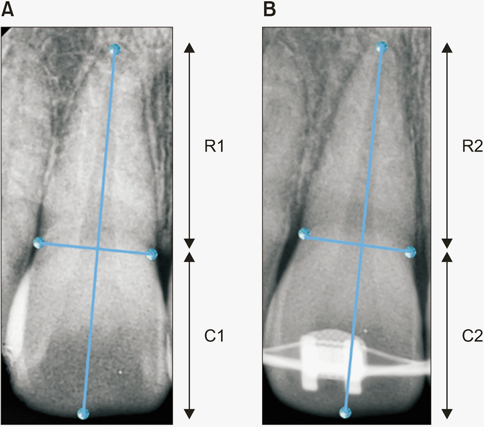 | Figure 2Measurements of total root and crown length at (A) T1: pre-treatment, (B) T2: post-treatment after 6 months were performed on intraoral periapical radiographs at the median point of the tooth axis.
R1, pre-treatment root length; R2, post-treatment root length; C1, pre-treatment crown length; C2, post-treatment crown length.
|
Image magnification between pre- and post-observational radiographs was rectified by exploiting the crown length as a reference, assuming crown lengths to be unaffected throughout the trial interval. Consequently, a correction factor (CF) was formulated by dividing the initial crown length (C1) by the post-treatment crown length (C2). The CF was computed to relate the pre-treatment (R1) and post-treatment (R2) root length of each tooth, as shown in the following equations. Then, the EARR per tooth was calculated in millimeters using the formula adapted from previous studies16,17:
The grading of apical root resorption was assessed separately by the principal investigator (A.A.S.) in the IOPA images by adapting the scoring system proposed by Levander and Malmgren.18
Sample size calculation
The sample size was calculated to detect a significant mean difference of 0.54 mm of root resorption with a standard deviation of 0.47 mm during the alignment stage of fixed appliance treatment after six months.19 The statistical power was set at 80%, with a confidence interval (CI) of 95%. A minimum of 12 participants for each group was required, and this number was increased to 15 per group considering a 20% dropout rate.
Randomization
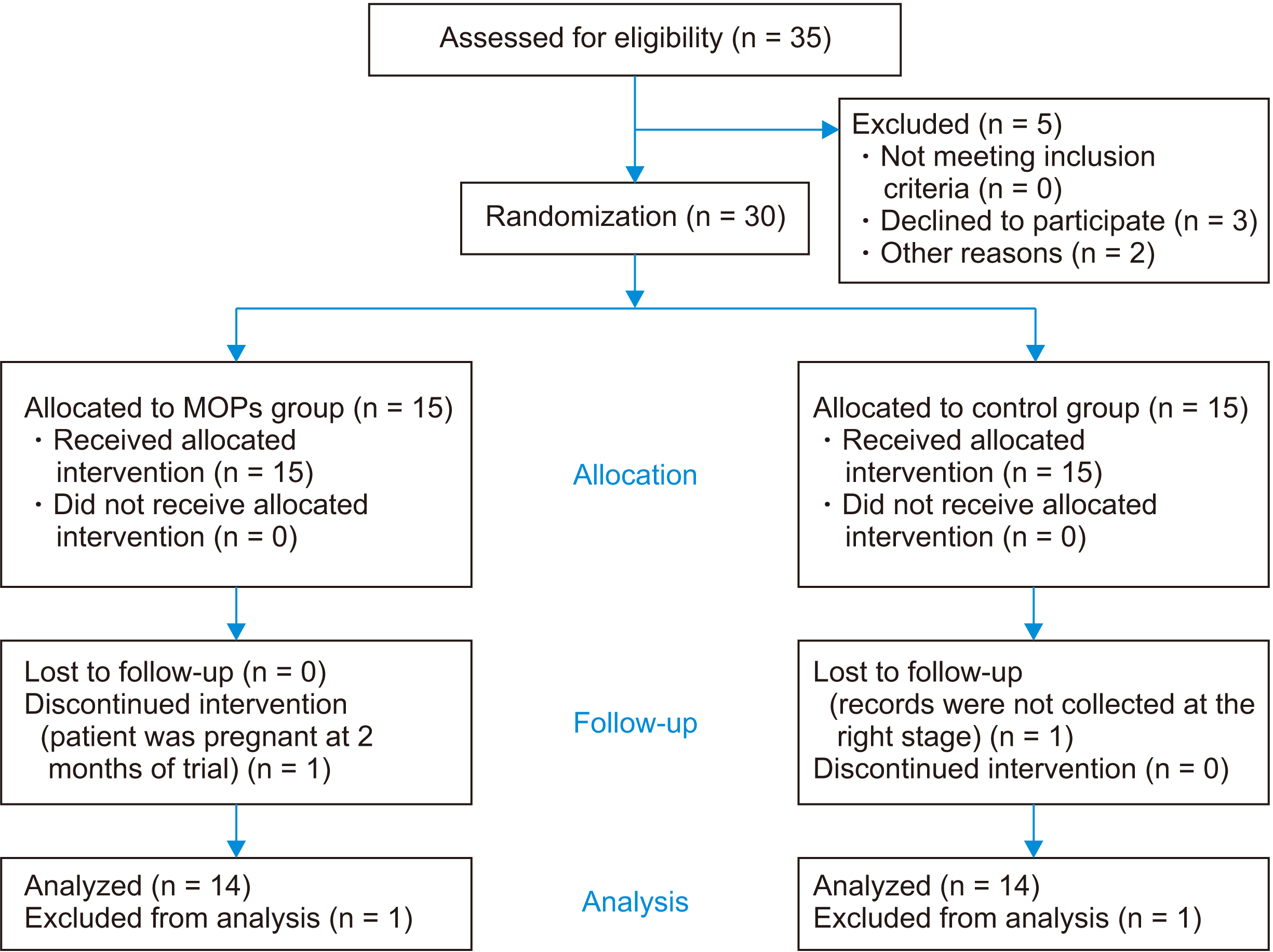
Once the participant agreed to the study protocol, written informed consent was obtained. Participants were allocated randomly to the MOP or control group in a 1:1 allocation ratio using block randomization of six numbers (three odd and three even numbers). Odd numbers were allocated to the MOP group while even numbers were assigned to the control group. Sequentially numbered opaque, sealed envelopes were used for concealed randomization. Cards were written with numbers and set in opaque sealed envelopes, which were kept by the central trial coordinator (N.H.N.), who was responsible for assigning the envelope numbers upon operator request (A.A.S.). The subjects’ participation protocol is shown in the Consolidated Standards of Reporting Trials flow diagram (Figure 3).
Blinding
Blinding of participants and the operator was not feasible due to the nature of the intervention. Thus, the outcome examiner was blinded to the subject’s assigned intervention. Patient information was concealed by the central trial coordinator (N.H.N.) prior to the measurement assessments.
Statistical analysis
Descriptive and analytical statistics were obtained using the SPSS software version 25.0 (IBM Corp., Armonk, NY, USA). The statistical significance level was prespecified at p < 0.05, with a 95% CI. Significant changes in EARR were analyzed at the patient level using repeated-measures analysis of variance to compare the mean differences in EARR among groups. Stata software version 13 (StataCorp, College Station, TX, USA) was used for kappa analysis.
Calibration for root resorption was performed on 15 IOPA radiographs to ensure the reliability of the measurement technique. The intra-rater reliability was excellent with an intra-class correlation coefficient of 0.82, with the 95% CI (0.65, 0.91), indicating a low level of random error. Further analysis with a paired t-test revealed no significant difference between the two readings (p = 0.242). Grading for the extent of EARR was also performed with an interval of two weeks on 15 IOPA radiographs, and the intra-rater reliability of Cohen’s kappa was 0.880 with 93.33% agreement (p < 0.001).
Go to :
RESULTS
Baseline characteristics
A total of 35 participants were eligible for this trial. However, five participants were excluded, including three patients who declined to participate and two who had moved away. The remaining 30 participants who met the inclusion criteria were enrolled for the trial. Subsequently, one pregnant participant dropped out from the MOP group, and one participant in the control group was lost with no follow-up records. An equal number of participants in both groups was maintained throughout the observation period. Thus, analysis as per protocol was undertaken to compare root resorption between the MOP and control groups. The mean age in the MOP and control groups was 22.80 ± 3.78 years and 22.50 ± 2.74 years, respectively, with no significant difference between the groups (p = 0.810) (Table 1).
Table 1
Demographic characteristics of the participants
| Variable | MOPs (n = 15) | Control (n = 15) | p-value | |
|---|---|---|---|---|
| Age (yr) | 22.80 ± 3.78 | 22.50 ± 2.74 | 0.810* | |
| Sex | Male | 2 (13.3) | 3 (20.0) | > 0.950† |
| Female | 13 (86.7) | 12 (80.0) | ||
| Incisor relationship | Class I | 6 (40.0) | 9 (60.0) | 0.564† |
| Class II/1 | 4 (26.7) | 3 (20.0) | ||
| Class II/2 | 2 (13.3) | 0 (0) | ||
| Class III | 3 (20.0) | 3 (20.0) |
![]()
Root resorption
A comparison of the changes in root length (Table 2) showed no significant difference between the MOP and control groups (p = 0.238 and p = 0.225, respectively). Further analysis between groups also did not show a significant difference (p = 0.419).
Table 2
Intergroup comparison of change in the root length (mm) between groups in six months duration
![]()
Approximately 50% of the teeth in both groups showed no root resorption in the first six months. Most of the roots in the MOP and control groups (42.86% and 52.38%, respectively) showed only mild resorption. Less than 8% of the roots in both groups (7.14% in the MOP group and 4.76% in the control group) showed moderate resorption (Table 3).
Go to :
DISCUSSION
Prevalence of external apical root resorption
Mild resorption is defined as some alteration of the root apex, while moderate resorption indicates less than 2 mm of root resorption.18 In an extensive study for developing an index for root resorption with an observation period of six to nine months after orthodontic treatment, Levander and Malmgren18 reported that 66% of the teeth showed zero or minor resorption, 33% showed moderate resorption, 17% showed resorption, and 1% showed extreme resorption. Similarly, in their study of 302 patients, Smale et al.20 assessed root resorption for 4 incisors over 6 months after initial orthodontic alignment and reported that 47.1% of the patients showed no root resorption, 29.9% and 19.4% showed mild and moderate resorption, respectively, and only 3.6% showed severe root resorption. Overall, almost half (47.62%) of the measured teeth showed mild root resorption and only 5.95% showed moderate root resorption. The remaining teeth showed no incidence of severe or extreme resorption.
Effects of micro-osteoperforations on external apical root resorption
The MOP and the control groups showed no significant differences in mean root resorption (p = 0.419), and the changes in root length over the six-month period within groups were not significant respectively (p = 0.238 MOP and p = 0.225 in control groups). The present study is the first to compare the EARR on the anterior maxillary teeth during initial alignment with that after MOP application. Our results are consistent with those reported by Alkebsi et al.21 for a three-month trial of MOP-assisted orthodontic canine retraction. They found that the difference in mean root resorption between the intervention and control groups, with EARRs of 0.61 mm and 0.73 mm, respectively, was not significant. Similarly, the split-mouth study by Aboalnaga et al.,22 also found no significant difference in EARR in the four-month canine retraction period. The latter study used adjusted cone-beam computed tomography (CBCT) for measurement of root resorption. In contrast, a microcomputed tomography (micro-CT) RCT on the extracted first maxillary premolars following 28 days of buccal tipping with a force of 150 g reported that MOPs significantly increased EARR by 0.17 mm.23 However, the results of that study should be considered with caution, since the clinical relevance of that technique may be debatable in general clinical practice.
The lesser extent of EARR exhibited by the MOP group is in agreement with the findings of a previous trial.9 This is due to regional acceleratory phenomenon activity induced by the disruption of the alveolar bone by MOPs, which causes intensification of osteoclastic activity and reduced bone density. Such a reaction subsequently reduces the possibility of hyalinization necrosis and resorption of the tooth root. Additionally, the increased cortical bone porosity simultaneously reduces mechanical resistance for OTM and EARR.9
Diagnostic tools
Most EARR diagnoses are based on radiographic evaluation, with apical blunting or shortening of the root length appearing as the first noticeable signs. Root resorption is measured by using IOPA radiographs with a CF adapted from previous studies to compensate for image magnification.17,24 One of the limitations of using IOPA is the tendency to underestimate apical root resorption. This is justified by a study performed by Dudic et al.25 that aimed to validate the accuracy of IOPA against a micro-CT scanner method in the diagnosis of EARR on extracted teeth. IOPA detected only 55% of EARRs in the experimental teeth whereas the micro-CT scans detected 86% of the EARRs on similar teeth. The most reliable and valid tool to assess root resorption is CBCT. It allows the roots to be visualized and evaluated on any surface and eradicates the image superimposition. It also ensures accuracy in the measurement and ensures better comparability. Nevertheless, CBCT exposes patients with a higher dose of radiation and is more expensive.26 For these reasons, CBCT endorsement in orthodontics is limited to specific indications.
The effectiveness of detecting EARR by using two-dimensional intraoral radiographs is arguable. Root resorption is a three-dimensional process that can occur at any of the root surfaces. Our EARR evaluation used two-dimensional long-cone periapical radiographs, which could limit the accuracy of root resorption calculation and its occurrence. Even though this approach shows limitations in evaluating all surfaces and tends to underestimate root resorption, the clinical relevance of the undetected EARR on plain radiographic images is debatable. The severity of such EARR is usually clinically insignificant. Moreover, both CBCT and IOPA have been shown to detect a comparable percentage of moderate and severe EARR. Thus, the protocol using IOPA radiographs in this study reflected contemporary orthodontic practice, yielding a meaningful trial outcome pertinent to routine clinical practice.
In general, mild to moderate EARR rarely shows clinical significance unless it is accompanied by severe or extreme resorption. However, root resorption of more than quarter of its length may adversely affect root longevity and yield an unfavorable crown-root ratio.27 Remarkably, Levander and Malmgren28 suggested that long-term tooth survival was not compromised even in cases with major EARR. Sondeijker et al.29 developed a comprehensive clinical practice guideline that recommended different management approaches for EARR before, during, and after orthodontic treatment. Even though the guidelines were established principally from a universal agreement between clinicians with limited existing evidence, they permitted clinicians to act in response to EARR based on present expertise. The guidelines include a strong recommendation to inform patients about the risk of EARR prior to orthodontic treatment, with greater caution to be exercised in extraction cases, which are associated with a greater possibility of severe EARR. They also strongly recommend reassessment and revision of the treatment aims and plan when EARR equal to or more than 2 mm is apparent during fixed appliance treatment.29
Adverse effects
No root perforation or soft tissue scarring were identified throughout the trial period in all participants. No accidental adverse effects were reported.
Limitations
Root resorption is a three-dimensional process that can occur at any of the root surfaces. Our EARR evaluations were performed using two-dimensional IOPA, which could limit the accurate calculation of resorption, since its occurrence is three-dimensional. Future studies should investigate the effect of MOPs throughout the treatment duration to assess its complete effectiveness.
The clinical effectiveness of MOPs on the rate of tooth movement over a broader range of orthodontic treatments remains to be investigated. The alignment duration and pain experienced by the patients with the additional MOPs are described in another part of the study.
Generalizability
The generalizability of this study may be limited by the single-center nature of the clinical trial. However, these findings are representative of general clinical practice, where standard mechanotherapy is performed in conventional orthodontic treatment for adults.
Go to :
 XML Download