Introduction
Reflux nephropathy (RN) is renal scarring that is presumably induced by vesicoureteral reflux (VUR) of infected urine into the renal parenchyma [1]. The diagnosis is suspected in children with urinary tract infection (UTI) or findings of hydronephrosis on prenatal ultrasound. Extensive renal scarring impairs renal function and can result in renin-mediated hypertension, chronic kidney disease (CKD) or end-stage renal disease.
Asymptomatic bacteriuria (ASB) is uncommon in infant and boys and occurs in about 1% to 3% of healthy girls. However, it is common in children who have urologic abnormalities associated with VUR. Although there may be an increased risk for symptomatic UTI in children with bacteriuria, there had been no evidence of an increased risk for subsequent renal scarring or renal insufficiency [2]. In addition, recent evidences have shown no benefit and often harm with the use of antibiotics to treat pediatric ASB [3].
We report a case of 10-year-old boy with high grade VUR and CKD who was referred to the pediatric department for incidentally detected ASB.
Go to : 
Case
A 10-year-old boy was referred to our pediatric department because of pyuria and ASB. He had no relevant past medical or surgical history. His height was 137.1 cm (25–50 percentile), body weight was 37.2 kg (50–75 percentile) and blood pressure was 110/60 mmHg (50–90 percentile). Vital signs were stable and physical examinations revealed no abnormal findings. Urinalysis showed specific gravity of 1.009, pH 6.0, pyuria (3+), nitrite (1+), proteinuria (trace), and hematuria (trace), RBC 0–2/HPF, WBC many/HPF. Urine culture collected by voided urine showed Klebsiella pneumonia >100,000 (CFU/mL).
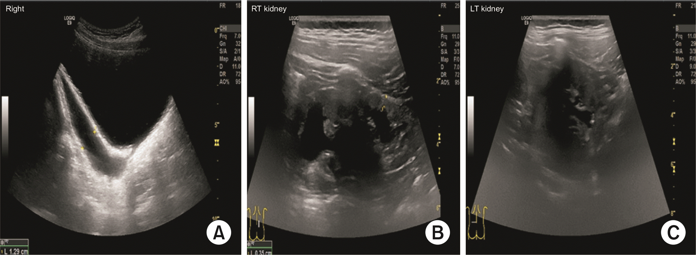
Abdominal ultrasonography revealed both hydroureteronephrosis with parenchymal scarring and asymmetric hypoplasia of right kidney (Fig. 1). He was presumably diagnosed with RN and was hospitalized for further evaluation.
Laboratory findings were as follows: hemoglobin 12.1 g/dL, WBC count 4,500/μL (segmental neutrophil 38.3%, lymphocyte 52.8%), platelet count 248,000/μL, albumin 3.8 g/dL, total protein 6.7 g/dL, BUN 16 mg/dL, creatinine 1.10 mg/dL, sodium 143 mmol/L, potassium 4.3 mmol/L, chloride 108 mmol/L, calcium 9.2 mg/dL, phosphorus 4.3 mg/dL, and CRP 4.45 mg/dL.
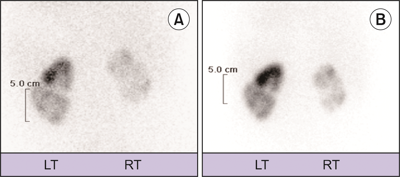
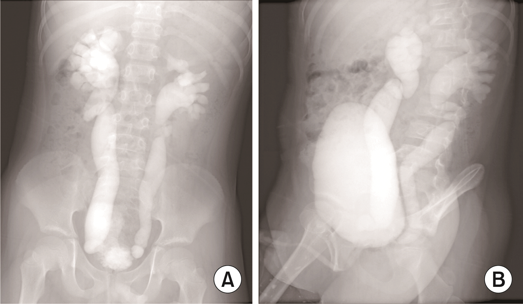
Urologic evaluation was completed with a technetium-99m-labeled dimercaptosuccinic acid (99mTC-DMSA) scan and voiding cystourethrography. 99mTC-DMSArenal scan revealed multiple cortical defects in both kidneys. The differential kidney function was 25% for right and 75% for the left kidney (Fig. 2A). Voiding cystourethrography revealed grade V VUR on both kidneys and distended bladder (Fig. 3). He had irregular voiding habit along with withholding maneuver and constipation. Based on the result of the laboratory tests, imaging study, and voiding history, we diagnosed as severe RN of both kidneys and CKD stage II with bladder and bowel dysfunction. We consulted to urology department and ureteroneocystostomy was performed on the patient on the second day of hospitalization. Postoperative cystography revealed complete resolution of VUR.
 | Fig. 2Technetium-99m-labeled dimercaptosuccinic acid (99mTC-DMSA) scan. (A) Initial 99mTC-DMSA scan showed multiple cortical defects in both kidneys. The differential kidney function was 25% for right (RT) and 75% for the left (LT) kidney. (B) After 6 months, 99mTC-DMSA scan showed small sized right kidney with decreased uptake and multiple cortical defects in both kidneys. The differential kidney function was 21% for right and 79% for the left kidney. |
After 6 months, follow-up 99mTC-DMSA scan revealed small sized right kidney with decreased uptake and multiple cortical defects in both kidneys. The differential kidney function was 21% for right and 79% for the left kidney (Fig. 2B). During the outpatient follow-up for 3 years, his height was 163.7 cm (50–75 percentile), body weight was 60 kg (75–90 percentile) and blood pressure was 126/77 mmHg (90–95 percentile). Laboratory profile showed as following: BUN 21 mg/dL, creatinine 1.23 mg/dL, uric acid 7.1 mg/dL, albumin 4.7 g/dL, total protein 7.1 g/dL, serum total calcium 10.1 mg/dL, phosphorus 5.0 mg/dL, cystatin-C 1.64 mg/L, and parathyroid hormon 43.8 pg/mL. Urinalysis showed no proteinuria, hematuria, or pyuria. Additional laboratory findings were added for the next three years (Table 1).
Table 1
Laboratory data on admission and follow-up
He was prescribed osmotic laxative treatment for 6 months after ureteroneocystostomy. He had been on regular intake of angiotensin receptor blocker and phosphate binder (sevelamer).
Go to : 
Discussion
VUR, a congenital anomaly characterized by either a unilateral or bilateral reflux of urine from the bladder to the kidneys, is common in young children. About 30% of children under 5 years of age with VUR were identified by routine voiding cystourethrogram after UTI, and 9% to 20% of prenatal hydronephrosis with VUR are detected postnatally [4]. For most, VUR resolves spontaneously. About 20% to 30% will have further infections, and only a few will experience long-term renal sequelae.
It is well known that severity of renal scarring is associated with the severity of VUR. Chen et al. [5] identified that older age of VUR diagnosis (≥5 years), higher grade of VUR, and higher number of UTI were risk factors for renal scarring. Similarly, Swerkersson et al. [6] showed direct relationship among male sex, high-grade VUR and renal dysplasia. Thus, efforts to prevent renal scarring should be directed towards a rapid diagnosis and treatment of VUR.
Few reports have focused on prevalence and risk factors for deteriorating renal function associated with VUR. According to data from North American Pediatric Renal Trials and Collaborative Studies, there is an estimative of 3.5% to 5.2% of the children in renal replacement therapy because of VUR nephropathy [7].
In addition to the prevalence of CKD among VUR patients, findings regarding the predictive risk factors for the development and progression of CKD in children with VUR have been conflicting and it is debated whether VUR is a benign or nonbenign condition [8,9]. Silva et al. [10] determined that grade V VUR, bilateral renal damage, and a delay in the diagnosis of VUR of 12 months after UTI were independent predictors of CKD. Also, a report by Novak et al. [7] suggested that older age, higher CKD stage, and history of UTI are significant risk factors for CKD progression in children with VUR. Sjostrom et al. [11] mentioned that there was a tendency of renal function deterioration in inverse proportion to increasing degree of VUR and bladder dysfunction. Contrary to previous studies, Chen et al. [5] found that a younger age of VUR diagnosis, renal scarring, and history of acute pyelonephritis were risk factors for deteriorating renal function. Meanwhile, there is another opinion that VUR and CKD is not related. Ishikura et al. [12] reported that the main cause of kidney dysfunction and its progression in VUR children might be the presence of hypoplastic/dysplastic kidney, not VUR.
These discrepancies in clinical studies may be explained by differences in patient population, study design, data analyses and also in difference of medical insurance. Therefore, understanding of the many possible risk factors for renal scarring and deteriorating renal function can be useful in ethiological/pathogenic insights and therapeutic approach for VUR.
Interestingly, our patient was presented as ASB without previous febrile UTI at age of 10. He was diagnosed with high grade VUR, CKD and bladder and bowel dysfunction. This rare case can enlighten our insight of VUR and CKD correlation should not be overlooked until older ages.
Finally, it is likely to be reasonable to investigate a thorough radiological imaging studies in male patient with ASB to discriminate urogenital tract abnormality such as VUR. Based on this case report, older age of male children with ASB, bilateral high-grade VUR, at presentation should monitor to ensure that the condition will not progress to CKD.
Go to : 


 PDF
PDF Citation
 Print
Print


 XML Download