

 PDF
PDF Citation
 Print
Print


I. Introduction
Salivary glands sialolithiasis is a common oral disease that occurs more commonly than minor salivary gland sialolithiasis (MSGS). However, MSGS has been found to have a higher incidence than originally believed1-3.
Many MSGS cases, being asymptomatic or small in size, may be missed during an intraoral examination1. In addition, some cases may be misdiagnosed as a common mucocele or a fibroma due to irritation or as a diapneusia3. Consequently, the patient may not undergo a histopathological examination, which is needed to establish the MSGS diagnosis.
In this paper, a case of MSGS located in the upper lip mucosa, primarily diagnosed as a mucocele, is reported.
Go to : 
II. Case Report
A 65-year-old male visited our private clinic for evaluation of a submucosal nodule on the left side of the upper lip.
The patient had undergone surgical excision of a similar mass in the same location two months prior and reported recurrence after a few weeks.
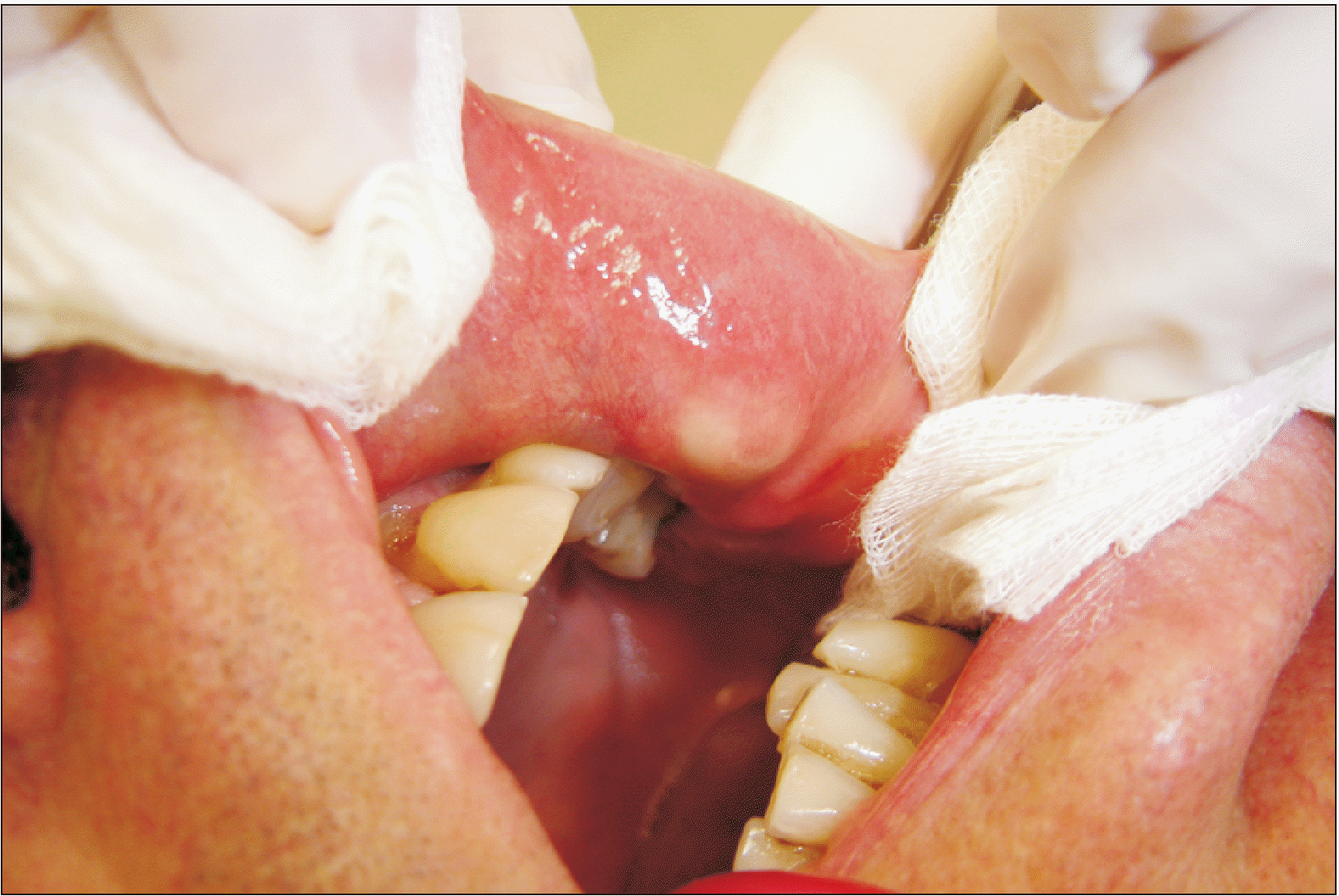
Intraoral clinical examination revealed a movable asymptomatic submucosal nodule, about 2 cm in diameter, on the left side of the upper lip, fluctuant in palpation. A whiteish color was visible through the mucosa, under pressure.(Fig. 1) The patient was edentulous, having undergone no dental procedure in the recent past and no injury due to mechanical irritation of the upper labial mucosa. His medical history was unremarkable. An initial diagnosis of possible mucocele was based on clinical examination and lesion appearance.
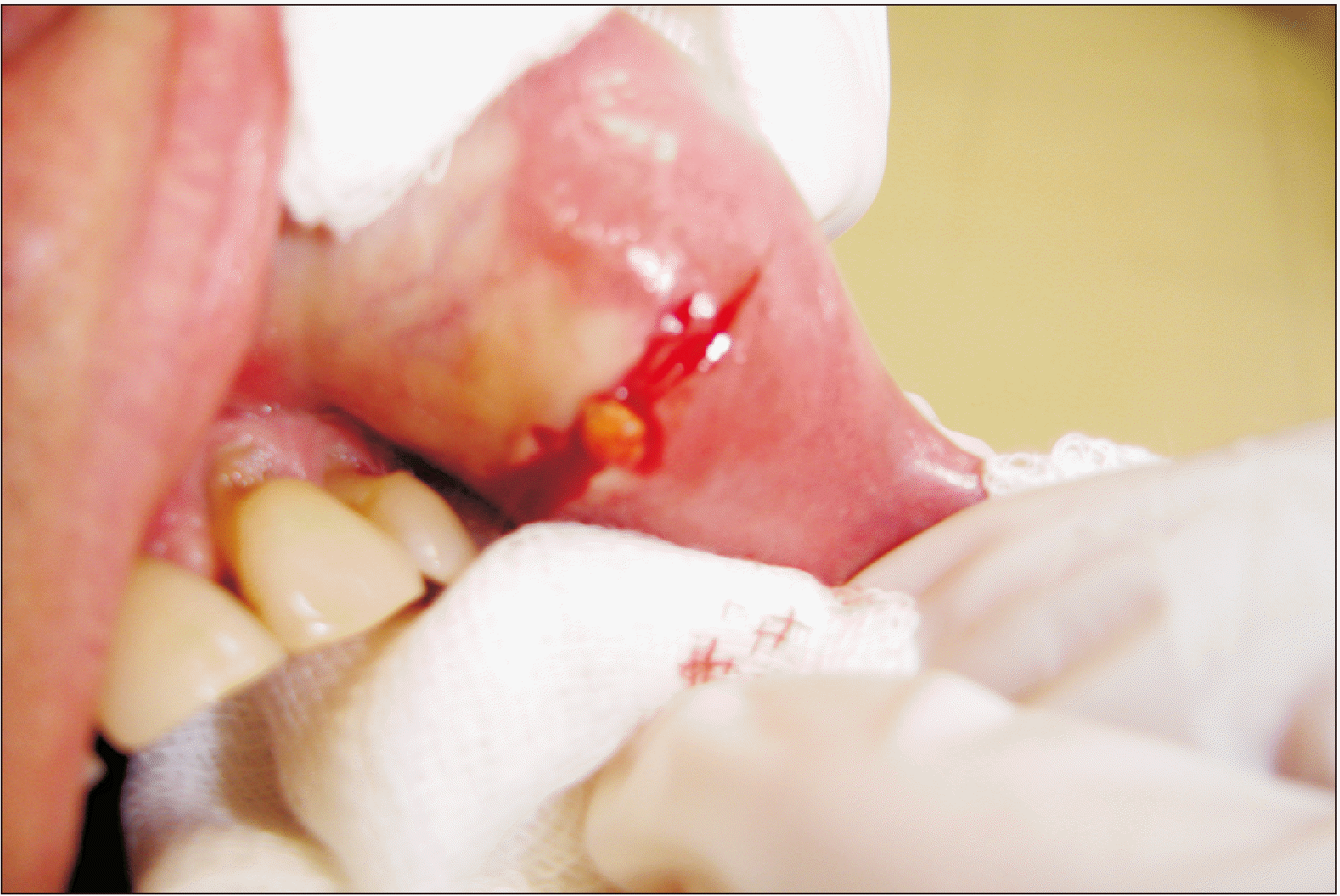
An excision was made under local anesthesia to reveal the lesion. A spherical, whitish sialolith was avulsed.(Fig. 2) The cyst-like lesion was removed in total, along with the adjacent minor salivary glands. Closure was accomplished with black silk sutures that were removed after one week. Healing was uneventful, and there has been no recurrence over two years.
Histological examination revealed chronic sialadenitis of minor salivary glands, with severe dilatation of the ducts, lobular atrophy, and severe lymphocytic infiltration in the stroma with lymph follicle formation.(Fig. 3, 4)
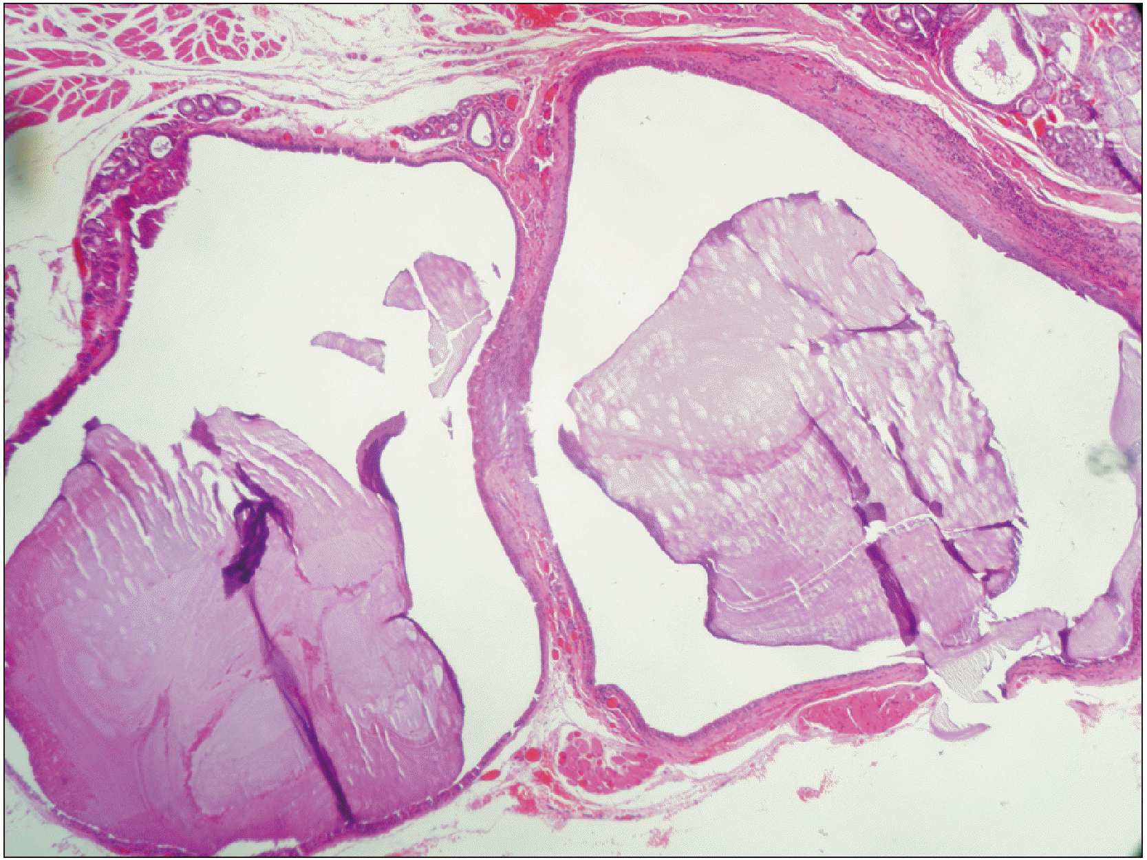
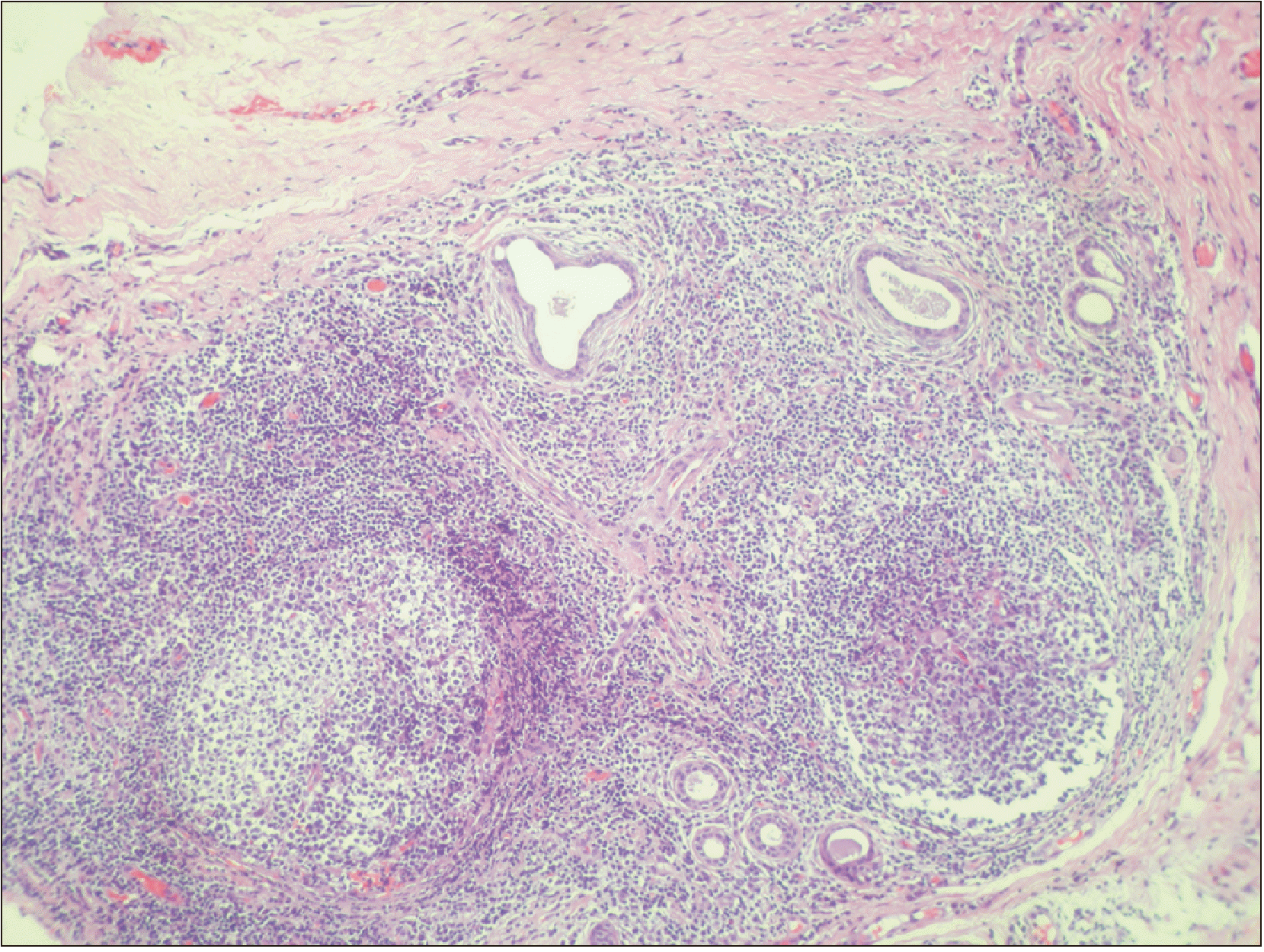
The sialolith existence with histopathological findings established the MSGS diagnosis.
Go to :
III. Discussion
A case of MSGS, initially diagnosed as an upper lip mucocele, is described. In the medical literature, the first MSGS case was reported by Papin (1864-1865)2,3. However, the first histopathologically established MSGS case was published much later by Lighterman in 19552,3. Since then, some solitary MSGS cases are described as a small, hard, firm or movable asymptomatic submucosal nodule4. Holst published the first extensive report of nine cases, including the first reported tongue–located case, as well as the first with nodules in multiple locations in the same patient (two locations in the upper lip).
These data suggest that MSGS is not the uncommon entity it was believed to be1. However, misdiagnosis is rather common, as the clinical appearance of this particular disease is asymptomatic. Additionally, a sialolith may not be present during surgical excision. So, it is essential that the pathologist is informed about the specific oral mucosal lesion so that he will not overlook the existence of a sialolith or particles of it1. The existence of a micro sialolith in the acinar parenchyma or in the acinar duct may lead to development of a retention mycocyst.
Pullon and Miller2 performed the first MSGS review along with report of 10 new cases, for a total of 55 cases in the medical literature. The majority of these lesions was located in the buccal mucosa (24 cases), followed by the upper lip (22 cases). The male:female ratio was 2:1.
Anneroth and Hansen5 describe 49 new cases with similar findings. Among them, the upper lip is the most common location. The male:female ratio is similar to that reported by previous authors, with a slight predilection for males.
The largest review of 239 MSGS cases by Ben Lagha et al.3 confirms that (1) the most common lesion location is the upper lip (49.2%), followed by the buccal mucosa (37.3%); (2) MSGS affects both males and females (58.3% and 41.7%), with a slight predilection for males; and (3) the mean age of affected patients is 50 years (range, 9-90 years).
Development of sialoliths is a multifactoral event. Sialoliths are composed of a central core of organic material, covered by a layered calcified cortex. That calcified cortex consists of complicated salts of calcium, phosphate, and especially hydroxyapatite8. Salivary dysfunction or disturbances of secretion may contribute to creation of sialoliths. Sialolith formation occurs through gradual calcification of an organic matrix of glycoproteins or of a foreign body. Lee and Wong8 reported an extensive study of the pathogenesis of sialolithiasis in minor salivary glands.
Concerning establishment of the diagnosis, radiographs are not essential as many of the calculi (sialolith) can be radiotransparent3. In addition, an avulsion of calculus (calculi) is possible.
Diagnostic ultrasound may help in establishing the diagnosis, as it is used to indicate major salivary gland sialoliths9. This tool may also contribute to removal of calculus or such particles with use of an ultrasound-guided needle9.
Nevertheless, ultrasound use seems to be limited in MSGS diagnosis since there is only one related paper in the literature.
Differential diagnosis includes mucocele6,10, swellings due to local irritation like fibroma and diapneusia, chronic abscess of the oral mucosa, and neoplasms either benign (lymphangioma, pleiomorphic adenoma) or malignant11,12.
Final diagnosis is clinically established in cases with sialolith avulsion noted during the surgical procedure, as in our case, but the diagnosis can be confirmed only histopathologically. Among the histological findings is the presence of a sialolith in the minor salivary gland duct or fragments of sialoliths in different stages of calcification13,14.
Stasis of saliva, which accompanies MSGS, usually results in minor salivary gland inflammation with a chronic sialadenitis appearance, as in our case. The salivary duct is dilated and surrounded by lymphocytes, plasma cells, and eosinophils8,11,14. Squamous metaplasia and pseudostratified columnar metaplasia in duct epithelium are additional characteristic histological features13.
MSGS tends to be a painless lesion but can become painful when the salivary gland parenchyma or excretory duct becomes infected, with or without pus. In that case, antibiotic medications are recommended. An antibiotic with high diffusion in saliva, such as macrolide, is preferable3.
Surgical excision is the treatment of choice. Lithotripsy, the treatment of choice in sialolithiasis of major salivary glands15, is not applicable in MSGS. To prevent recurrence, the lesion ought to be removed surgically, along with all affected minor salivary glands.
In conclusion, MSGS is not an uncommon oral clinical entity, can mimic a mucocele, and must always be included in the differential diagnosis of swelling with or without fluctuation, especially in the upper lip. Surgical excision with affected minor salivary glands is the treatment of choice, and histological examination is needed to establish the clinical diagnosis of MSGS.
Go to :
 XML Download