

 PDF
PDF Citation
 Print
Print


I. Introduction
Nasal septal abscess (NSA) with a dental origin is uncommon1. In 1920, McKenzie2 reported a case of NSA originating from an alveolar abscess of the right upper lateral incisor. Since then, six articles have been published in the English literature1-6.
NSA is defined as a collection of pus located between the nasal bony or cartilaginous septum and the mucoperichondrium or mucoperiosteum6,7. Although NSA is uncommon, immediate therapy is indicated to avoid complications such as facial deformity or intracranial infection8. Treatments include antibiotic coverage and surgical drainage including diagnostic needle aspiration8. In 1945, Beck9 classified the etiology of NSAs into three categories: nasal trauma, sinonasal or dental infection, and spontaneous occurrence without an underlying cause. Among these, the most common cause of NSA is an infection of the nasal septal hematoma after accidental or surgical nasal trauma8-11. Herein, we report a case of uncommon presentation of NSA with a dental origin in a patient with a history of left upper central incisor extraction 4 days prior.
Go to : 
II. Case Report
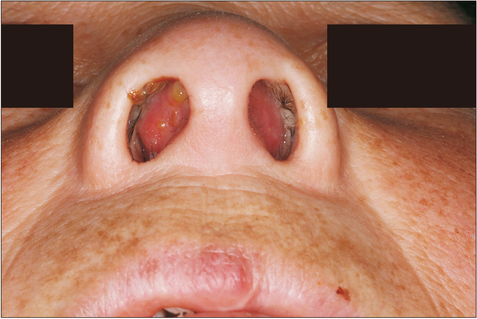
A 64-year-old female patient with no immunodeficiency or history of trauma visited Jeonbuk National University with the chief complaint of nasal cavity swelling. The patient had no other underlying disease except controlled diabetes mellitus for 13 years with medication. On the first visit, laboratory testing for diabetes indicated the following: HbA1c, 6.5% and serum glucose, 158 mg/dL. Nasal obstruction had begun 4 days previous, when her left upper central incisor was extracted. On physical examination, reddish bulging around the nasal septum was observed. The patient reported discomfort with nasal breathing due to the obstruction.(Fig. 1) There was no pus discharge around the extracted tooth. On needle aspiration of the nasal septal area, a relatively transparent brownish fluid was observed.
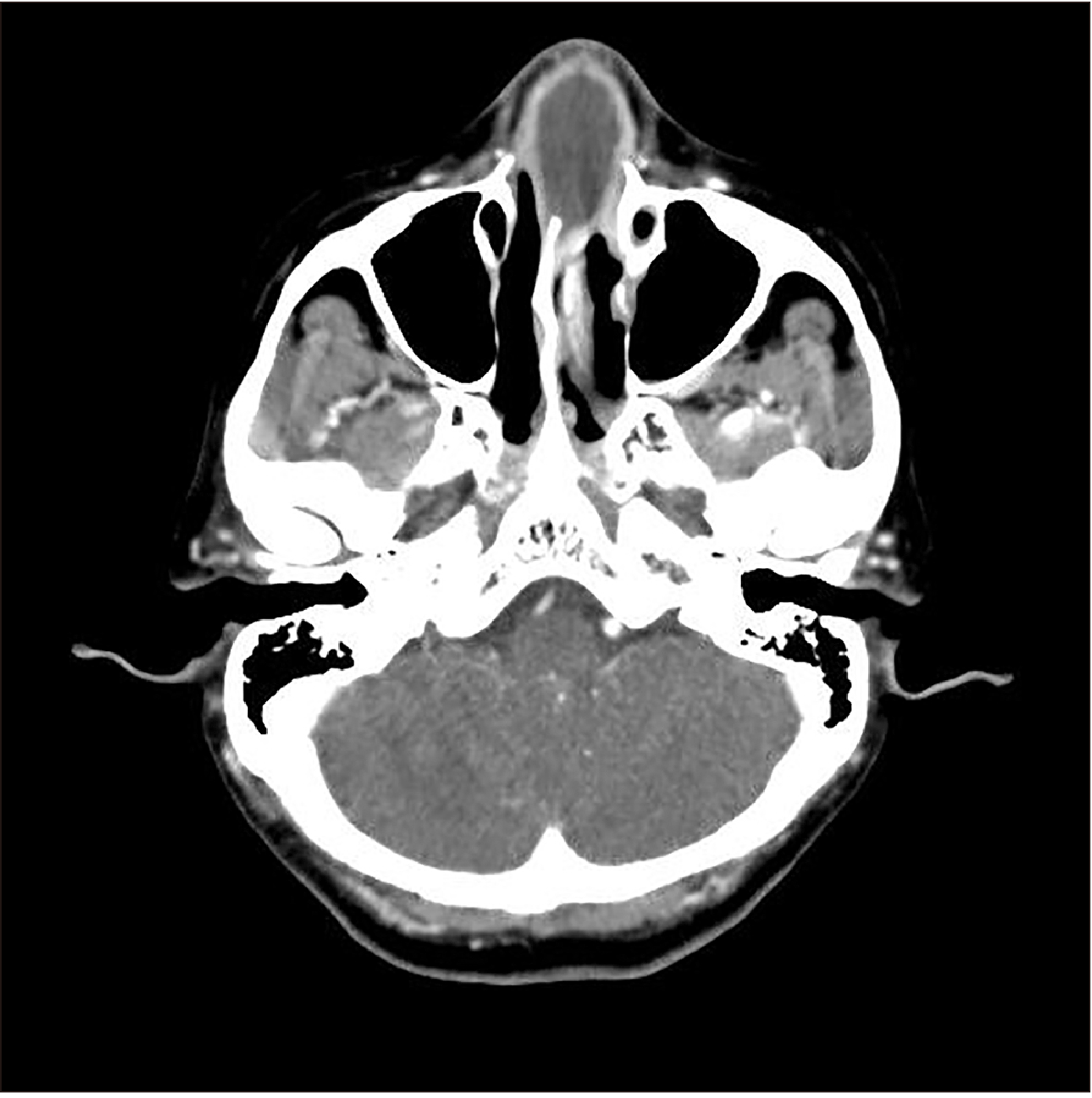
As a result of facial computed tomography (CT) with enhancement (Fig. 2), she was diagnosed with NSA. Triple antibiotic therapy with piperacillin sodium/sulbactam sodium, metronidazole, and amikacin was immediately initiated after admission. Medication dosage and method used were as follows: combicin 3 g (Samsung, Hwaseong, Korea; two vials, three times a day, intravenously), metronidazole 0.5 g/100 mL/Bag (CJ HealthCare, Seoul, Korea; one vial, three times a day, intravenously), amikacin 250 mg/2 mL/vial (Shinpoong, Ansan, Korea; one vial, three times a day, intravenously) The patient’s body temperature was 36.8°C, white blood cell (WBC) count was 18,150 leukocytes/µL, high sensitivity C-reactive protein (hsCRP) level was 74 mg/L, and erythrocyte sedimentation rate (ESR) was 104 mm/hr. Over 2 days, nasal septal swelling persisted, and daily aspiration and pus culture were performed.
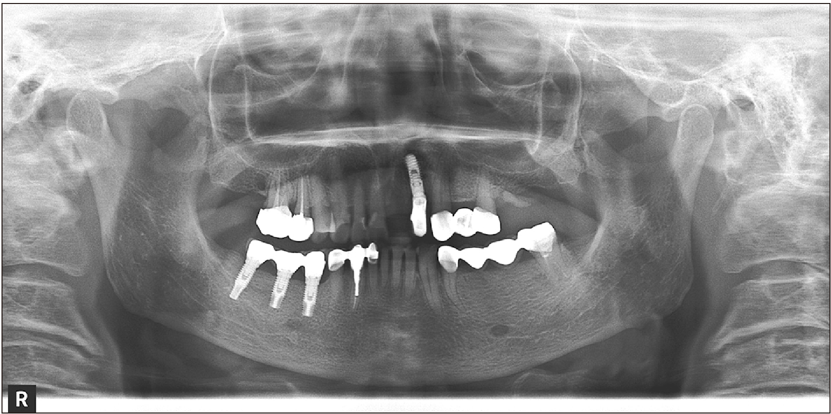
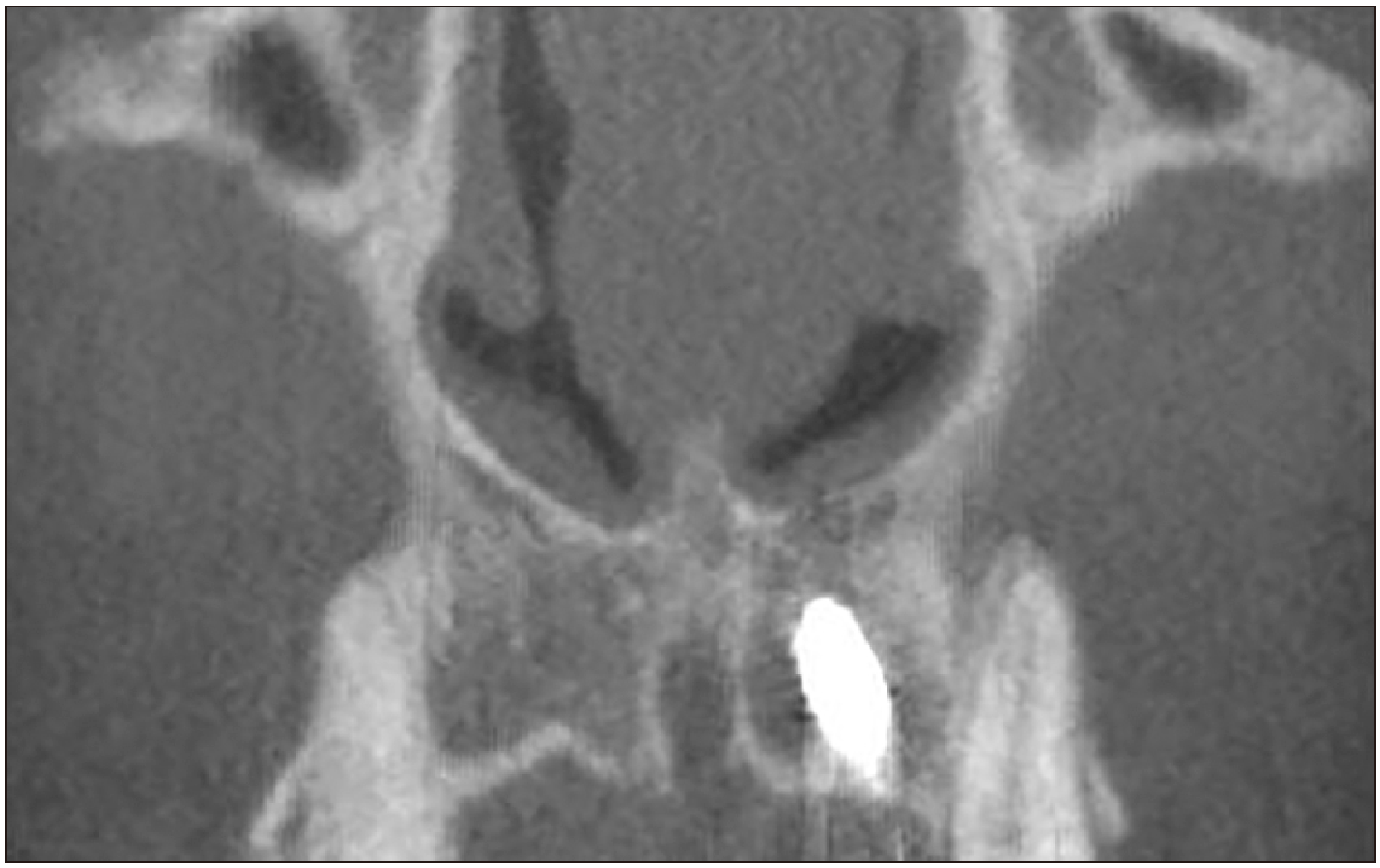
Panoramic view (Fig. 3) and additional cone-beam CT (CBCT) (Fig. 4) were performed. Using panoramic view, an extracted left upper central incisor was confirmed. CBCT revealed chronic inflammation and localized osteomyelitis of the left upper lateral implantation apex area, as well as partial bone destruction of the nasal floor. When the patient visited our department, the left upper central incisor had been extracted. On the 3rd day of admission, intra-oral incision and drainage were performed and removed moderate yellowish pus. Klebsiella pneumoniae was detected in the pus culture. Despite antibiotic therapy and surgical drainage, laboratory test results were not remarkably improved, and the patient’s nasal breathing remained obstructed. On the 10th day of admission, incision and drainage through the nasal septal mucosa were performed. Biopsy results revealed chondroid tissue with hemorrhagic necrosis (necrotizing inflammation). Body temperature remained within the normal range, except on the 2nd day of admission when it reached 37.8°C. The patient was admitted to our department for 2 weeks of triple antibiotic therapy. On completion of the therapy, blood cell counts were improved: WBC count was 4,810 leukocytes/µL, hsCRP was 0.4 mg/L, and ESR was 51 mm/hr. However, the patient was referred to the otorhinolaryngology department because of continuous nasal septal swelling and obstruction.
Approximately 1 cm×1 cm-sized pinkish-colored, round edematous swelling was found in both anterior portions of the nasal septum. The patient was referred to the otorhinolaryngology department and diagnosed with nasal septal hematoma. Under local anesthesia, curettage, irrigation, trans-septal suture, and merocele packing were performed via an intranasal approach. During surgery, a partial defect of the anterior nasal septal cartilage was found. Also, a large amount of serous bloody-colored fluid was discharged; however, the result of culture showed “no growth for two days”. The patient was discharged after 1 week of hospitalization with patency of the bilateral nasal airway.
After a 2-month follow-up, the patient showed no evidence of infection or nasal deformity.
In the present study, two authors (D.H.L. and S.M.L.) independently searched PubMed for relevant articles regardless of year and country using the terms: (“nasal septal abscess”) OR (“nasal septum abscess”). The search was conducted on June 1, 2019. Initially, 229 articles were identified from the database, of which 13 related articles were selected via title and abstract screening. All articles were case reports. Two articles unrelated to the field of dentistry were excluded through full text readings. All discrepancies related to the qualification criteria were resolved through discussion. Articles that were not in English (Chinese=1, French=2, Portuguese=1) were excluded. Additionally, two articles were included via reference lists. Finally, a total of nine articles (10 patients) was selected. After adding the present case, 10 articles (11 patients) were chosen for detailed review and data extraction.
The following data were independently extracted from each eligible article by the two authors (D.H.L. and S.M.L.): first author, year of publication, age, sex, previous dental history or findings, pathology, treatment or medication, follow-up period, and complications1-6,12-14.(Table 1)
Table 1
Previous studies of nasal septal abscess from dental origin cases
| Study |
Age (yr)/ sex |
Dental history or findings | Pathology | Treatment and medication |
F/U period |
Complication |
|---|---|---|---|---|---|---|
| McKenzie2 (1920) | 19/M |
#12 alveolar abscess (1 week prior, intraoral pus discharge) |
- |
I&D (intranasal, intraoral) |
- | - |
| Chopra and Desai3 (1969) | 26/M |
Swelling on #21, #22 areas #21 tenderness, #22 mobility, non-vital Apical abscess of #21, #22 |
- | #21, #22 extraction, curettage, intraoral I&D | - | Previous depressed bridge of the nose |
| da Silva et al.4 (1982) | 13/M |
No dental pain Severe caries of #21 |
Coagulase (+)Staphylococcus |
I&D (intranasal) #21 extraction Cloxacillin |
- | No |
| 7/F |
#24 extraction (4 days prior, due to infection) |
Coagulase (+)Staphylococcus |
I&D (intranasal, intraoral) Cloxacillin |
1 yr | Saddle nose | |
| Özan et al.1 (2005) | 21/F |
#22 root canal therapy (2 weeks prior) Periapical lesion on root apex |
- |
Tooth apical resection, Granulation tissue curettage Amoxicillin+clavulanic acid |
1 mo | No |
| Cho et al.5 (2006) | 20/M |
#11 severe caries Impacted tooth with dentigerous cyst on apex of #11 |
Streptococcus constellatus |
Cyst enucleation Impacted tooth extraction Amoxicillin, sulbactam |
- | Slightly depressed nose |
| Aremu et al.12 (2011) | 41/M | #22 RCT (10 days prior) | - |
Intranasal needle aspiration Amoxicillin/clavulanic acid 625 mg, metronidazole 500 mg |
1 mo | No |
| Maeda et al.13 (2016) | 69/F | #12, #11, #21 extraction | - |
I&D (intraoral) Sequestrectomy, necrotic septal cartilage removal |
7 mo | No |
| Ben-Shlomo et al.6 (2018) | 12/M |
Partial fracture of #21 (2 years prior) Apical cystic lesion of #21 |
Mixed bacterial flora |
#21 extraction I&D of nasal septum Amoxicillin+clavulanic acid |
No | |
| Hyo et al.14 (2019) | 69/M | #11 area trauma (2 weeks prior) | Veillonella parvula, Peptostreptococcus sp. |
I&D (intranasal) Piperacillin, clindamycin, cefmetazole, imipenem, minocycline, levofloxacin |
- | - |
| This study (2019) | 64/F | #21 extraction (4 days prior) | Klebsiella pneumoniae |
I&D (intraoral) Pus aspiration on septal area Curettage on nasal hematoma Piperacillin/sulbactam, metronidazole, amikacin |
2 mo | No |
![]()
The age of the patients ranged from 7 to 69 years (mean, 32.82 years; standard deviation, ±23.86 years). The sex composition of the patients was males (n=7; 63.6%), females (n=4; 36.4%). Previous dental history findings related to NSAs included periapical lesions (n=2; 18.2%), dental caries (n=2; 18.2%), previous tooth extraction (n=3; 27.3%), previous root canal therapy (n=2; 18.2%), cystic lesions (n=1; 9.1%), and dental trauma (n=1; 9.1%). The distribution of the tooth related to NSA were maxillary incisor (n=9; 81.8%), maxillary premolar (n=1; 9.1%), and impacted tooth of the premaxillary area (n=1; 9.1%), revealing clear dominance of the maxillary incisor.(Table 2)
Table 2
Summary of reviewed articles regarding nasal septal abscess from dental origin cases including the present case
![]()
Go to :
III. Discussion
Although NSA can occur as a result of infection of any neighboring structure, the most frequent cause is infection of the nasal septal hematoma after accidental or surgical trauma9-11. If NSA is not diagnosed and promptly treated, it can lead to severe and life-threatening complications. Possible complications include cavernous sinus thrombosis, meningitis, brain abscess, and periorbital cellulitis/abscess as well as deformity of the nasal area6,11. Trauma is a common cause of septal hematoma, which can develop into NSA if not diagnosed or properly treated in time. Up to 85% of NSAs are caused by an infected septal hematoma secondary to nasal trauma11,15. Less common causes, apart from nose and sinus infection, are dental infection and complication of nasal surgery11,16.
In the present case, it was presumed that the inflammation present in the apex of the left upper central incisor was diffused into the nasal septal area by the pressure of the local anesthesia injection at the time of extraction and subsequently developed into NSA. CBCT revealed that the apices of the extracted left upper central incisor and left upper lateral implant were extremely close, which might have deteriorated the underlying chronic inflammation and osteomyelitis.
Nontraumatic or spontaneously occurring NSA has been reported mainly in immunocompromised patients10,17,18. Most case are pediatric because of frequent nasal trauma in children19. Nasal trauma causes rupture of the small vessels to the nasal septum, and blood collects to separate the cartilage and mucoperichondrium. Ischemia and increased tissue pressure destroy the cartilage. Further, blood accumulation provides an ideal medium for bacterial growth, and infection of the hematoma can occur within 3 days and cause cartilage necrosis within 24-48 hours10,11,19. Eventually, this leads to nasal cartilage destruction and septal perforation, which can lead to cosmetic sequelae such as septal deformity and saddle nose10,16. It can cause significant damage in relation to nasal patency and growth7. Staphylococcus aureus is the most common infectious microorganism, found in about 70% in of cases. Other relatively common microorganisms include Haemophilus influenzae, group A β-hemolytic streptococcus, Streptococcus pneumoniae, and other Streptococcus sp.11. Less frequently observed microorganisms are Klebsiella pneumoniae, Enterobacteriaceae, Streptococcus milleri, and anaerobic bacteria11.
The purpose of NSA treatment is to prevent infectious complications and avoid destruction of the nasal septal cartilage10. Therefore, if NSA is diagnosed, rapid decompression of subperiochondrial blood collection should be performed through surgical incision and drainage10. In addition, empirical systemic antibiotics should be administered immediately, starting with broad-spectrum antibiotics. In general, augumentin, penicillin, cloxacillin, and cefuroxime are used for the most common pathogens in NSAs. Furthermore, culture-based antibiotics are recommended when patients show no improvement or deterioration despite the use of common antibiotics. Some clinicians recommend the use of gentamycin to cover gram-negative bacteria, whereas others recommend the use of metronidazole in the presence of infections with a dental origin11. If nose deformity is present, further reconstruction surgical repair might be required.
In conclusion, NSA is most commonly caused by trauma, followed by dental infection and spontaneous occurrence without an underlying cause. Early diagnosis and treatment are required because NSA can lead to serious potential complications. In the present case, there was no history of facial trauma except extraction of the maxillary right central incisor 4 days prior. NSA was diagnosed by aspiration of the nasal septal area and enhanced CT. It is believed that inflammation around the teeth existed before extraction and spread to the nasal septum by factors such as the pressure caused by the injection at the time of extraction and subsequently developed into NSA. If NSA is suspected in patients without facial trauma, careful examination of the teeth, especially the maxillary incisor area, and the possibility of dental origin should be considered.
Go to :
 XML Download