Journal List > Korean J Gastroenterol > v.77(2) > 1146796

삼킴곤란의 진단: 고해상도 내압 검사 및 엔도플립

Abstract

Esophageal motility disorders were re-defined when high-resolution manometry was employed to better understand their pathogenesis. Newly developed parameters including integrated relaxation pressure (IRP), distal contractile integral, and distal latency showed better diagnostic yield compared with previously used conventional parameters. Therefore, Chicago classification was formulated, and its diagnostic cascade begins by assessing the IRP value. However, IRP showed limitation due to its inconsistency, and other studies have tried to overcome this. Recent studies showed that provocative tests, supplementing the conventional esophageal manometry protocol, have improved the diagnostic yield of the esophageal motility disorders. Therefore, position change from supine to upright, solid or semi-solid swallowing, multiple rapid swallows, and the rapid drink challenge were newly added to the manometry protocol in the revised Chicago classification version 4.0. Impedance planimetry enables measurement of bag cross-sectional area at various locations. The functional lumen imaging probe (FLIP) has been applied to assess luminal distensibility. This probe can also measure pressure, serial cross-sectional areas, and tension-strain relationship. The esophagogastric junction’s distensibility is decreased in achalasia. Therefore, EndoFLIP can be used to assess contractility and distensibility of the esophagus in the patients with achalasia, including repetitive antegrade or retrograde contractions. EndoFLIP can detect achalasia patients with relatively low IRP, which was difficult to diagnose using the current high-resolution manometry. EndoFLIP also provides information on the contractile activity and distensibility of the esophageal body in patients with achalasia. The use of provocative tests, newly added in Chicago classification 4.0 version, and EndoFLIP can expand understanding of esophageal motility disorders.

서 론

2000년대에 들어서 Clouse 등에 의한 고해상도 내압 검사법이 식도 운동 질환의 진단에 도입된 이래, 여러 가지 많은 발전이 있었으며 특히 2008년 시카고 Northwestern 대학의 Kahrilas 등1에 의해서 시카고 진단기준이 제시되었으며, 이후 식도 운동 질환에 대한 여러 연구가 이루어지고 있다.2,3 특히 2015년 제3차 개정판4 이후 여러 가지 연구들이 있었으며 이를 반영한 4차 개정판5이 2021년에 발표되었다. 또한, 시카고 그룹에 의해서 식도내압 검사로는 미처 진단이 쉽지 않은 여러 가지 식도 운동 질환에 대해서 EndoFLIP (functional lumen imaging probe [FLIP], 상품명 EndoFLIP)이라는 검사법이 도입되었으며, 특히 여러 연구에 따르면 내압 검사에서는 정상이었으나 EndoFLIP에서는 비정상으로 관찰되는 식도운동 질환도 일부 있는 것으로 알려져 있다.6-8 본고에서는 삼킴곤란의 진단에 있어, 고해상도 내압 검사 및 EndoFLIP에 초점을 맞춰서 알아보고자 한다.

본 론

1. 고해상도 내압 검사

1) 고해상도 내압 검사에 의한 진단

고해상도 내압 검사는 기존에 1970년대에부터 쓰이던 고식적 검사법에 비해서 좀 더 촘촘하게 배열된 압력 센서(주로 1 cm 간격)를 통하여 측정된 압력을 다시 MATLAB® (MathWorks, Natick, MA, USA) 등의 공학용 소프트웨어 프로그램을 통하여 0.1 cm 간격의 압력으로 외삽(extrapolation)하여 압력이 높은 부위는 붉은색 계열로, 압력이 낮은 부위는 푸른색 계열로 표시하여 과거의 고식적 내압 검사법에서 놓치기 쉬웠던 하부식도조임근의 거짓 이완(pseudo-relaxation)을 감별할 수 있어 특히 아칼라지아 같은 질병의 진단율을 높일 수 있었다.9 또한 과거에 쓰이던 하부식도조임근의 이완기 압력보다 진단에 있어 민감도와 특이도가 더 높은 적분된 이완압력(integrated relaxation pressure, IRP)을 제시하였다.10 그리고 이전에는 고전적 아칼라지아(classic achalasia) 및 강력한 아칼라지아(vigorous acha-lasia)로 막연하게 구분하던 아칼라지아를 적분된 이완압력 수치는 비정상적으로 높으나 식도 체부 운동은 전혀 관찰되지 않는 1형, 식도 체부의 돌림근육층(circular muscle layer)의 운동은 떨어져 있으나 세로근육층(longitudinal muscle layer)의 운동만 일부 남아서 전식도 압력(panesophageal pressurization)을 20% 이상의 삼킴에서 관찰되는 2형 그리고 식도 하부 체부의 수축을 20% 이상의 삼킴에서 관찰되는 3형으로 새롭게 구분하였고, 특히 2형의 경우 치료 이후 증상 개선이 다른 1형이나 3형에 비해서 유의하게 높다는 것이 알려졌다.11-13
고식적 내압 검사에서부터 쓰였던 호두까기식도(nutcracker esophagus)의 경우, 식도 체부의 압력이 높을수록 삼킴곤란이나 비심인성 흉통 등이 증가할 것이라는 막연한 추측에서 만들어진, 식도내압 검사 결과만으로 판정하는 진단으로써, 비교적 평균 나이 25세의 젊은 무증상 일반인 결과치를 바탕으로 하여 2표준편차 이상일 때 증상이 있을 것으로 추측하여 120 mmHg 이상으로 정의하였으나, 이후 식도 이완능이 다소 떨어진다고 알려진 고령층을 포함한 95명의 무증상 일반인 결과치를 바탕으로 하여 180 mmHg 이상으로 정의하였으나 여전히 무증상인 경우에도 180 mmHg 이상인 경우가 발견되었다.14-17 후속 연구에서는 3표준편차 이상으로 기준을더 올렸음에도 불구하고 여전히 증상과 연관성은 떨어지는 문제점이 지적되었다.18,19 따라서 정확하게 식도의 운동성을 측정할 수 있는 새로운 지표의 개발이 요구되었다. 이후 시카고 기준에서는 식도근육의 운동성을 나타내는 지표로서 단순히 식도의 압력만이 아닌, mmHg로 대표되는 압력과 그 압력이 지속되는 시간(sec) 및 식도 운동성을 길이(cm)의 적분을 통하여 좀 더 식도근육의 운동성을 명확하고 자세하게 보여주는 distal contractile integral (DCI; 단위: mmHg·s·cm)을 제시하였고 무증상과 환자군 연구를 통하여, 5,000 mmHg·s·cm 이상 또는 20% 이상 삼킴에서 8,000 mmHg·s·cm 이상일 경우 실제 증상이 있는 환자군과 관련성이 높음을 보여주었다.1,20 또한 후속 연구에 단순하게 DCI 값만 높은 것 외에 multipeak이나 카오스 수축이 있는 경우에 증상과 더 관련성이 높음을 보여주었다.21-23
또한 과거에 고식적 검사법으로는 진단이 쉽지 않았고 복잡하였던 식도 하부 경련(distal esophageal spasm)의 진단을 distal latency (DL)가 10회 삼킴 중에서 2회 이상의 4.5초 이하로 짧아져 있는 경우로 정의하였다.24 2015년에 나온 시카고 3차 개정판에서는 적분된 이완압력을 바탕으로 하여 아칼라시아 또는 위식도 접합부 출구 폐쇄(esophagogastric outlet obstruction, EGJOO)로 대변되는 하부식도조임근 이완불가능 질환을 먼저 감별하고, 그 외 DL과 DCI값에 따라서 식도 하부 경련 혹은 Jackhammer esophagus 등을 감별하게 하였다.4
하지만 연구가 거듭됨에 따라서 적분된 이완압력이 정상이거나 오히려 낮으면서도 아칼라시아로 진단되는 환자들이 있다는 것이 알려졌으며, 이를 보완하기 위해서 시카고 그룹에서는 classification and regression tree model 분석을 통하여, 1형 아칼라시아의 경우 기존의 15 mmHg가 아니라 더 낮은 적분된 이완압력을 기준으로 제시하기도 하였다.25
적분된 이완압력을 기준으로 하여 계단식으로 내려가면서 진단하는 시카고 분류의 특성상 적분된 이완압력이 낮아도 아칼라시아가 있을 수 있다는 것은 임상에서 식도내압 검사 외 여러 가지 보완적인 검사들, 특히 EndoFLIP과 같은 검사의 필요성을 제시하였다. 또한, 기존의 시카고 기준은 Medtronics 사의 카테터와 진단 소프트웨어를 바탕으로 측정한 75명의 무증상 일반인과 400명의 환자군을 대상으로 한 것이므로 그 외 널리 쓰이고 있는 Diversatek이나 Laborie 같은 회사의 제품으로 측정한 결과값에서는 다소 높은 값을 보인다는 것이 후속 연구들에서 속속 밝혀졌다.3,26,27 또한, 시카고 분류에서 여러 가지 이유들에 의해서 단순히 적분된 이완압력만 높지만 식도연동운동은 정상에 가까운 위식도 접합부 출구 폐쇄는 시카고 진단기준에서 그 임상적 해석에 대해서 의문이 있었는데, 최근 연구들에 의하면 검사 시 자세 변경, 즉 앙와위(supine)에서 높았던 적분된 이완압력이 역위(upright)에서 떨어진다면 식도 내강의 구조물 등에 카테터가 닿아서 적분된 이완압력이 올라간 것으로 해석 가능하다는 주장이 나오고 있어, 검사 시 자세 변화가 중요한 이슈로 대두되었다.28

2) 식도 고해상도 내압 검사의 스트레스 유발검사(provocation test)

식도 운동 능력 평가에 있어 유발검사의 중요성도 대두되고 있는데, 연구에 의하면 2 mL의 액체를 2-3초 간격으로 5회가량 연속해서 삼키게 하는 multiple rapid swallows를 통하여 식도 체부의 수축 여력(contractile reserve)을 알아볼 수 있다고 알려졌다.29-31 특히 항역류 수술 이후 삼킴곤란 발생 여부를 예측하는 데 있어 도움이 된다.29 또한, 식도내압 검사에 일반적으로 널리 쓰이는 액체류가 아닌 점액질의 액체류나 기타 음식 등을 삼키게 한 후 내압 검사를 시행함으로써 진단율을 높일 수 있었다는 보고가 있다.32-34 그리고 200 mL의 액체를 연달아서 삼키게 하는 rapid drink challenge는 위식도경계부(esophagogastric junction)의 기능을 파악하는 데 도움이 된다고 하며, 특히 적분된 이완압력이 낮거나 애매한 경우에서 rapid drink challenge를 시행하여 아칼라시아 등의 진단에 도움이 된다고 한다.35-38 이러한 provocation test는 단순히 연구 목적에서만 시행되는 것이 아니라, 최근 발표된 시카고 기준 4차 개정판에서는 검사 프로토콜에 정식으로 포함되어 있어, 진단에 꼭 필요한 검사로 권장되고 있다.5

3) 마약성 식도 이상 운동

서구에서는 마약류 사용의 증가에 따라 마약성 진통제에 의한 식도 운동 이상도 알려지고 있다. 이러한 마약성 진통제가 작용하는 수용체는 크게 3가지로 나눠지는데, μ, δ, κ 수용체가 알려져 있다.39 식도에서는 주로 μ 수용체가 작용하는데, 만성적으로 마약성 진통제를 꾸준하게 복용하면 하부 식도 조임근의 이완이 떨어지며, 하부 식도 체부의 압력 및 속도가 증가하면서 동시성 수축을 보이고 위식도 경계부의 압력이 증가하면서 마치 아칼라시아 3형 또는 2형이나 위식도 접합부 출구 폐쇄처럼 보이게 된다.40-44 이런 환자들에서 해당 약제를 중단하면 이러한 이상 소견이 일부 호전되는 것이 마약성 식도 기능 이상의 특징이라고 할 수 있다.40 이러한 마약성 식도 기능 이상 소견은 마약성 진통제 사용이 상대적으로 많은 서구 뿐만 아니라 국내에서도 보고가 있었으며 최근에 서울아산병원에서도 경험하여 소개하는 바이다(Fig. 1).45 따라서 약제복용력에 대해서도 자세하게 확인하는 것이 중요하며, 특히경구 내시경적 식도 근육 절제술(per oral endoscopic myotomy, POEM)과 같은 비가역적인 시술이 점점 더 늘어나고있는 현실에서 POEM 등의 비가역적 시술을 시도하기 전에,환자의 약제 과거력을 평가하고 만약 마약성 진통제 성분을장기 복용하고 있었다면 약제를 중단하고, 용량을 줄인 이후에도 계속해서 식도 운동의 이상이 관찰된다면 다음 단계의비가역적 치료를 고려하는 것이 적절한 치료 결정으로 생각된다.46,47 아직까지 그 정확한 발병 기전은 알기 어려우나 산화질소(nitric oxide)가 관여할 것으로 추측된다.46,48
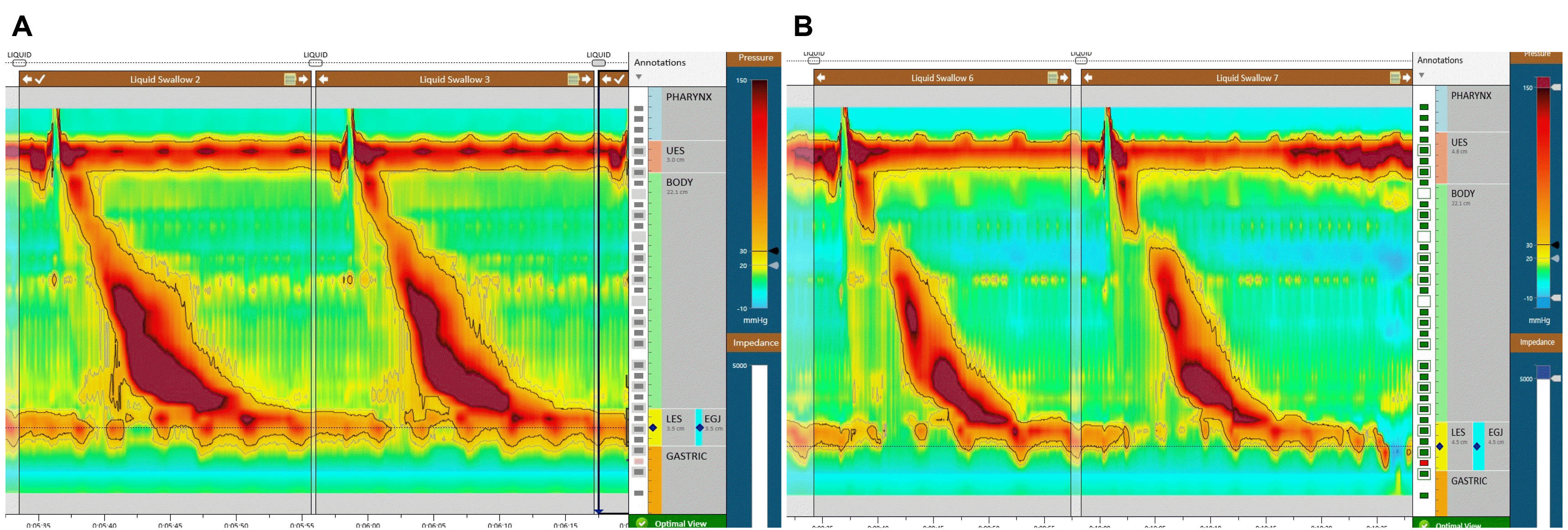
4) 임피던스 고해상도 내압 검사

고해상도 내압 검사 센서에 부착된 임피던스 센서를 통하여 내압 검사에서는 정상이나 실제로 증상이 있는 환자들의 원인을 파악하기 위한 노력이 고해상도 내압 검사법이 도입된 이후 꾸준하게 지속되었으며, 특히 식도 체부의 상부는 수의적으로 움직임이 가능한 골격근으로 이루어져 있으며, 식도 체부의 하부는 불수의근인 평활근으로 이루어져 있는데, 그 경계 부위, transition zone의 범위가 넓을수록 삼킴곤란이 있을 것으로 추측하였기 때문이다.49,50 하지만 여러 연구들에서 이러한 경계부위와 삼킴곤란의 정도를 명확하게 연결시키는 데는 많은 어려움이 있었다.51,52 최근 연구들에서는 임피던스 검사를 활용하여 비효율적 식도 운동(ineffective esoph-ageal motility)이나 시카고 기준에서는 정상으로 진단되었으나 삼킴곤란이 있는 환자들에서 의미 있는 임상 지표를 알아내기 위한 연구들이 이루어지고 있다.53,54 특히 임피던스 적분지수의 삼킴 전후 비율을 통한 연구에서 삼킴곤란 설문을 통한 삼킴곤란 정도와 의미 있는 결과를 얻었다는 보고가 있었고, 임피던스 값의 뒤집은 전도도(conductance) 값의 적분을통한 연구에서 바륨조영술과 유의한 일치율의 결과를 얻었다는 보고가 있다.54-57 이는 식도 내 식괴 이동(bolus transit)의측정에 있어서 임피던스 지표가 인체에 방사능 노출이 되는바륨조영술을 대신할 수 있음을 암시하고 있다. 실제 임상적적용을 위해서 임피던스를 이용한 지표들을 통해 식도 내 식괴 이동의 불완전성의 진단기준을 결정할 수 있는 추가적인연구가 필요할 것이다.

5) 시카고 기준 4차 개정판

최근 시카고 4차 개정판이 나와서 소개되고 있는데, 기존 3차 개정판과 크게 달라진 것으로는 일부 연구 목적으로만 사용되었던 provocation test가 정식 프로토콜로 채택이 되었으며, 적분된 이완압력이 카테터 및 시스템 제조 회사에 따라 정상값이 다름을 인정하여, Medtronic 사의 제품은 앙와위에서 15 mmHg가 기준이지만, Diversatek이나 Laborie 사 제품은 22 mmHg를 기준으로 제시하고 있고 upright에서는 Medtronic 사 제품은 12 mmHg, Diversatek/Laborie는 15 mmHg로 제시하고 있다.5 특히, 위식도 접합부 출구 폐쇄 진단에 있어서 4차 개정판에서는 앙와위 및 입위 양 자세에서 모두 적분된 이완압력이 높은 경우로 정의하고 있다.58 또한, 식도 하부 경련도 삼킴곤란 또는 비심인성 흉통 등의 증상이 있으면서 고해상도 내압 검사에서 정상 적분된 이완압력 및 20% 이상 삼킴에서 DL이 4.5초 이하로 짧아진 것으로 정의하고 있다.5 또한 과거 Jackhammer 식도라고 불리었던 과수축성(hypercontractile) 식도는 정상 적분된 이완압력 및 20% 이상의 과수축이 있으면서 삼킴곤란 또는 비심인성 흉통이 있는 경우로 정의하고 있다.5

2. EndoFLIP

임피던스 면적측정(impedance planimetry)을 이용한 식도 기능 연구는 1990년대부터 실험적으로 이루어져 왔으나, 실제 임상에서 그 이용이 가능해진 것은 Gregersen 등에 의해서 EndoFLIP 이라는 형태로 발전된 이후이다.59,60 이를 상용화한 것이 EndoFLIP이며 이를 임상에서 활용한 연구는 2010년대 초반부터 시카고 노스웨스턴 그룹에서 시작하였다.61 특히, EndoFLIP에서는 임피던스 면적 측정을 바탕으로 내강의 단면적 및 압력을 측정할 수 있고, 이를 바탕으로 하여 내강 또는 조임근의 팽창능을 측정할 수 있다.62 흔히 식도에서 쓰이는 EndoFLIP 카테터는 8 cm (16개의 임피던스 센서가 0.5 cm 간격 배열)와 16 cm (16개의 임피던스 센서가1 cm 간격 배열) 두 종류가 있는데, 8 cm 카테터는 위식도경계부의 팽창능과 단면적 측정이 가능하고, 16 cm 카테터는그 외 추가적으로 식도내강의 2차 연동운동도 파악이 가능하다.62 식도 내강에 식괴가 놓였을 때 정상적인 식도라면 식도하부로 내려보내는 앞방향(antegrade)으로 2차 연동운동이일어나겠지만, 아칼라시아처럼 정상적인 식도 운동이 소실된경우라면 오히려 역방향(retrograde)으로 2차 연동운동이 일어날 수 있다.62-64 8 cm 카테터는 위식도경계부에 풍선 중간부위를 놓고 20 mL부터 시작해서, 30초 간격으로 30, 40, 50 mL까지 풍선에 액체를 주입하여 관찰하고, 16 cm 카테터는 풍선이 위식도경계부를 지나친 후 2-3개의 센서가 위식도경계부 아래에 놓이게 한 후, 30 mL부터 시작해서 60초 간격으로40, 50, 60, 70 mL까지 주입하여 관찰하는 것을 권고하고 있다.62
특히, 아칼라시아에서 적분된 이완압력이 정상이거나 오히려 낮은 경우에 있어 EndoFLIP이 효과적임이 알려져있다.65-67 또한 최근에 EndoFLIP을 이용하여 2차 연동운동의 파형을 분석한 panometry에서는 삼킴곤란으로 내원한 환자들 중에서 고해상도 내압 검사에서 정상이었던 환자의 50%에서 비정상 EndoFLIP 소견을 보여서 진단이 가능하였다는 연구 결과도 있다.67 최근 연구들을 종합하면 팽창능을 기준으로, 2 mm2/mmHg 이하라면 비정상으로 간주되며 2-3 mm2/mmHg 사이는 경계부, 3 mm2/mmHg 이상은 정상으로 여겨진다.62 또한, 아칼라시아 중에서도 앞방향 2차 연동운동 등이 일부 살아있는 경우에는 POEM 등 시술 이후 식도체부 운동능이 일부 회복되거나 증상 개선의 여지가 좀 더 있을 것으로 예측하는 일부 소규모 연구가 있으나, 이에 대해서는 대규모의 전향적 연구가 필요할 것으로 보인다.63

결 론

최근 제시되고 있는 식도 운동 질환 검사법에 대한 충분한 이해를 통하여 삼킴곤란을 주소로 내원하는 환자들을 진단한다면 그에 맞는 올바른 치료를 할 수 있을 것으로 생각된다. 요약하자면 고해상도 식도 내압 검사와 시카고 분류 4.0 버전에 새로 추가된 유발검사 및 EndoFLIP의 사용은 식도 운동 장애의 진단 및 이해를 넓혀주었다. 또한, 마약성 식도 등 기존에 비교적 잘 알려지지 않은 질병에 대한 이해를 통하여 감별 진단에 도움이 될 것으로 보인다. 식도 내 식괴 이동(bolus transit) 고해상도 내압 검사의 임피던스 지표 연구가 진행 중이며, 추후 연구 결과가 기대된다.

Notes

Financial support

None.

Conflict of interest

None.

REFERENCES

1. Kahrilas PJ, Ghosh SK, Pandolfino JE. 2008; Esophageal motility disorders in terms of pressure topography: the Chicago Classification. J Clin Gastroenterol. 42:627–635. DOI: 10.1097/MCG.0b013e31815ea291. PMID: 18364587. PMCID: PMC2895002.
pmid pmc
2. Clouse RE, Staiano A, Alrakawi A, Haroian L. 2000; Application of topographical methods to clinical esophageal manometry. Am J Gastroenterol. 95:2720–2730. DOI: 10.1111/j.1572-0241.2000.03178.x. PMID: 11051340.
crossref pmid
3. Pandolfino JE, Ghosh SK, Rice J, Clarke JO, Kwiatek MA, Kahrilas PJ. 2008; Classifying esophageal motility by pressure topography characteristics: a study of 400 patients and 75 controls. Am J Gastroenterol. 103:27–37. DOI: 10.1111/j.1572-0241.2007.01532.x. PMID: 17900331.
crossref pmid
4. Kahrilas PJ, Bredenoord AJ, Fox M, et al. 2015; The Chicago Classification of esophageal motility disorders, v3.0. Neurogastroenterol Motil. 27:160–174. DOI: 10.1111/nmo.12477. PMID: 25469569. PMCID: PMC4308501.
crossref pmid
5. Yadlapati R, Kahrilas PJ, Fox MR, et al. 2021; Esophageal motility disorders on high-resolution manometry: Chicago classification version 4.0©. Neurogastroenterol Motil. 33:e14058. DOI: 10.1111/nmo.14058. PMID: 33373111.
6. Donnan EN, Pandolfino JE. 2020; EndoFLIP in the esophagus: assessing sphincter function, wall stiffness, and motility to guide treatment. Gastroenterol Clin North Am. 49:427–435. DOI: 10.1016/j.gtc.2020.04.002. PMID: 32718562. PMCID: PMC7387752.
pmid
7. Acharya S, Halder S, Carlson DA, et al. 2021; Assessment of esophageal body peristaltic work using functional lumen imaging probe panometry. Am J Physiol Gastrointest Liver Physiol. 320:G217–G226. DOI: 10.1152/ajpgi.00324.2020. PMID: 33174457.
crossref
8. Hirano I, Pandolfino JE, Boeckxstaens GE. 2017; Functional lumen imaging probe for the management of esophageal disorders: expert review from the clinical practice updates Committee of the AGA Institute. Clin Gastroenterol Hepatol. 15:325–334. DOI: 10.1016/j.cgh.2016.10.022. PMID: 28212976. PMCID: PMC5757507.
crossref pmid pmc
9. Fox MR, Bredenoord AJ. 2008; Oesophageal high-resolution manometry: moving from research into clinical practice. Gut. 57:405–423. DOI: 10.1136/gut.2007.127993. PMID: 17895358.
crossref pmid
10. Ghosh SK, Pandolfino JE, Rice J, Clarke JO, Kwiatek M, Kahrilas PJ. 2007; Impaired deglutitive EGJ relaxation in clinical esophageal manometry: a quantitative analysis of 400 patients and 75 controls. Am J Physiol Gastrointest Liver Physiol. 293:G878–G885. DOI: 10.1152/ajpgi.00252.2007. PMID: 17690172.
crossref
11. Pandolfino JE, Kwiatek MA, Nealis T, Bulsiewicz W, Post J, Kahrilas PJ. 2008; Achalasia: a new clinically relevant classification by high-resolution manometry. Gastroenterology. 135:1526–1533. DOI: 10.1053/j.gastro.2008.07.022. PMID: 18722376. PMCID: PMC2894987.
crossref pmid pmc
12. Ghoshal UC, Rangan M. 2011; A review of factors predicting outcome of pneumatic dilation in patients with achalasia cardia. J Neurogastroenterol Motil. 17:9–13. DOI: 10.5056/jnm.2011.17.1.9. PMID: 21369487. PMCID: PMC3042226.
crossref pmid pmc
13. Jung HK, Hong SJ, Lee OY, et al. 2020; 2019 Seoul consensus on esophageal achalasia guidelines. J Neurogastroenterol Motil. 26:180–203. DOI: 10.5056/jnm20014. PMID: 32235027. PMCID: PMC7176504.
crossref pmid pmc
14. Benjamin SB, Gerhardt DC, Castell DO. 1979; High amplitude, peristaltic esophageal contractions associated with chest pain and/or dysphagia. Gastroenterology. 77:478–483. DOI: 10.1016/0016-5085(79)90008-8. PMID: 456842.
crossref pmid
15. Drane WE, Johnson DA, Hagan DP, Cattau EL Jr. 1987; "Nutcracker" esophagus: diagnosis with radionuclide esophageal scintigraphy versus manometry. Radiology. 163:33–37. DOI: 10.1148/radiology.163.1.3823454. PMID: 3823454.
crossref pmid
16. Jung KW, Jung HY, Yoon IJ, et al. 2010; Basal and residual lower esophageal pressures increase in old age in classic achalasia, but not vigorous achalasia. J Gastroenterol Hepatol. 25:1452–1455. DOI: 10.1111/j.1440-1746.2010.06298.x. PMID: 20659237.
crossref
17. Jung KW, Jung HY, Myung SJ, et al. 2015; The effect of age on the key parameters in the Chicago classification: a study using high-resolution esophageal manometry in asymptomatic normal individuals. Neurogastroenterol Motil. 27:246–257. DOI: 10.1111/nmo.12482. PMID: 25521290.
crossref pmid
18. Agrawal A, Hila A, Tutuian R, Mainie I, Castell DO. 2006; Clinical relevance of the nutcracker esophagus: suggested revision of criteria for diagnosis. J Clin Gastroenterol. 40:504–509. DOI: 10.1097/00004836-200607000-00008. PMID: 16825932.
crossref pmid
19. Jung KW, Jung HY, Yoon IJ, et al. 2010; New diagnostic criteria for nutcracker esophagus using conventional water-perfused manometry: a comparison between nutcracker esophagus with and without gastroesophageal reflux disease. J Gastroenterol Hepatol. 25:1239–1243. DOI: 10.1111/j.1440-1746.2010.06301.x. PMID: 20594250.
crossref
20. Roman S, Pandolfino JE, Chen J, Boris L, Luger D, Kahrilas PJ. 2012; Phenotypes and clinical context of hypercontractility in high-resolution esophageal pressure topography (EPT). Am J Gastroenterol. 107:37–45. DOI: 10.1038/ajg.2011.313. PMID: 21931377. PMCID: PMC3641840.
crossref pmid
21. Xiao Y, Carlson DA, Lin Z, Alhalel N, Pandolfino JE. 2018; Jackhammer esophagus: assessing the balance between prepeak and postpeak contractile integral. Neurogastroenterol Motil. 30:e13262. DOI: 10.1111/nmo.13262. PMID: 29193439. PMCID: PMC6317707.
crossref
22. Xiao Y, Carlson DA, Lin Z, Pandolfino JE. 2020; Chaotic peak propagation in patients with Jackhammer esophagus. Neurogastroenterol Motil. 32:e13725. DOI: 10.1111/nmo.13725. PMID: 31532046. PMCID: PMC7082896.
crossref
23. de Bortoli N, Gyawali PC, Roman S, et al. 2021; Hypercontractile esophagus from pathophysiology to management: proceedings of the pisa symposium. Am J Gastroenterol. 116:263–273. DOI: 10.14309/ajg.0000000000001061. PMID: 33273259.
crossref pmid
24. Pandolfino JE, Roman S, Carlson D, et al. 2011; Distal esophageal spasm in high-resolution esophageal pressure topography: defining clinical phenotypes. Gastroenterology. 141:469–475. DOI: 10.1053/j.gastro.2011.04.058. PMID: 21679709. PMCID: PMC3626105.
crossref pmid pmc
25. Lin Z, Kahrilas PJ, Roman S, Boris L, Carlson D, Pandolfino JE. 2012; Refining the criterion for an abnormal integrated relaxation pressure in esophageal pressure topography based on the pattern of esophageal contractility using a classification and regression tree model. Neurogastroenterol Motil. 24:e356–e363. DOI: 10.1111/j.1365-2982.2012.01952.x. PMID: 22716041. PMCID: PMC3616504.
crossref
26. Herregods TV, Roman S, Kahrilas PJ, Smout AJ, Bredenoord AJ. 2015; Normative values in esophageal high-resolution manometry. Neurogastroenterol Motil. 27:175–187. DOI: 10.1111/nmo.12500. PMID: 25545201.
crossref pmid
27. Rengarajan A, Rogers BD, Wong Z, et al. 2020; Oct. 31. High-resolution manometry thresholds and motor patterns among asymptomatic individuals. Clin Gastroenterol Hepatol. [Epub ahead of print]. DOI: 10.1016/j.cgh.2020.10.052. PMID: 33144149.
crossref
28. Triggs JR, Carlson DA, Beveridge C, et al. 2019; Upright integrated relaxation pressure facilitates characterization of esophagogastric junction outflow obstruction. Clin Gastroenterol Hepatol. 17:2218–2226.e2. DOI: 10.1016/j.cgh.2019.01.024. PMID: 30708108. PMCID: PMC6663640.
pmid pmc
29. Shaker A, Stoikes N, Drapekin J, Kushnir V, Brunt LM, Gyawali CP. 2013; Multiple rapid swallow responses during esophageal high-resolution manometry reflect esophageal body peristaltic reserve. Am J Gastroenterol. 108:1706–1712. DOI: 10.1038/ajg.2013.289. PMID: 24019081. PMCID: PMC4091619.
crossref pmid pmc
30. Carlson DA, Roman S. 2018; Esophageal provocation tests: are they useful to improve diagnostic yield of high resolution manometry? Neurogastroenterol Motil. 30:e13321. DOI: 10.1111/nmo.13321. PMID: 29603510.
crossref
31. Min YW, Shin I, Son HJ, Rhee PL. 2015; Multiple rapid swallow maneuver enhances the clinical utility of high-resolution manometry in patients showing ineffective esophageal motility. Medicine (Baltimore). 94:e1669. DOI: 10.1097/MD.0000000000001669. PMID: 26448010. PMCID: PMC4616734.
crossref
32. Blonski W, Hila A, Jain V, Freeman J, Vela M, Castell DO. 2007; Impedance manometry with viscous test solution increases detection of esophageal function defects compared to liquid swallows. Scand J Gastroenterol. 42:917–922. DOI: 10.1080/00365520701245702. PMID: 17613920.
crossref pmid
33. Wong U, Person EB, Castell DO, von Rosenvinge E, Raufman JP, Xie G. 2018; Improving high-resolution impedance manometry using novel viscous and super-viscous substrates in the supine and upright positions: a pilot study. J Neurogastroenterol Motil. 24:570–576. DOI: 10.5056/jnm18010. PMID: 30122029. PMCID: PMC6175560.
crossref pmid pmc
34. Araujo IK, Roman S, Napoléon M, Mion F. 2020; Diagnostic yield of adding solid food swallows during high-resolution manometry in esophageal motility disorders. Neurogastroenterol Motil. e14060. DOI: 10.1111/nmo.14060. PMID: 33314459.
crossref
35. Ang D, Hollenstein M, Misselwitz B, et al. 2017; Rapid drink challenge in high-resolution manometry: an adjunctive test for detection of esophageal motility disorders. Neurogastroenterol Motil. 29(1):DOI: 10.1111/nmo.12902. PMID: 27420913.
crossref
36. Marin I, Caballero N, Guarner-Argente C, Serra J. 2018; Rapid drink challenge test for the clinical evaluation of patients with Achalasia. Neurogastroenterol Motil. 30:e13438. DOI: 10.1111/nmo.13438. PMID: 30101425.
crossref
37. Hsing LC, Jung KW. 2020; Role of the rapid drink challenge test in esophageal motility disorder diagnosis. J Neurogastroenterol Motil. 26:167–168. DOI: 10.5056/jnm20035. PMID: 32235024. PMCID: PMC7176506.
crossref pmid pmc
38. Krause AJ, Su H, Triggs JR, et al. 2020 ; Oct. 11. Multiple rapid swallows and rapid drink challenge in patients with esophagogastric junction outflow obstruction on high-resolution manometry. Neurogastroenterol Motil. [Epub ahead of print]. DOI: 10.1111/nmo.14000. PMID: 33043557. PMCID: PMC7902305.
crossref
39. Wood JD, Galligan JJ. 2004; Function of opioids in the enteric nervous system. Neurogastroenterol Motil. 16 Suppl 2:17–28. DOI: 10.1111/j.1743-3150.2004.00554.x. PMID: 15357848.
crossref pmid
40. Kraichely RE, Arora AS, Murray JA. 2010; Opiate-induced oesophageal dysmotility. Aliment Pharmacol Ther. 31:601–606. DOI: 10.1111/j.1365-2036.2009.04212.x. PMID: 20003176. PMCID: PMC3092396.
crossref pmid
41. Ratuapli SK, Crowell MD, DiBaise JK, et al. 2015; Opioid-induced esophageal dysfunction (OIED) in patients on chronic opioids. Am J Gastroenterol. 110:979–984. DOI: 10.1038/ajg.2015.154. PMID: 26032150.
crossref
42. Ravi K, Murray JA, Geno DM, Katzka DA. 2016; Achalasia and chronic opiate use: innocent bystanders or associated conditions? Dis Esophagus. 29:15–21. DOI: 10.1111/dote.12291. PMID: 25604060.
crossref pmid
43. Jung KW, Kraichely RE, Arora AS, Katzka DA, Romero Y, Murray JA. 2011; Manometric characteristics of opioid esophageal dysmotility disorder by high-resolution manometry. Gastroenterology. 140:S–229. DOI: 10.1016/S0016-5085(11)60925-6.
crossref
44. Kim GH, Jung KW. 2019; Emerging issues in esophageal motility diseases. Korean J Gastroenterol. 73:322–326. DOI: 10.4166/kjg.2019.73.6.322. PMID: 31234622.
crossref
45. Jung KW, Myung SJ, Jung HY. 2012; A patient with dysphagia associated with opioid medication. J Neurogastroenterol Motil. 18:220–221. DOI: 10.5056/jnm.2012.18.2.220. PMID: 22523734. PMCID: PMC3325310.
crossref pmid pmc
46. Kim GH, Jung KW. 2019; The role of opioids and alcohol in the development of achalasia type III and esophagogastric junction outflow obstruction. J Neurogastroenterol Motil. 25:177–178. DOI: 10.5056/jnm19047. PMID: 30982237. PMCID: PMC6474699.
crossref pmid pmc
47. Kim GH, Jung KW, Jung HY, et al. 2019; Superior clinical outcomes of peroral endoscopic myotomy compared with balloon dilation in all achalasia subtypes. J Gastroenterol Hepatol. 34:659–665. DOI: 10.1111/jgh.14616. PMID: 30695124.
crossref pmid
48. Murray JA, Ledlow A, Launspach J, Evans D, Loveday M, Conklin JL. 1995; The effects of recombinant human hemoglobin on esophageal motor functions in humans. Gastroenterology. 109:1241–1248. DOI: 10.1016/0016-5085(95)90584-7. PMID: 7557091.
pmid
49. Ghosh SK, Janiak P, Fox M, Schwizer W, Hebbard GS, Brasseur JG. 2008; Physiology of the oesophageal transition zone in the presence of chronic bolus retention: studies using concurrent high resolution manometry and digital fluoroscopy. Neurogastroenterol Motil. 20:750–759. DOI: 10.1111/j.1365-2982.2008.01129.x. PMID: 18422907.
crossref pmid
50. Pohl D, Ribolsi M, Savarino E, et al. 2008; Characteristics of the esophageal low-pressure zone in healthy volunteers and patients with esophageal symptoms: assessment by high-resolution manometry. Am J Gastroenterol. 103:2544–2549. DOI: 10.1111/j.1572-0241.2008.02062.x. PMID: 18684179.
crossref pmid
51. Ghosh SK, Pandolfino JE, Kwiatek MA, Kahrilas PJ. 2008; Oesophageal peristaltic transition zone defects: real but few and far between. Neurogastroenterol Motil. 20:1283–1290. DOI: 10.1111/j.1365-2982.2008.01169.x. PMID: 18662328. PMCID: PMC2886597.
crossref pmid pmc
52. Jung KW, Jung HY, Romero Y, Katzka D, Murray JA. 2011; Impact of display alternatives in the determination of bolus handling: a study using high-resolution manometry with impedance. Am J Gastroenterol. 106:1854–1857. DOI: 10.1038/ajg.2011.233. PMID: 21979209.
crossref pmid
53. Rommel N, Van Oudenhove L, Tack J, Omari TI. 2014; Automated impedance manometry analysis as a method to assess esophageal function. Neurogastroenterol Motil. 26:636–645. DOI: 10.1111/nmo.12308. PMID: 24447538.
crossref pmid
54. Singendonk MJ, Lin Z, Scheerens C, et al. 2019; High-resolution impedance manometry parameters in the evaluation of esophageal function of non-obstructive dysphagia patients. Neurogastroenterol Motil. 31:e13505. DOI: 10.1111/nmo.13505. PMID: 30426609.
crossref
55. Lin Z, Nicodème F, Lin CY, et al. 2014; Parameters for quantifying bolus retention with high-resolution impedance manometry. Neurogastroenterol Motil. 26:929–936. DOI: 10.1111/nmo.12346. PMID: 24750336. PMCID: PMC4120956.
crossref
56. Carlson DA, Beveridge CA, Lin Z, et al. 2018; Improved assessment of bolus clearance in patients with achalasia using high-resolution impedance manometry. Clin Gastroenterol Hepatol. 16:672–680.e1. DOI: 10.1016/j.cgh.2017.11.019. PMID: 29155168. PMCID: PMC5911237.
crossref pmid pmc
57. Gong EJ, Choi K, Jung KW, et al. 2020; New parameter for quantifying bolus transit with high-resolution impedance manometry: a comparison with simultaneous esophagogram. Neurogastroenterol Motil. 32:e13847. DOI: 10.1111/nmo.13847. PMID: 32299145.
crossref
58. Yadlapati R, Pandolfino JE, Fox MR, Bredenoord AJ, Kahrilas PJ. 2021; What is new in Chicago Classification version 4.0? Neurogastroenterol Motil. 33:e14053. DOI: 10.1111/nmo.14053. PMID: 33340190.
crossref
59. Rao SS, Gregersen H, Hayek B, Summers RW, Christensen J. 1996; Unexplained chest pain: the hypersensitive, hyperreactive, and poorly compliant esophagus. Ann Intern Med. 124:950–958. DOI: 10.7326/0003-4819-124-11-199606010-00002. PMID: 8624062.
crossref pmid
60. Gregersen H, Vinter-Jensen L, Juhl CO, Dajani EZ. 1996; Impedance planimetric characterization of the distal oesophagus in the Goettingen minipig. J Biomech. 29:63–68. DOI: 10.1016/0021-9290(95)00016-X. PMID: 8839018.
crossref pmid
61. Kwiatek MA, Pandolfino JE, Hirano I, Kahrilas PJ. 2010; Esophagogastric junction distensibility assessed with an endoscopic functional luminal imaging probe (EndoFLIP). Gastrointest Endosc. 72:272–278. DOI: 10.1016/j.gie.2010.01.069. PMID: 20541755. PMCID: PMC3019759.
crossref pmid pmc
62. Savarino E, di Pietro M, Bredenoord AJ, et al. 2020; Use of the functional lumen imaging probe in clinical esophagology. Am J Gastroenterol. 115:1786–1796. DOI: 10.14309/ajg.0000000000000773. PMID: 33156096.
crossref pmid
63. Carlson DA, Lin Z, Kahrilas PJ, et al. 2015; The functional lumen imaging probe detects esophageal contractility not observed with manometry in patients with achalasia. Gastroenterology. 149:1742–1751. DOI: 10.1053/j.gastro.2015.08.005. PMID: 26278501. PMCID: PMC4663149.
pmid pmc
64. Carlson DA, Lin Z, Rogers MC, Lin CY, Kahrilas PJ, Pandolfino JE. 2015; Utilizing functional lumen imaging probe topography to evaluate esophageal contractility during volumetric distention: a pilot study. Neurogastroenterol Motil. 27:981–989. DOI: 10.1111/nmo.12572. PMID: 25898916. PMCID: PMC4478241.
crossref pmid pmc
65. Teitelbaum EN, Soper NJ, Pandolfino JE, et al. 2015; Esophagogastric junction distensibility measurements during Heller myotomy and POEM for achalasia predict postoperative symptomatic outcomes. Surg Endosc. 29:522–528. DOI: 10.1007/s00464-014-3733-1. PMID: 25055891. PMCID: PMC4343529.
crossref pmid
66. Ponds FA, Bredenoord AJ, Kessing BF, Smout AJ. 2017; Esophagogastric junction distensibility identifies achalasia subgroup with manometrically normal esophagogastric junction relaxation. Neurogastroenterol Motil. 29(1):DOI: 10.1111/nmo.12908. PMID: 27458129.
crossref
67. Carlson DA, Kahrilas PJ, Lin Z, et al. 2016; Evaluation of esophageal motility utilizing the functional lumen imaging probe. Am J Gastroenterol. 111:1726–1735. DOI: 10.1038/ajg.2016.454. PMID: 27725650. PMCID: PMC5224528.
crossref pmid pmc

Fig. 1
A case of opioid-induced esophageal dysmotility. A 48-year-old male attended the clinic with complaints of persistent dysphagia, noting that symptoms onset approximately 3 year prior. His upper endoscopy with esophageal biopsy produced non-specific findings. (A) His initial esophageal manometry showed a median integrated relaxation pressure (IRP) of 21 mmHg and a mean distal contractile integral (DCI) of 6,269 mmHg․s․cm. Four out of 10 swallows showed a DCI of more than 8,000 mmHg․s․cm. Based on the manometry findings, an initial suspected diagnosis of hypercontractile esophagus, including jackhammer esophagus, was given. However, the patient indicated that he had been using a 5 ug buprenorphine patch for 3 years to treat chronic back pain. Therefore, opioid-induced esophageal dysmotility disorder was presumed, and halting all opioid medication was advised. (B) His dysphagia disappeared after stopping the opioid medication. His 3-week opioid cessation follow-up manometry showed a median IRP of 17 mmHg and a mean DCI of 4,988 mmHg․s․cm. Just one in 10 swallows showed more than 8,000 mmHg․s․cm, and manometry findings were within normal finding.
kjg-77-2-64-f1.tif
TOOLS
Similar articles