Journal List > Korean J Gastroenterol > v.77(4) > 1146791

Successful Medical Treatment for Peritonitis Resulting from a Large Pyogenic Liver Abscess Rupture

Abstract

The rupture of a pyogenic liver abscess (PLA) with peritonitis is a rare occurrence but a surgical emergency with a high mortality rate in the case of gas-forming PLA. Rare cases of ruptured PLA that recovered completely with only medical treatment have been reported. This paper reports a case of a large PLA rupture with peritonitis. In this case, surgical intervention was too risky because of the patient’s age and poor general condition. The patient recovered fully with appropriate antibiotic therapy and sufficient percutaneous drainage. Therefore, medical treatment may be considered an alternative option in cases of a ruptured large PLA with peritonitis if surgical intervention is too risky.

INTRODUCTION

A pyogenic liver abscess (PLA) is a space-occupying lesion in the liver caused by a bacterial infection.1 The annual incidence of PLA is estimated to be 2.3 cases per 100,000 population and is higher among men than women.2 PLA is an important clinical problem with a significant mortality rate, ranging from 8% to 31% in the 1990s. Over the past few decades, the mortality associated with PLA has decreased gradually because of highly effective broad-spectrum antibiotics and the advent of cross-sectional CT and ultrasound imaging.3 In general, the treatment of PLA includes antibiotic therapy and percutaneous or surgical drainage. A rupture of PLA with peritonitis is rare but has a high mortality rate in the case of gas-forming PLA.4,5 In a retrospective study conducted in Korea, the prevalence of PLA rupture was 3.8%.6 The rupture of PLA is considered a surgical emergency. On the other hand, rare cases of ruptured PLA have recovered successfully with only medical treatment. The authors reported a similar case in 2018. Moreover, a 72-year-old patient with ruptured PLA was recently reported in Japan.7,8 Here, this paper presents a rare case of successful medical treatment for peritonitis resulting from a larger PLA rupture.

CASE REPORT

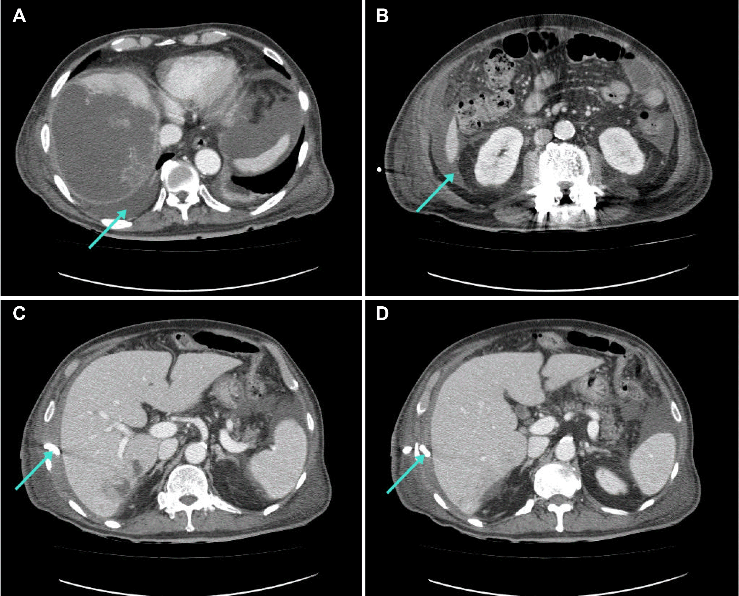
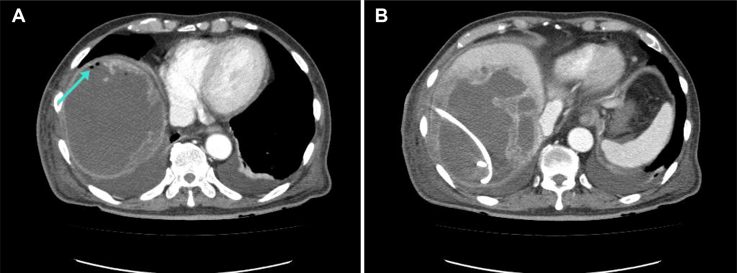
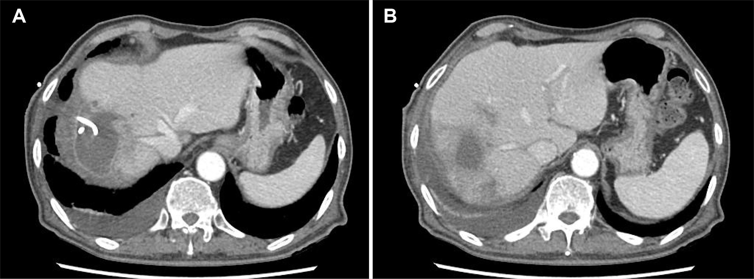
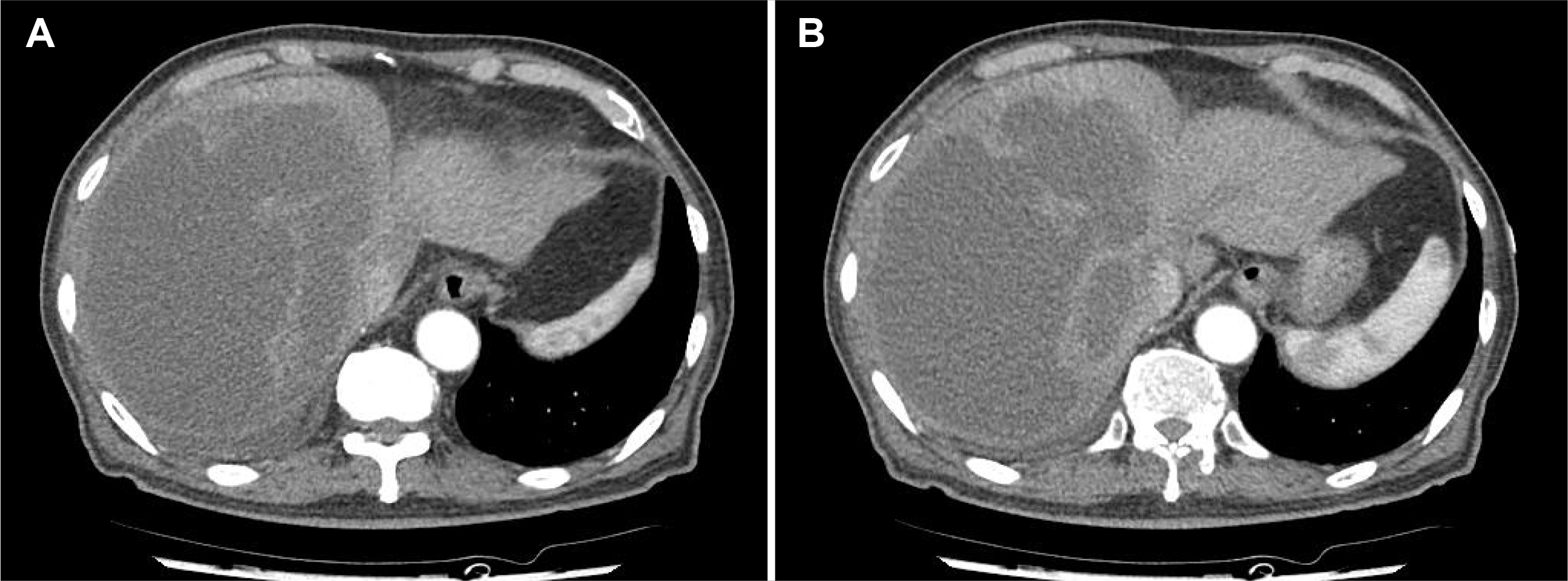
An 85-year-old man visited the Seoul Paik Hospital because of general weakness and right upper quadrant abdominal pain that began 10 days prior. He was diagnosed with sigmoid colon cancer and underwent a laparoscopic anterior resection 10 years earlier. A physical examination revealed a blood pressure of 132/78 mmHg, a body temperature of 36.6℃, and a heart rate of 90 beats/min. The laboratory data revealed a hemoglobin level of 9.1 g/dL, white blood cell count of 9,470/mm3, platelet count of 144,000/mm3, AST of 57 U/L, ALT of 73 U/L, ALP of 250 U/L, albumin of 2.7 g/dL, PT of 15.3 seconds, total bilirubin level of 1.54 mg/dL, CRP of 19.4 mg/dL, and procalcitonin level of 2.69 ng/mL. Abdominal contrast-enhanced CT showed a 17 cm-sized liver abscess with internal septa in the right lobe (Fig. 1). The patient underwent ultrasound-guided percutaneous catheter drainage (PCD) and empirical antibiotic therapy. On hospital day 2, the patient complained of abdominal pain with increased severity. A physical examination revealed abdominal tenderness and rebound tenderness of the entire abdomen, with a body temperature of 38.5°C. An abdominal CT scan showed leakage of the liver abscess in the peritoneal cavity, pleural space, and pararenal space (Fig. 2A, B). The PCD tip was located in the perihepatic space, not the abscess (Fig. 2C, D). Surgical treatment was considered risky because of the patient's age and performance status (European Cooperative Oncology Group scale 4). The PCD was repositioned, and imipenem/cilastatin was maintained as empirical antibiotics. Consecutive drained pus cultures were positive for Klebsiella pneumoniae (K. pneumoniae), and the strain was sensitive to imipenem. Follow-up CT showed that the abscess decreased in size from 17 cm to 13 cm (Fig. 3). The patient’s abdominal pain and tenderness improved as the PCD and intravenous antibiotics were continued. As observed through the blood test results, the levels of CRP and procalcitonin decreased gradually. On hospital day 44, an abdominal CT revealed a significant decrease in abscess size and reduced fluid collection in the peritoneal cavity and pararenal space (Fig. 4). On hospital day 55, the PCD was removed, and the patient had recovered fully from peritonitis and PLA.
DISCUSSION

The common pathogens of PLA are Escherichia coli (E. coli), K. pneumoniae, Bacteroides, Streptococci, and Staphylococci.9 Before the 1980s, E. coli was the most common pathogen causing PLA in Korea.10 On the other hand, during the past 2 decades, highly virulent strains of K. pneumoniae have emerged as the predominant cause of PLA in Asian countries, along with the United States and Europe. K. pneumoniae is associated with a higher likelihood of hematogenous spread and potential for metastatic infections.11 Compared to other bacteria, PLA infected with K. pneumoniae has a higher risk of rupture.12 In addition to a K. pneumoniae infection, diabetes mellitus, gas-forming liver abscess, large-sized abscess (>8 cm), and left lobe involvement are suggested risk factors for the spontaneous rupture of liver abscess.6 In the present case, the drained pus cultures were positive for K. pneumoniae, and the initial CT showed a 17 cm-sized liver abscess. The rupture might not have been caused by the PLA itself but by the catheter inserted initially. Air bubbles on follow-up CT may have been caused by the inserted catheter (Fig. 3A). Even if it was an iatrogenic PLA rupture, this case is meaningful in that it was treated only with antibiotics and PCD.
The rupture of PLA worsens the prognosis compared to non-ruptured PLA. According to Chung et al.10, the mean duration of hospital stay in patients with ruptured PLA was significantly longer than that in those with non-ruptured PLA. Furthermore, patients with ruptured PLA had more septic metastases, including the eyes, central nerve system, spinal cord, lungs, psoas muscle, and gluteal muscle.10 In the past, the mortality rate of the rupture of PLA was up to 43.5%, and a recent study reported a mortality rate of 25.9%.4,13 The mortality associated with the rupture of PLA is still high.
In general, non-surgical methods are preferred for the treatment of PLA. Surgical intervention is the sole option for the following patients: could not be treated by PCD, had an unsuccessful treatment, multiple abscess cavities, are believed to have a perforated abscess, or have additional abdominal pathology requiring laparotomy.14 The rupture of PLA is considered a surgical emergency. In particular, ruptures resulting in peritonitis would require urgent surgical intervention.15 Previously, the authors reported a case in which a ruptured PLA measuring 10 cm recovered completely with only medical treatment.8 In another case, ruptured PLA measuring 12 cm ×14 cm×7 cm was treated with antibiotics and PCD; however, it was localized in the neighboring tissues.16 In the present case, the abscess size was 17 cm in diameter. Although the abscess size was large and was accompanied by peritonitis with rupture, antibiotic therapy and PCD were administered instead of surgical treatment, considering the patient’s age and poor general condition. Traditionally, surgical intervention is required in large, multi-loculated abscesses and ruptures, resulting in peritonitis.17 The present patient recovered with medical treatment due to the early diagnosis of the ruptured PLA, appropriate antibiotic selection, and sufficient drainage of pus via PCD. Therefore, medical treatment can be considered in cases of a ruptured large liver abscess with peritonitis if surgical intervention is too risky.

Notes

Financial support

None.

Conflict of interest

None.

REFERENCES

1. Elia C, Bracco C, et al. erraino C. 2018; Characteristics and management of pyogenic liver abscess: a european experience. Medicine (Baltimore). 97:e0628. DOI: 10.1097/MD.0000000000010628. PMID: 29742700. PMCID: PMC5959441.
2. Abbas MT, Khan FY, Muhsin SA, Al-Dehwe B, Abukamar M, Elzouki AN. 2014; Epidemiology, clinical features and outcome of river abscess: a single reference center experience in Qatar. Oman Med J. 29:260–263. DOI: 10.5001/omj.2014.69. PMID: 25170406. PMCID: PMC4137581.
pmid pmc
3. Sohn SH, Kim KH, Park JH, Kim TN. 2016; Predictors of mortality in Korean patients with pyogenic liver abscess: a single center, retrospective study. Korean J Gastroenterol. 67:238–244. DOI: 10.4166/kjg.2016.67.5.238. PMID: 27206434.
crossref pmid
4. Chou FF, Sheen-Chen SM, Lee TY. 1995; Rupture of pyogenic liver abscess. Am J Gastroenterol. 90:767–770.
5. Thng CB, Tan YP, Shelat VG. 2018; Gas-forming pyogenic liver abscess: a world review. Ann Hepatobiliary Pancreat Surg. 22:11–18. DOI: 10.14701/ahbps.2018.22.1.11. PMID: 29536051. PMCID: PMC5845606.
crossref pmid pmc
6. Jun CH, Yoon JH, Wi JW, et al. 2015; Risk factors and clinical outcomes for spontaneous rupture of pyogenic liver abscess. J Dig Dis. 16:31–36. DOI: 10.1111/1751-2980.12209. PMID: 25385432.
crossref pmid
7. Motoyama T, Ogasawara S, Chiba T, et al. 2013; Successful non-surgical treatment of ruptured pyogenic liver abscess. Intern Med. 52:2619–2622. DOI: 10.2169/internalmedicine.52.0980. PMID: 24292751.
crossref pmid
8. Lee KJ, Ryu SH. 2018; Ruptured gas-forming pyogenic liver abscess into the peritoneal cavity treated successfully with medical treatment. Korean J Gastroenterol. 71:45–48. DOI: 10.4166/kjg.2018.71.1.45. PMID: 29361813.
crossref pmid
9. Kong H, Yu F, Zhang W, Li X. 2017; Clinical and microbiological characteristics of pyogenic liver abscess in a tertiary hospital in East China. Medicine (Baltimore). 96:e8050. DOI: 10.1097/MD.0000000000008050. PMID: 28906397. PMCID: PMC5604666.
crossref
10. Chung DR, Lee SS, Lee HR, et al. 2007; Emerging invasive liver abscess caused by K1 serotype Klebsiella pneumoniae in Korea. J Infect. 54:578–583. DOI: 10.1016/j.jinf.2006.11.008. PMID: 17175028.
crossref pmid
11. Liu Y, Wang JY, Jiang W. 2013; An increasing prominent disease of Klebsiella pneumoniae liver abscess: etiology, diagnosis, and treatment. Gastroenterol Res Pract. 2013:258514. DOI: 10.1155/2013/258514. PMID: 24194749. PMCID: PMC3806164.
12. Lübbert C, Wiegand J, Karlas T. 2014; Therapy of liver abscesses. Viszeralmedizin. 30:334–341. DOI: 10.1159/000366579. PMID: 26287275. PMCID: PMC4513824.
crossref pmid pmc
13. Desai N, Parmar N, Negi S. Savani. C Soni D. 2015; Management of ruptured liver abscess: a study of 54 cases. IJSR. 4:1651–1654.
14. Önder A, Kapan M, BöyüK A, et al. 2011; Surgical management of pyogenic liver abscess. Eur Rev Med Pharmacol Sci. 15:1182–1186.
pmid
15. Chong VH, Zainal-Abidin Z, Hassan H, Chong CF. 2010; Rare complications of pyogenic liver abscess. Singapore Med J. 51:e169–e172.
16. Zizzo M, Zaghi C, Manenti A, Luppi D, Ugoletti L, Bonilauri S. 2016; Abdominal wall abscess secondary to spontaneous rupture of pyogenic liver abscess. Int J Surg Case Rep. 25:110–113. DOI: 10.1016/j.ijscr.2016.06.026. PMID: 27351622. PMCID: PMC4925907.
crossref pmid pmc
17. Chung YF, Tan YM, Lui HF, et al. 2007; Management of pyogenic liver abscesses - percutaneous or open drainage? Singapore Med J. 48:1158–1165.
pmid

Fig. 1
Computed tomography (CT) scan of the abdomen. (A, B) Initial abdominal CT shows massive, multiple abscesses in the right lobe of the liver.
kjg-77-4-190-f1.tif
Fig. 2
Computed tomography (CT) scan of the abdomen. (A, B) On hospital day 2, a CT scan revealed rupture of the liver abscess in the peritoneal cavity, pleural space, and pararenal space (arrow). (C, D) The percutaneous catheter drainage tip is located in the perihepatic space (arrow).
kjg-77-4-190-f2.tif
Fig. 3
Computed tomography (CT) scan of the abdomen. (A, B) On hospital day 6, the CT scan showed air bubbles (arrow of A) and a decrease in abscess size.
kjg-77-4-190-f3.tif
Fig. 4
Computed tomography (CT) scan of the abdomen. (A, B) On hospital 44, the CT scan shows a significant size reduction of the abscess and diminished fluid collection.
kjg-77-4-190-f4.tif
TOOLS
Similar articles