

 PDF
PDF Citation
 Print
Print


Introduction
Alveolar rhabdomyosarcoma (ARMS) is a rare subtype of rhabdomyosarcoma (RMS), which is identified as an aggressive tumor arising in mesenchymal tissues. In general, RMS is mostly diagnosed in the pediatric population. predominantly in patients younger than 10 years of age [1]. RMS is believed to arise from cells of skeletal muscle lineage. In particular, elderly patients are rarely diagnosed with this type of tumor, which results in very poor prognosis. RMS is differentiated into three types: embryonal, alveolar, and pleomorphic. As this type of tumor occurs mostly in the pediatric population, adult RMS constitutes a very small fraction of soft tissue sarcomas, between 2% to 5%. [2] As this case consists of a very rare case of alveolar-type RMS, which especially arose in gynecologic regions, this special case of ARMS of the uterine corpus in an elderly patient can be helpful to further our understanding of this rare type of tumor.
Case report
A 90-year-old Korean woman visited our emergency department because of dyspnea and intermittent vaginal spotting. The symptoms first started with scanty vaginal spotting, which gradually increased over several months. She first visited a local practitioner for general work-up, including endometrial biopsy, which identified a malignant tumor with myogenic differentiation. The patient decided not to receive any surgical intervention because of her age and general condition. As her symptoms of abdominal distension and dyspnea increased due to compression of the mass, the patient was transferred to our hospital.
Physical examination disclosed a large mass in the pelvic and abdominal cavities, and blood analysis showed mild anemia with a hemoglobin count of 9.7 g/dL (normal range 12.0-16.0 g/dL). The C-reactive protein level was elevated to 21.7 mg/dL (normal range <0.5 mg/dL), with serum CA-125 level increased to 111.0 U/mL (normal range <35 U/mL). Aspartate aminotransferase (normal range ≤32 U/L) and alanine aminotransferase level (normal range ≤33 U/L) were in the normal range, while blood urea nitrogen increased to 38.7 (normal range 6-23 mg/dL), and creatinine increased to 1.18 (normal range 0.50-0.90 mg/dL).
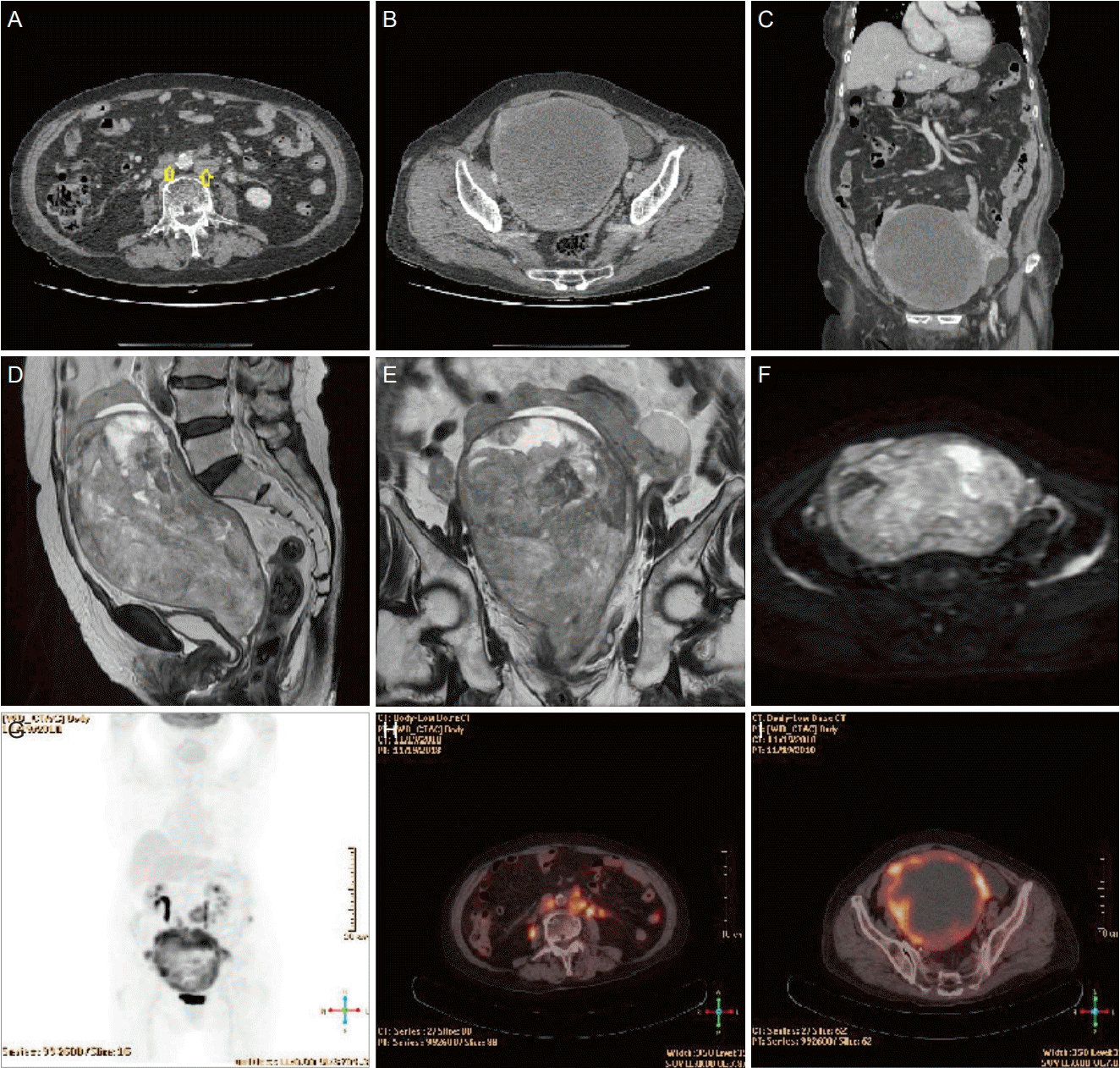
Computed tomography (CT), pelvic magnetic resonance imaging, and positron emission tomography from a previous clinic showed an approximately complex echogenic uterine mass of 19 cm in the pelvic cavity. Along with multiple enlarged aortocavals, and para-aortic lymph nodes, and suspected metastasis to the bilateral adnexa could be a clue of suspected malignancy. Based on these laboratory and imaging test results, we suspected this mass to be a possible advanced endometrial cancer, or any other types of uterine corpus malignancy (Fig. 1).
After extensive discussion with the patient and her family, written informed consent was obtained for the operation. The physicians decided to perform diagnostic laparotomy, possibly to the extent of the clinical debulking operation. Laparotomy revealed a large uterine mass with lesions highly suggestive of necrotic uterine tissue. Total hysterectomy, bilateral salpingo-oophorectomy, and para-aortic and bilateral pelvic lymphadenectomy were performed. The patient was admitted to the surgical intensive care unit for postoperative care and hemodynamic monitoring. Three days after surgery, the patient developed dyspnea. Decreased kidney function and aggravation of pulmonary edema suggested multiple organ failure. Metabolic acidosis worsened despite medical intervention and the patient expired on the eighth postoperative day.
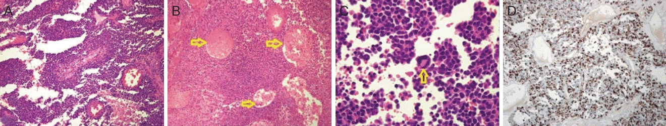
On microscopic examination, the histopathologic findings suggested a papillary growth pattern and several necrotic spots in the low-power view (Fig 2A, B). The tumor was mostly composed of poorly differentiated cells, indicating the formation of irregular alveolar spaces (Fig. 2B, marked with yellow arrows). Multinucleated giant cells with frequent mitotic changes within the nucleus were observed in the alveolar spaces (Fig. 2C). These findings are typical for ARMS. Immunoperoxidase staining was positive for desmin, indicating rhabdomyoblasts (Fig. 2D) [3]. The department of pathology has concluded that this specimen resulted in CK (-); therefore, no further evaluation on keratin staining was required.
Discussion
RMS has four different varieties, including alveolar (with botryoid subtype), embryonal, and pleomorphic variants [4]. Embryonal RMS is the most common type of RMS in the pediatric population. This type of tumor usually originates in skeletal tissues, especially in extremities [2]. The morphological characteristics of each type of RMS vary according to their type. Histopathologically, embryonal RMS appears as edematous and hypocellular with a typical hypercellular zone with rarely observed rhabdomyoblasts. Pleomorphic RMS contains hyperchromatic cells with scant eosinophilic cytoplasm and significant variations in nuclear size and shape. As a microscopic result of this case report, the ARMS is composed of small round hyperchromatic cells separated by fibrous septa, appearing as alveolar structure. This type of tumor shows positive staining for desmin, myogenin, and myo-D1 with negative hormone receptor staining.
As shown in this case report, this subtype of RMS, especially occurred in elderly patients distinctly from its general nature, has a very aggressive clinical course [3]. Median progression-free survival and disease-specific survival was 9 months and 21 months, respectively. Five-year disease specific survival was only 29% [5]. It should be considered that the old age of the patient contributed towards aggravating the already aggressive clinical course of this type of tumor.
To the best of our knowledge, only a few cases of ARMS arising in the uterine corpus have been reported. with an extremely poor prognosis, resulting in mortality. As in the review by Chiarle et al. [6], in older patients, where the clinical presentation, and the rapid progression to death with widespread metastases are in line with the reported literature on pure RMS of the uterus as well as with the outcome of the ARMS of the female genitourinary tract. A recent study by Gerber et al. [7] investigated the adult patients with RMS consisting of variable subtypes over a 10-year period, which resulted in 45% of five-year overall survival for non-metastatic patients and 26% for metastatic patients, and treatment failure correlated with a poor prognosis. According to a report by Little et al. [8], patients with alveolar RMS had inferior outcome, with significantly poorer distant metastasis-free survival rates than any other subtype, with a higher incidence of lymph node metastasis. In another case of uterine ARMS, neither chemotherapy nor radiotherapy could be started before the patient died of fulminant neoplastic dissemination 17 days postoperatively [9].
Due to the paucity of cases, a standard treatment protocol or optimal treatment options have not been established in the literature for adult patients [10]. In a higher incidence pediatric population, treatment involves a multimodal approach with surgery, chemotherapy, and radiotherapy, which has improved over the decades [9]. For ARMS, debulking surgery could be considered an optimal treatment, possibly followed by adjuvant therapy. The chemotherapy regimen for the pediatric population of RMS has been well established by using combinations of multiple agents including vincristine, dactinomycins, and cyclophosphamides. However, the treatment regimen for the adult population has not yet been standardized. Adaptation of the pediatric regimen, with relatively high response rates, is widely applied to patients [5]. Although chemotherapy could first be considered a non-surgical option, we decided to perform a diagnostic laparotomy to relieve the patient from the symptoms of dyspnea, with severe abdominal distension caused by the huge pelvic mass. In addition, we evaluated her general condition and concluded that she could not tolerate systemic chemotherapy for several cycles.
In conclusion, ARMS of the uterus is an exceedingly rare disease that progresses with aggressive characteristics, thus resulting in a poor prognosis with a short period of diseasefree survival rate. Early diagnosis followed by pertinent treatment is mandatory. The standard treatment options and fixed regimen are unclear for systemic chemotherapy. Hence, further studies should be performed on adult RMS to offer patients the best chance of survival.
 XML Download